Cell lines & reagents
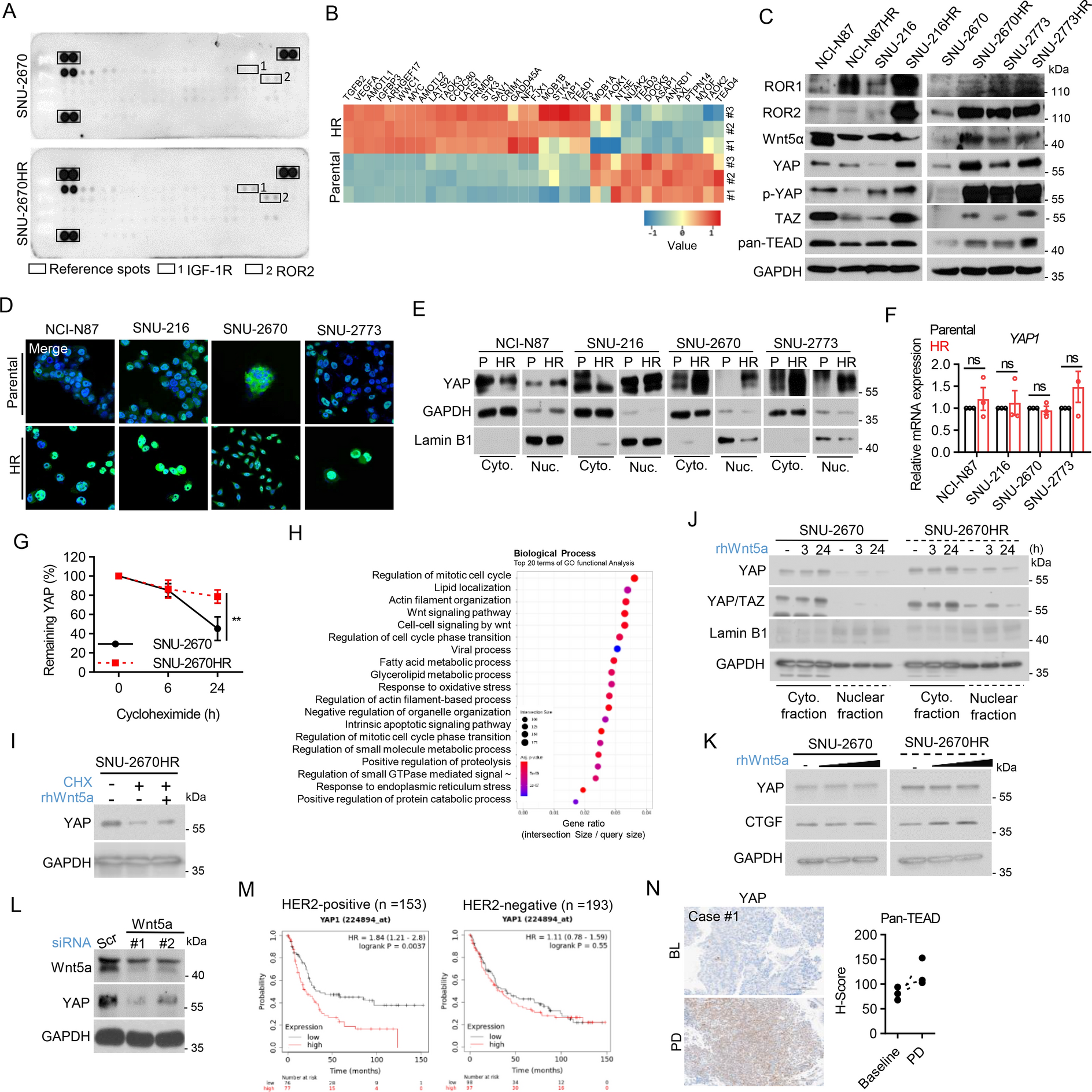
Four human cancer cell lines with HER2 amplification were used in this study. NCI-N87 and SNU-216 cells (GC cell lines) were purchased from the Korean Cell Line Bank (Seoul, Korea). Patient-derived biliary tract cancer (BTC) cell lines with HER2 amplification (SNU-2670 and SNU-2773) were established as previously described [24]. HR cell lines derived from the above-mentioned cell lines (SNU-2670HR and SNU-2773HR) were generated as previously described [25]. Cell lines were maintained in RPMI medium (Welgene Inc. Gyeongsan, Korea), 10% FBS, and 10 µg/ml gentamycin at 37 °C with 5% CO2. Verteporfin (S1786), CA3 (CIL56; #S8661), and Ozuriftamab (#S8758) were purchased from Selleck Chemicals (Houston, TX, USA). Recombinant Human/Mouse Wnt-5a Protein (#645-WN-010) and cycloheximide (#C7698) were purchased from R&D Systems (Minneapolis, MN, USA) and Sigma-Aldrich (St. Louis, MO, USA), respectively.
Immunoblotting
Immunoblot assays were performed as previously described [26]. Briefly, protein samples were prepared from cell lysates using SDS sample loading gel electrophoresis (SDS-PAGE). Separated proteins were transferred onto a nitrocellulose blotting membrane and blocked with 1% nonfat milk/bovine serum albumin (BSA)-supplemented Tris-buffered saline and Tween 20 buffer. Primary and secondary antibody incubations were performed, followed by chemiluminescence detection. Information of antibodies used in this study are as follows; Santa Cruz Biotechnology (Dallas, TX, USA); anti-GAPDH (#sc-25778), anti-Cyclin E (#sc-247), Cell Signaling Technology (Danvers, MA, USA); anti-ROR1 (#cst-4102), anti-ROR2 (#cst-4105), anti-Phospho-YAP (Ser127) (#cst-13008), anti-YAP (#cst-14074), anti-Wnt5a (#cst-2392), anti-TAZ (#cst-83669), anti-Pan-TEAD (#cst-13295), anti-Bcl-xL (#cst-2764), anti-PD-L1 (#cst-13684), anti-Phospho-Stat3 (#cst-9131), anti-IRF1 (#cst-8478), anti-Phospho-Stat1 (Tyr701) (#cst-9167), anti-CTGF (#cst-86641), anti-Cyclin D1 (#cst-2978), anti-p27 (#cst-3686), anti-PARP (#cst-9532), anti-XIAP (#cst-14334), anti-Bax (#cst-14796), anti-β-Actin (#cst-3700), Invitrogen (Waltham, MA, USA); anti-CD63 (#MA1-19281), Abcam (Cambridge, UK); anti-Lamin B1 (#ab16048).
Immunofluorescence
Cells seeded in glass-bottom culture dishes were fixed with 4% paraformaldehyde (Biosesang, Yongin, Korea) and permeabilized with 0.5% Triton X-100 (Sigma-Aldrich, #X100). It was then blocked with 2% BSA/PBS, followed by incubation with primary antibody, anti-YAP antibody (Cell Signaling Technology, #cst-14074), and incubated overnight. After secondary antibody incubation with Alexa Fluor 488 goat anti-rabbit IgG (Invitrogen, #A-11008), nuclei were counterstained with DAPI and subjected to fixation with an antifade mounting solution (Vector Labs, Newark, CA, USA, #H-1000-10). Images were acquired using a STELLARIS 5 confocal microscope (Leica Microsystems).
Cell viability assay
Cells were incubated for a predetermined period in 96-well plates and treated with 3-(4,5-dimethylthiazol-2yl)-2,5-diphenyltetrazolium bromide (MTT) solution (Tokyo Chemical Industry Co., Ltd., Tokyo, Japan; #D0801) for 4 h. Post-incubation, to determine cell viability, MTT formazan was dissolved in DMSO and colorimetric absorbance was measured at 540 nm using a Multiskan GO spectrophotometer (Thermo Fisher Scientific, Waltham, MA, USA).
The siRNA transfection
The cells were transfected with 50 nM siRNA targeting Wnt5a or YAP1 (Genolution, Seoul, Korea) using Lipofectamine 2000 (Thermo Fisher Scientific) in a serum-free medium for 6 h. The medium was then replaced with 10% FBS culture medium and the cells were incubated for 24 h with siYAP1. For siWnt5a, this process was repeated, resulting in a total incubation time of 48 h. Transfected cells were harvested and reseeded for subsequent experiments. Sequence information on siRNAs are as follows; negative control: sense 5′-AAU UCU CCG AAC GUG UCA CGU UU-3′, anti-sense 5′- ACG UGA CAC GUU CGG AGA AUU UU-3’, siYAP#1: sense 5′-GAU GGA UAC AGG UGA UAC UUU-3′, anti-sense 5′-AGU AUC ACC UGU AUC CAU CUU-3′, siYAP#2: sense 5′-GUA UUG CUG ACC UCU UUC AUU-3′, anti-sense 5′-UGA AAG AGG UCA GCA AUA CUU-3’, siWnt5a#1: sense 5’-GAA ACU GUG CCA CUU GUA UUU-3′, anti-sense 5′-AUA CAA GUG GCA CAG UUU CUU-3′, and siWnt5a#2: sense 5′-CAA AGA AUG CCA GUA UCA AUU-3′, anti-sense 5′-UUG AUA CUG GCA UUC UUU GUU-3′.
RT-qPCR
Total RNA was separated using TRIzol (Thermo Fisher Scientific, #15,596,018) and chloroform (Sigma-Aldrich, #288,306), and precipitated with isopropanol. Using purified RNA, cDNA was synthesized using the ImProm-II™ Reverse Transcription System (Promega, Madison, WI, USA, #A3800) following the manufacturer’s instruction. Real-time PCR was performed using TOPreal SYBR Green qPCR PreMIX (Enzynomics, Korea, #RT500M) on a QuantStudio 3 Real-Time PCR Instrument (Applied Biosystems, Waltham, MA, USA). Primers used for the RT-qPCR are as follows; YAP: sense 5′-GGC TGA AAC AGC AAG AAC TG-3′, anti-sense 5′-GAA GAC ACT GGA TTT TGA GTC-3′; AREG: sense 5′-GAG CCG ACT ATG ACT ACT CAG A-3′, anti-sense 5’-TCA CTT TCC GTC TTG TTT TGG G-3′; CTGF: sense 5′-CAG CAT GGA CGT TCG TCT G-3′, anti-sense 5′- AAC CAC GGT TTG GTC CTT GG-3′; CYR61: sense 5′-GGT CAA AGT TAC CGG GCA GT-3′, anti-sense 5′-GGA GGC ATC GAA TCC CAG C-3′.
Cell cycle analysis
Cells collected by trypsinization were fixed with 70% ethanol for at least 48 h. The cells were subjected to flow cytometric analysis of cell-cycle distribution following the sequential addition of RNase A and propidium iodide using the BD FACSCanto II system (BD Biosciences, Franklin Lakes, NJ, USA).
Wound-healing assay
Confluent cell monolayers seeded in 6-well plate were gently scratched using a 200 µl pipet tip and treated with vehicle (PBS) or verteporfin After 24 or 48 h of incubation, cell migration into the scratched area was observed under a CKX41 inverted microscope (Olympus, Tokyo, Japan). The extent of wound closure was determined with 10 measurements in each experiment using the ImageJ software.
ELISA
The extracellular release of CCL5, CXCL10, and HMGB1 was measured using the Human CCL5/RANTES Quantikine ELISA Kit (R&D Systems, #DRN00B), Human CXCL10/IP-10 Quantikine ELISA Kit (R&D Systems, #DIP100), and Human HMGB1 ELISA Kit (Assay Genie, #HUFI00660). All procedures were performed according to the manufacturer’s instructions.
Flow cytometry
Cells were collected by trypsinization, and 1 × 10⁶ cells were analyzed using the BD FACSCanto II system (BD Biosciences) after 30 min of antibody incubation in cell staining buffer (BioLegend, #420201). The antibodies used were as follows: APC anti-human CD274 (B7-H1, PD-L1) antibody (BioLegend, #329708) and Calreticulin antibody (1G6A7) [FITC] (Novus Biologicals, #NBP1-47518F).
Caspase-3 activity
Caspase-3 activity was assessed using the EnzChek® Caspase-3 Assay Kit #1 (Thermo Fisher Scientific, #E-13183) according to the manufacturer’s instructions. Briefly, cancer cells pre-treated with verteporfin for 48 h were co-cultured with PBMCs for additional 72 h. After co-culture, cells were harvested and lysed with 1 × Cell Lysis Buffer. Lysates were centrifuged to remove cellular debris, and 50 µL of the supernatant from each sample was transferred to a 96-well plate. Subsequently, 50 µL of a 2 × substrate solution containing Z-DEVD–AMC was added to each well. Following incubation for 30 min at room temperature in the dark, fluorescence was measured using a microplate reader.
Phospho-RTK array
The Proteome Profiler Human Phospho-RTK Array Kit (R&D Systems, #ARY001B) was used for the phospho-RTK array. Reagents and samples were prepared according to the manufacturer’s instructions. Briefly, arrays were incubated for 1 h at room temperature. Cell lysates were prepared and added to the arrays, followed by overnight incubation at 4 °C. The arrays were then washed three times with wash buffer for 10 min each. The anti-phospho-tyrosine-HRP detection antibody was added and it was incubated for 2 h. Chemiluminescence detection was performed by adding the Chemi Reagent Mix and exposing the membranes to an X-ray film.
Bulk RNA-Seq
Total RNA was extracted using the TRIzol reagent and verified using a TapeStation RNA Screentape (Agilent, #5067-5576). Samples with an RNA integrity number (RIN) > 7.0 were used for library construction using the TruSeq Stranded mRNA Sample Prep Kit (Illumina, #RS-122-2101). Libraries were quantified using KAPA Library Quantification kits and assessed using a TapeStation D1000 ScreenTape (Agilent, #5067-5582) before sequencing on an Illumina NovaSeq platform. Raw reads were trimmed using Trimmomatic 0.38 and aligned to the Homo sapiens reference genome (GRCh38) using HISAT v2.1.0. Transcripts were assembled and read counts per gene were calculated using StringTie v2.1.3b. Differentially expressed genes (DEGs) were identified with DESeq2 (|fold change|≥ 2, raw p < 0.05). Hierarchical clustering was performed using complete linkage and Euclidean distances. Functional and pathway analyses were conducted using gProfiler.
Exosomal PD-L1 analysis
Total exosomes were isolated using the Total Exosome Isolation Reagent (Invitrogen, #4478359) following the manufacturer’s instructions. Briefly, cell-free cell culture media were mixed with the reagent at a ratio of 2:1 and incubated for 24 h at 4 °C. After centrifugation at 10,000 × g for 1 h at 4 °C, pellet containing exosomes were resuspended in PBS for subsequent immunoblotting.
PBMC co-culture experiments
Peripheral blood mononuclear cells (PBMCs) were isolated from peripheral blood using Ficoll gradient centrifugation (Ficoll–Paque Plus, Cytiva, #17-1440-03) and stored in the deep freezer until use. Cancer cells pre-treated with verteporfin for 48 h were harvested and reseeded in a 100 mm culture dish. After 24 h, PBMCs were thawed and co-cultured with cancer cells at a ratio of 5:1 (effector to target cells) for additional 72 h with and without the CD3/CD28 T Cell Activator (#10991, STEMCELL Technologies). Flow cytometry was used to determine the proportion of T cells and PD-1 expression in CD8+ T cells. The antibodies used are as follows: CD3 monoclonal antibody (UCHT1), APC (Invitrogen, #17-0038-42), CD4 monoclonal antibody (RPA-T4), PE (Invitrogen, #12–0049-42), CD8a monoclonal antibody (RPA-T8), FITC (Invitrogen, #11-0088-42), CD279 (PD-1) monoclonal antibody (MIH4), PE (Invitrogen, #11-9969-42), Pacific Blue™ anti-human/mouse granzyme B recombinant antibody (Biolegend, #372,218), APC/Cyanine7 anti-human CD14 antibody (BioLegend, #325620), PE anti-human CD11c antibody (BioLegend, #337206), CD80 (B7-1) antibody, FITC (eBioscience™, Invitrogen, #11-0809-42), and APC-anti-human HLA-DR antibody (Biolegend, #307610).
Mouse tumor xenograft
Animal experiments were conducted at the Institute for Experimental Animals, College of Medicine, Seoul National University (Seoul, Korea), following institutional guidelines, with prior approval from the Institutional Animal Care and Use Committee (No. SNU-211223-1-1). A xenograft tumor model was established administering a subcutaneous injection of 1 × 10⁷ SNU-2773 or SNU-2773HR cells into the right flank of a 4-week-old female BALB/c nude mice (Orient Bio Inc., Seongnam, Korea). When the tumors reached approximately 200 mm3, the mice were randomly divided into two intervention groups. Vehicle (PBS) or 50 mg/kg verteporfin was intraperitoneally administered twice a week for 3 weeks. Tumor volume (½ (Length × Width2) and weight were measured every other day until sacrifice.
Immunohistochemistry
The excised tumor tissues were fixed with 4% paraformaldehyde and embedded in paraffin blocks. After cutting and mounting the sections, they were deparaffinized and rehydrated following antigen retrieval. Immunohistochemical staining was performed using anti-Ki-67 (Invitrogen, #MA5-14,520), anti-YAP (Cell Signaling Technology, #14,074), and anti-PD-L1 (Cell Signaling Technology, #13,684). Each marker was detected using the OptiView DAB IHC Detection Kit (Venetana, #760-700). TUNEL assay was performed using the ApopTag® Plus Peroxidase In Situ Apoptosis Kit (#s7101, Millipore). All procedures were performed according to the manufacturer’s instructions.
Human sample collection and immunohistochemistry
Tumor tissues were obtained from three patients diagnosed with HER2-positive gastric cancer who experienced disease progression following trastuzumab-based therapy at the Seoul National University Hospital. Paired biopsies were collected before and after trastuzumab treatment and post-treatment biopsies were obtained at the time of disease progression. Paraffin-embedded tissue blocks were processed into sliced sections and subjected to antigen retrieval. IHC was performed using antibodies against YAP (Cell Signaling Technology, #14,074) and TEAD (Abcam, #ab97460) and visualization was performed using the OptiView DAB IHC Detection Kit (Venetana, #760-700). H-score analysis utilizing analyzer-assisted interpretation was employed to assess the extent of immunoreactivity.
Kaplan–Meier analysis
The prognostic significance of YAP1 in patients with GC stratified by HER2 subtype was assessed using the web-based Kaplan–Meier plotter database [27]. Survival data and log-rank p values were extracted from publicly available Gene Expression Omnibus (GEO) datasets, including GSE14210, GSE15459, GSE22377, GSE29272, and GSE51105.
Statistical analysis
Statistical analyses were performed using SigmaPlot version 10.0 or GraphPad Prism version 8.0. All statistical tests were two-sided unless otherwise indicated in the figure legends. Statistical significance was set at p < 0.05.

Comments (0)