Tissue samples and DNA extraction
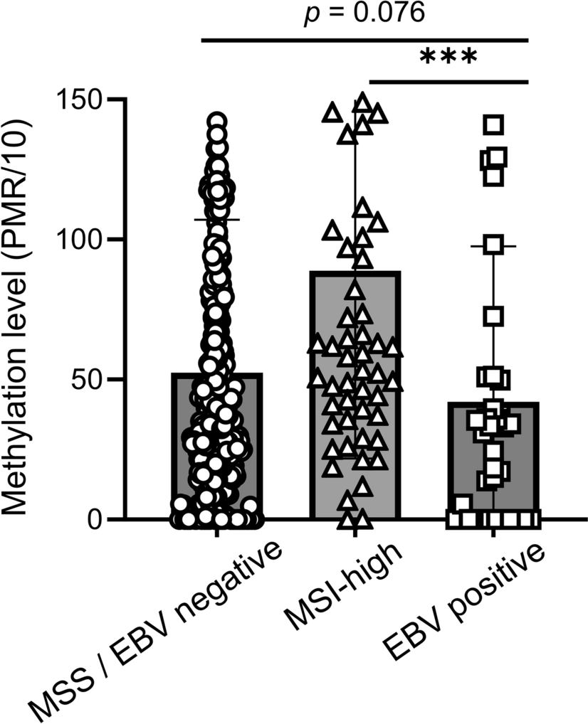
The Institutional Review Board of Seoul National University Hospital approved the study protocol (H-2412–139-1600). The study was conducted in accordance with the Declaration of Helsinki. From 483 patients with advanced GC who underwent surgery at Seoul National University Hospital between 2007 and 2008, archival tissue blocks with high tumor cellularity and representative histology were chosen by reviewing histological slides. MSI and EBV status data for this GC cohort were obtained from previous studies [15,16,17]. DNA was extracted from unstained tissue slides by scraping the tumor tissues into microtubes containing lysis buffer and proteinase K.
Bisulfite modification and the MethyLight assay
The genomic DNA was bisulfite modified using the EZ DNA Methylation Kit (Zymo Research, Irvine, CA, USA). The bisulfite-modified DNA was subjected to the MethyLight assay as previously described [18], and the percentage of the methylated ratio was determined for the ZNF793 promoter CpG island locus. The oligonucleotide sequences of the primers and probe are shown in Supplementary Table 1.
Cell culture
The non-EBV GC cell line (AGS, SNU1, SNU5, SNU620, and SNU638) and EBV GC cell lines (SNU719 and NCC24) were purchased from the Korean Cell Line Bank (Seoul, Korea). Another EBV GC cell line, YCCEL1, was kindly provided by Dr. Sun-Young Rha at the Songdang Institute for Cancer Research, Yonsei University College of Medicine (Seoul, Korea). All the cells were cultured in RPMI 1640 (Welgene, Gyeongsan-si, Gyeongsangbuk-do, Korea), which contained 10% FBS (Gibco, New York, USA) and 1% penicillin ‒ streptomycin solution (Welgene). The e-Myco™ plus Mycoplasma PCR Detection Kit (ver 2.0) (iNtRON Biotechnology, Seongnam-si, Gyeonggi-do, Korea) was used to ensure that all cell cultures were free of mycoplasma contamination.
Plasmid transfection and lentiviral transduction
Single-guide RNAs (sgRNAs) that target ZNF793 or the control were inserted into the LentiCRISPRv2 vector (#52,961, Addgene Inc., Cambridge, MA, USA) and used to transfect HEK293FT cells with ViraPower™ Packaging Mix (Invitrogen, Carlsbad, CA, USA) and Lipofectamine 2000 Transfection Reagent (Thermo Fisher Scientific, Waltham, MA, USA) to produce a lentivirus-containing supernatant. AGS and SNU719 cells were transduced with the lentiviruses for 48 h with 10 μg/ml polybrene (Merck, Darmstadt, Hesse, Germany). The transduced cells were then treated with 2 μg/ml puromycin (Sigma‒Aldrich, St. Louis, MO, USA) for 7 days for selection. Control and ZNF793 knockout cells were subsequently plated in 96-well plates as single cells in medium supplemented with puromycin for culture. The relevant sequences are shown in Supplementary Table 2.
The ZNF793 expression plasmid (ZNF793 (NM 001013659) Human Untagged Clone, Origene Technologies, Rockville, MD, USA) and the control plasmid (pCMV6-Entry, Origene Technologies) were utilized to induce the expression of ZNF793 in AGS and SNU719 cells. The transfection was conducted using Lipofectamine 2000 with an incubation period of 72 h. Then, the transfected cells were selected with G418 solution (Sigma‒Aldrich) at a concentration of 2 mg/ml for 7 days.
siRNA transfection
ZNF793 siRNA (h) (sc-97736, Santa Cruz Biotechnology, Inc., Dallas, TX, USA) and control siRNA (Fluorescein Conjugate)-A (sc-36869, Santa Cruz Biotechnology, Inc.) were used for the downregulation of ZNF793 in SNU620 cell. Transfection was conducted using Lipofectamine 2000 with an incubation period of 48 h.
RNA extraction and real-time qPCR
Following the manufacturer's instructions, we extracted RNA with the RNeasy Plus Mini Kit (Qiagen, Hilden, Germany) and synthesized cDNA with RevertAid Master Mix (Thermo Fisher Scientific). Real-time polymerase chain reaction (qPCR) was performed using Power SYBR Green PCR Master Mix (Applied Biosystems, Foster City, CA, USA) and HotStarTaq Plus DNA Polymerase (Qiagen) on a CFX384 Touch Real-Time PCR System (Bio-Rad, Hercules, CA, USA). GAPDH was used as an internal control, and relative expression was analyzed via the 2−ΔΔCT method. The sequences of primers used for RT‒qPCR are shown in Supplementary Table 3.
Western blotting
Cells and tissues were lysed in RIPA buffer (Merck, Darmstadt, Germany) supplemented with protease inhibitor (Roche, Mannheim, Germany) and phosphatase inhibitor cocktail (Roche) to extract total protein. The Pierce™ BCA Protein Assay Kit (Thermo Fisher Scientific) was used to measure the protein concentration. Proteins were quantified and run on 12% SDS‒polyacrylamide gels and then transferred to PVDF membranes (Merck Millipore, Darmstadt, Germany) by electrophoresis. The membranes were blocked with 5% skim milk (BD Difco, Franklin Lakes, NJ, USA) for 2 h at room temperature and then incubated overnight at 4 °C with the primary antibody. The primary antibodies used are described in Supplementary Table 4. After that, the membranes were incubated for 2 h at room temperature with diluted goat anti-mouse IgG (H + L)-HRP (GenDEPOT, Katy, Texas, USA) or mouse anti-rabbit IgG-HRP (Santa Cruz, Dallas, Texas, USA) as secondary antibodies. Clarity Western ECL Blotting Substrates (Bio-Rad) and a ChemiDoc Imaging System (Bio-Rad) were used to detect and image the protein bands. The intensities of the Western blot bands were quantified via ImageJ software and normalized to those of β-actin.
MTT cell proliferation assay
The 96-well plates were seeded with 2.5 × 10³ cells per well and cultured for 1‒6 days. Each well was treated with 0.5 mg/ml MTT stock solution (10 μl of 5 mg/ml + 90 μl of RPMI 1640) and incubated for 4 h at 37 °C. The formazan that formed was dissolved in 150 μl of DMSO (Sigma‒Aldrich), and the absorbance was measured at 490 nm with a Varioskan LUX Multimode Microplate Reader (Thermo Fisher).
Cell migration and invasion assays
The migration assay involved adding 2 × 10⁴ cells (AGS) or 5 × 10⁴ cells (SNU719) to each 8.0 μm diameter pore cell culture insert (Corning, New York, USA) and incubating them for 48 (AGS) or 96 (SNU719) hours in a cell culture incubator. The non-invasive cells were then gently removed with wet cotton-tipped swabs. The migrated cells were stained with crystal violet.
For the invasion assay, the Matrigel matrix (Corning) was diluted with PBS to 300 μg/ml, and each insert was coated with 0.2 ml of the mixture and then incubated at 37 °C for 2 h. The PBS was removed from the inserts without touching the basement membrane layer. The inside of each insert was seeded with 2 × 10⁴ cells (AGS) or 5 × 10⁴ cells (SNU719), and the lower well of the 24-well plate was filled with 600 μl of media containing 10% FBS. After culturing for 48 (AGS) or 96 (SNU719) hours in a cell culture incubator, noninvasive cells were gently removed using wet cotton-tipped swabs. Invaded cells were stained with crystal violet.
Apoptosis assay
Apoptosis was analyzed via Annexin V labeling using an Annexin V-FITC detection kit (Invitrogen, California, USA). Briefly, the cells were resuspended in binding buffer at a density of 3 × 10⁶/ml. The cell suspension was incubated with Alexa Fluor™ 488 Annexin V and propidium iodide for 15 min at room temperature in the dark and analyzed on a BD FACS Canto™ Clinical Flow Cytometry System (Biosciences, San Jose, CA, USA).
Tumor sphere formation assay
A 6-well plate with a low-attachment surface was seeded with 2 × 10⁴ cells in 3 ml of medium without FBS (RPMI-1640, 20 ng/ml EGF, 10 ng/ml basic fibroblast growth factor, and penicillin‒streptomycin). Each well was replenished with 500 μl of medium every week, and the cells were counted after 2–3 weeks.
Xenograft tumor growth assay
sgZNF793 or control cells were combined with Matrigel at a 1:1 ratio, and the final concentration of cells was 4 × 10⁷/ml. Then, 4 × 10⁶ cells per site were injected under the skin of BALB/c nude mice. The tumor size was periodically monitored until tumor removal. The animal experiments were approved by the Biomedical Research Institute of Seoul National University Hospital (approval no. 3520220102).
Statistical analysis
Statistical analyses were performed via GraphPad Prism version 10 (GraphPad Software, San Diego, CA). The data were first subjected to a normality test. Statistical significance was assessed via either Student’s t test or the Mann‒Whitney test. All the results are shown as the means ± SDs, and P < 0.05 indicates statistical significance.

Comments (0)