Participants and clinical samples
The scope of our study comprised 20 patients with IPF, who were admitted to Qilu Hospital at Shandong University (Jinan, China) between October 2022 and June 2023. Their diagnosis was confirmed using a comprehensive approach involving clinical evaluation, radiological imaging, and pathological examination according to the consensus criteria established by the American Thoracic Society/European Respiratory Society (Raghu et al. 2011). During the same period, we randomly selected 20 age-matched healthy people from the recruited volunteers as controls, excluding respiratory disease and smokers. Fresh peripheral blood samples were collected from all volunteers using a one-time vacuum sampling vessel containing EDTA (cat# 367,525, Corning, USA), and plasma was harvested after centrifugation. Total RNA was extracted from plasma samples by using TRIzol reagent (cat# 15,596,026, ThermoFisher, USA) following the guidelines provided by the manufacturer.
Bioinformatic analysis
The gene expression omnibus database was utilized to obtain microarray data related to IPF. The GSE102660 dataset contained circRNA expression data of 3 pairs of IPF cases and controls. The GSE21411 dataset contained miRNA expression data from 9 IPF cases/6 controls. The GSE53845 and GSE101286 datasets contained mRNA expression data from 40 IPF cases/8 controls and 7 IPF cases/3 controls, respectively. The threshold used to identify genes with differential expression was adjust P-value < 0.05 and |log2fold-change|≥ 1. The Circinteractome (https://circinteractome.nia.nih.gov/index.html) (Dudekula et al. 2016), RNAalifold (http://rna.tbi.univie.ac.at/cgi-bin/RNAWebSuite/RNAalifold.cgi), and circMIR (https://www.bio-inf.cn/circmir/) databases were used to predict the relationship between circRNAs and miRNAs. The multiMiR package of R (version 4.2.3) software and targetscan (https://www.targetscan.org/vert_80/) database were used to predict the relationship between miRNAs and mRNAs. The results were visualized using R software.
Cell culture
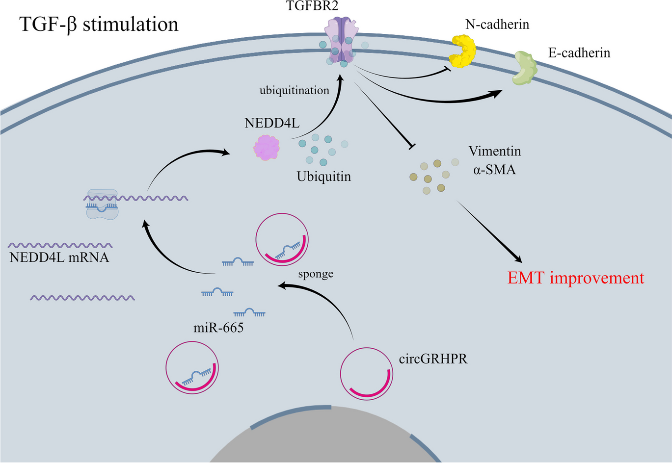
The cell line A549 (cat# CCL-185) represents human alveolar epithelial cells and the cell line Beas-2b (cat# CRL-3588) represents human bronchial epithelial cells, respectively. The A549 and Beas-2b cells were acquired from the American Type Culture Collection (USA) and cultured in Ham’s F-12K (Kaighn’s) Medium (cat# L450KJ, BasalMedia, CHN) and RMPI-1640 medium (cat# L210KJ, BasalMedia, CHN), respectively. All media were enhanced with a 10% concentration of fetal bovine serum (cat# A6901FBS, Invigentech, USA). Similar to previous studies (Li et al. 2020a; Wojcik-Pszczola et al. 2022), cells were starved overnight and further cultured in serum-depleted medium (1%) supplemented with TGF-β1 (10 ng/mL, cat# 10,804-HNAC, Sino Biological, CHN) for 48 h to induce the phenotype of EMT progression. All cells were incubated in a temperature-controlled environment at 37 °C with a CO2 concentration of 5%.
cDNA synthesis and quantitative real-time PCR (qRT-PCR)
As described in our previous study (Wu et al. 2021a; Wu et al. 2021b), total RNA from A549 and Beas-2b cells in 6-well plates was extracted using FastPure Cell/Tissue Total RNA Isolation Kit V2 (cat# RC112-01, Vazyme, CHN). The total RNA samples were quantified using an ultramicroscopic spectrophotometer (cat# DS11 + , DeNovix, USA) to determine their concentrations. For circRNAs and mRNAs, cDNA synthesis was performed using the Hifair® III 1st Strand cDNA Synthesis Kit (cat# 11139ES60, Yeasen, CHN). For miRNAs, the All-in-One™ miRNA First-Strand cDNA Synthesis Kit 2.0 was utilized for the performance of cDNA synthesis (cat# QP113, GeneCopoeia, USA). LightCycler 480 II Real-Time PCR System (Roche) was used for quantitative quantification analysis. The internal control for circRNA and mRNA was 18S ribosomal RNA (18S rRNA), while U6 served as the internal control for miRNA expression. The 2−ΔΔCt method was utilized to determine the relative expression levels. The sequences of primers are presented in Table 1.
Table 1 The sequence of primersGel electrophoresis and Sanger sequencing
The agarose (cat# 1110GR100, BioFroxx, GER), TAE buffer (cat# ST716, Beyotime, CHN), and gel red (cat# EZGR001, WSHTBio, CHN) were mixed, heated, and cooled to prepare a 1.5% agarose gel. The PCR products derived from complementary DNA (cDNA) and genomic DNA (gDNA) were subjected to electrophoresis at a voltage of 100 V and a duration of 40 min, followed by detection using an ultraviolet chemiluminescence instrument (cat# 1600, Tanon, CHN). GoldBand 100-bp DNA ladder (cat# 10507ES60, Yeasen, CHN) was used as a standard DNA marker for DNA size. TIANgel Midi Purification Kit (cat# DP219-03, Tiangen, CHN) was used to recover and purify PCR products separated in agarose gel, and further Sanger sequencing was performed by Genewiz Biotechnology Co. LTD (Tianjin, CHN).
Actinomycin D experiment
A549 and Beas-2b cells were exposed to actinomycin D at a concentration of 2 μg/ mL in a 6-well plate (cat# GC16866, GlpBio, USA) for 0/12/16/20/22/24 h, respectively. The expression levels of circGRHPR and linear GRHPR were analyzed by qRT-PCR.
RNase R treatment
Total RNA was isolated from harvested A549 and Beas-2b cells. Each 2 μg of total RNA underwent treatment with RNase R (3U/μg, cat# GE10003, GlpBio, USA) or enzyme-free water for a duration of 30 min at a temperature of 37 °C. The expression levels of circGRHPR and linear GRHPR were detected by qRT-PCR.
Cell transfection
The overexpressed plasmid of circGRHPR (OE-circGRHPR) based on pGCMV vector and its negative control were purchased from GenePharma Pharmaceutical Technology Co. LTD (Shanghai, China). The overexpressed plasmid of NEDD4L (OE-NEDD4L) based on pCMV vector and its negative control were purchased from MiaoLing Plasmid Platform (Wuhan, China). The short interfering RNAs (siRNA) (si-circGRHPR, si-NEDD4L), miR-665 mimic or inhibitor, and negative controls (si-NC, NC-inhibitor) were produced by GenePharma Pharmaceutical Technology Co. LTD (Shanghai, China), and the sequences of oligonucleotides are shown in Table 2. When cells in the 6-well plate were cultured to 70% confluence, jetPRIME (cat# 101,000,046, Polyplus, FRA) reagent was used for transfection. Each well contained a total of 2 μg plasmid or oligonucleotides with a final concentration of 50 nM.
Table 2 The sequence of siRNA, probes, miRNA mimic, and inhibitorFluorescence in situ hybridization
A549 and Beas-2b cells in a 24-well plate were fixed using 4% paraformaldehyde (cat# BL539A, Biosharp, CHN); then, the cells were pre-hybridized using a fluorescent in situ hybridization kit (GenePharma, CHN). Cy3-labeled circGRHPR probe (GenePharma, CHN) and FAM-labeled miR-665 probe (GenePharma, CHN) were hybridized with A549 and Beas-2b cells overnight at 37 °C. Then, after staining the 4′,6-diamidino-2-phenylindole (DAPI, cat# G1012, Servicebio, CHN) for a duration of 10 min, a positive fluorescence microscope (cat# Ni-U, Nikon, JPN) was used to observe and obtain images. The sequences of probes are presented in Table 2.
Biotin-labeled probe RNA pull-down assay
The circGRHPR probe labeled with biotin and the negative control oligonucleotide probe were synthesized by GenePharma Pharmaceutical Technology Co. LTD (Shanghai, China). Subsequently, they were incubated with streptavidin magnetic beads (cat# HY-K0208, MedChemExpress, USA) for a duration of 2 h at room temperature. About 1 × 10⁷ A549 and Beas-2b cells were harvested in a 10-cm culture dish and fixed with 1% formaldehyde. Following that, the lysates were incubated with magnetic beads coated with the probe for one night in a 4 ℃ environment. After the beads were eluted, the obtained RNA samples were further reversed and the expression levels of miRNAs were detected by qRT-PCR. The sequences of biotin-labeled probes are presented in Table 2.
Biotin-labeled probe RNA capture assay
The miR-665 probe labeled with biotin and the negative control oligonucleotide probe were synthesized by GenePharma Pharmaceutical Technology Co. LTD (Shanghai, China) and transfected into A549 and Beas-2b cells in culture dishes, respectively. Cells were harvested 48 h later, and the cell lysates were subjected to an overnight incubation with streptavidin magnetic beads (cat# HY-K0208, MedChemExpress, USA) in 4 °C condition. The expression level of circGRHPR was evaluated by qRT-PCR. The sequences of biotin-labeled probes are presented in Table 2.
Dual-luciferase reporter assay
Dual-luciferase vector GP-miRGLO (GenePharma, CHN) was utilized to clone the wild-type and mutant binding sites of miR-665 in circGRHPR and the 3′ UTR of NEDD4L. The miR-665 mimics or negative control (GenPharma, CHN) was transfected into A549 and Beas-2b cells along with the dual-luciferase reporter plasmids. After 48 h, the luciferase activity of Renilla and Firefly was detected by Assay Kit (cat# 11402ES60, Yeasen, CHN).
Western blot
As described in our previous study (Wu et al. 2021a), the lysates of A549 and Bsab-2b cells were prepared with the BCA protein quantification kit (cat# P0010, Beyotime, CHN) for protein samples. Relevant protein samples were subjected to SDS-PAGE electrophoresis, followed by subsequent transfer onto polyvinylidene fluoride membranes. The membranes were incubated with primary antibodies at 4 ℃ overnight. Primary antibodies include E-cadherin (1:500, cat# ET1607-75, Huabio, CHN), N-cadherin (1:1000, cat# ET1607-37, Huabio, CHN), Vimentin (1:5000, cat# ET1610-39, Huabio, CHN), α-smooth muscle actin (α-SMA, 1:1000, cat# ET1607-53, Huabio, CHN), NEDD4L (1:1000, cat# ET1611-42, Huabio, CHN), TGFBR2 (1:2000, cat# ER1917-66, Huabio, CHN), and glyceraldehyde-3-phosphate dehydrogenase (GAPDH, 1:10,000, cat# AB0037, Abway, CHN). After incubating with HRP-conjugated antibody (1:50,000, cat# ET1610-39, Huabio, CHN) for 60 min in normal temperature environment, the signal of protein band membrane was scanned by chemiluminescence instrument (cat# 5200, Tanon, CHN).
Coimmunoprecipitation (Co-IP)
A549 and Beas-2b cells were lysed using Cell Lysis Buffers for Western and IP (cat# P0013J, Beyotime, CHN). The supernatants were incubated with TGFBR2 antibody (1:100, cat# 66,636–1-Ig, Proteintech, CHN) or IgG antibody (1:100, cat# AC011, Abclonal, CHN) for a duration of 6 h at room temperature. The mixtures were further mixed with Protein A/G Magnetic Beads (cat# HY-K0202, MedChemExpress, USA) and subjected to overnight incubation at 4 ℃. After that, the beads were washed and the antigen–antibody complex was eluted for western blotting to evaluate the expression of ubiquitin (1:1000, cat# ET1609-21, Huabio, CHN).
Statistical analysis
All data were independently presented as the mean ± standard deviation (SD) from a minimum of three repetitions. The data was subjected to statistical analysis using software packages such as GraphPad Prism (version 9.0, USA) and R (version 4.2.3) software. The Student t test and one-way analysis of variance with Bonferroni correction were used for the analysis. The diagnostic value of circGRHPR was assessed using the receiver operating characteristic (ROC) curve. A statistically significant difference was determined when the P-value < 0.05.

留言 (0)