Cell culture
The ATC cell line C643 was purchased from Procell Life Science & Technology Co., Ltd., and OCUT-2C cells were purchased from iCell Bioscience Inc., Shanghai. C643 cells were cultured in RPMI 1640 (Gibco), and OCUT-2C cells were cultured in DMEM (Gibco), medium with 10% fetal bovine serum (FBS), and 1% penicillinstreptomycin solution (100 μg/ml, Leagene Biotech Co., Ltd.). The cells were incubated at 37 °C in a humidified 5% CO2 atmosphere. Ibuprofen (purity ≥ 99.93%) was purchased from MedChemExpress and was directly dissolved in RPMI 1640 or DMEM. Ibuprofen was dissolved the day before, and the solution was put in a water bath at 37 ℃ to aid in the dissolving process.
CCK8 assays
C643 and OCUT-2C cells were seeded in 96-well plates (5 × 10³ cells/well) in a complete medium and cultured overnight. Next, different concentrations of ibuprofen were administered to the different groups for 48 h. Then, 10 µl of Cell Counting Kit-8 (CCK-8) assay reagent (Dojindo, Kumamoto, Japan) was added to each well, and the 96-well plates were cultured at 5% CO2 at 37 °C for 2 h. The absorbance was measured at 450 nm using a microplate reader. The experiment was repeated three times for each well. The viability rate of control cells was set as 100% for the analysis.
Transwell
The 24-well culture plate, including transwell upper chamber inserts, was used for transwell invasion assays (Corning, New York, USA). First, 1 × 10⁴ cells or cells transfected with OE-RNA and/or control vectors were mixed with 100 ml serum-free RPMI-1640 or DMEM with different concentrations of ibuprofen (C643 at 0, 0.5, 1, and 1.5 mM, and OCUT-2C at 0, 1, 2, and 3 mM) added to the upper chamber. Then, 600 ml of medium containing 10% FBS with the same concentrations of ibuprofen was added to the lower chamber. After culture at 5% CO2 at 37 °C for 48 h, the chamber was removed from the plates and fixed with 4% paraformaldehyde for 30 min. The cells that traversed through the membrane pores were stained with crystal violet. Ultimately, the number of cells passing from the upper chamber to the lower chamber was observed through an inverted microscope (Olympus, Tokyo, Japan).
Wound healing assay
The cells were seeded in a 6-well plate at 4 × 10⁵ cells or transfected with OE-RNA and/or control vectors cultured overnight. Then, the cell surface was scratched using a 200 µl sterile pipette when the cells reached > 95% confluence. Then, the suspended cell fragments were removed by washing with PBS buffer, and the cells were cultured in serum-free RPMI-1640 or DMEM for 48 h with different concentrations of ibuprofen. Finally, the ability of cells to migrate was assessed using an inverted microscope (Olympus, Tokyo, Japan).
Flow cytometry
Apoptosis was analyzed via flow cytometry. The cells or cells transfected with OE-RNA and/or control vectors were spread in a 6 cm dish at a density of 4 × 10⁵ cells/well and cultured overnight. Next, the cells were treated with different concentrations of ibuprofen for 48 h. Then, the cells floating in the medium were collected, washed three times with PBS buffer, and digested with EDTA-free trypsin. Then, the obtained cells were labeled with PI and Annexin V (Jiangsu Keygen Biotech Corp, Ltd). Finally, a flow cytometer was used for analysis. Double-positive (PI and Annexin V) cells were considered apoptotic.
LDH
Cells were seeded in a 6 cm dish at a density of 30 × 104 cells/well. Then, the cells were cultured with ibuprofen at different concentrations for 48 h. The supernatant of cells or cells transfected with OE-RNA and/or control vectors with different concentrations of ibuprofen was added to a 96-well plate. Distilled water, pyruvate standard solution, matrix buffer, and coenzyme I were added to the sample test well and the control well, and the cells were cultured at 37 °C for 15 min. After, 2,4-dinitrophenylhydrazine was added to each well and cultured at 37 °C for 15 min. Then, 0.4 mol/l NaOH solution was added, and the cells were cultured at 37 °C for 5 min. The absorbance at 450 nm was measured using a microplate reader. Then, the LDH content difference was calculated according to the obtained results.
Transmission electron microscopy (TEM)
Cells were seeded in a 10 cm dish at a density of 80 × 104 cells/well. Then, cells were treated with different concentrations of ibuprofen (C643 at 0.1.5 mM and OCUT-2C at 0.3 mM) for 48 h. Next, the cells were fixed with 2.5% glutaraldehyde according to the manufacturer’s protocol (Beijing Solarbio Science & Technology Co., Ltd.). Then, the samples were dehydrated with acetone and embedded in epoxy resin and hardener. Finally, the samples were cut into ultrathin sections via a Recheron ultrathin microtome, stained with uranium acetate, and observed by electron microscopy.
Immunofluorescence
Cells were seeded in a 96-well plate at a density of 5 × 10³ cells/well. First, the cells were treated with different concentrations of ibuprofen (C643 at 0.1.5 mM and OCUT at 0.3 mM) for 48 h. Next, the cells were fixed with 4% paraformaldehyde and then permeabilized with 0.5% Triton X-100. Then, the cells were incubated with the primary antibody at 4 °C overnight, and then the secondary antibody was added and incubated for 1 h. Immunofluorescence was performed with the following antibodies: rabbit anti‐ASC (Servicebio, GB115270), rabbit anti‐NLRP3 (SAB, 29125), rabbit anti‐GSDMD (Proteintech, 20770-1-AP), goat Anti-Rabbit IgG (abbkine, A23220) for Dylight 488, and Goat Anti-Rabbit IgG (abbkine,A23620) for Dylight 649. DAPI (Solarbio, S2110) was used as a nuclear counterstain. The images were observed with a Nikon Ti Eclipse Confocal Microscope and an LSM 980 using basic operating techniques.
OE-RNA transfection
The lentiviral OE-GSDMD vectors and their control vectors were constructed by Shanghai GenePharma Co., Ltd. All transfections were performed according to the manufacturer’s instructions. C643 and OCUT-2C cells were plated in six-well plates (1 × 10⁵ cells/well). Then, 5 µl of OE-GSDMD or control vectors was transfected with Lipofectamine 3000 (Invitrogen) based on the manufacturer’s protocol. After 24 h, transfected C643 and OCUT-2C cells were treated with ibuprofen and subjected to subsequent analyses.
Western blotting
RIPA buffer was used to lyse cells for protein extraction at 4 °C. Equal amounts of protein (20 μl) were added to 10% SDS/PAGE and transferred to a PVDF membrane. Then, the cells were blocked with a quick block solution for 15 min at room temperature and washed three times with TBST buffer. The membrane was incubated with the primary antibodies at 4 °C overnight, and then the secondary antibody was added and incubated for 1 h. Western blotting was performed with the following antibodies: rabbit anti‐ASC (Servicebio, GB115270), rabbit anti‐NLRP3 (SAB, 29125), rabbit anti‐GSDMD (Proteintech, 20770-1-AP), rabbit anti‐GAPDH (Good here, AB-P-R001), and goat anti‐rabbit IgG (Dingguo, IH-0011). The blots were visualized with an enhanced chemiluminescence kit (Servicebio) based on the manufacturer’s instructions.
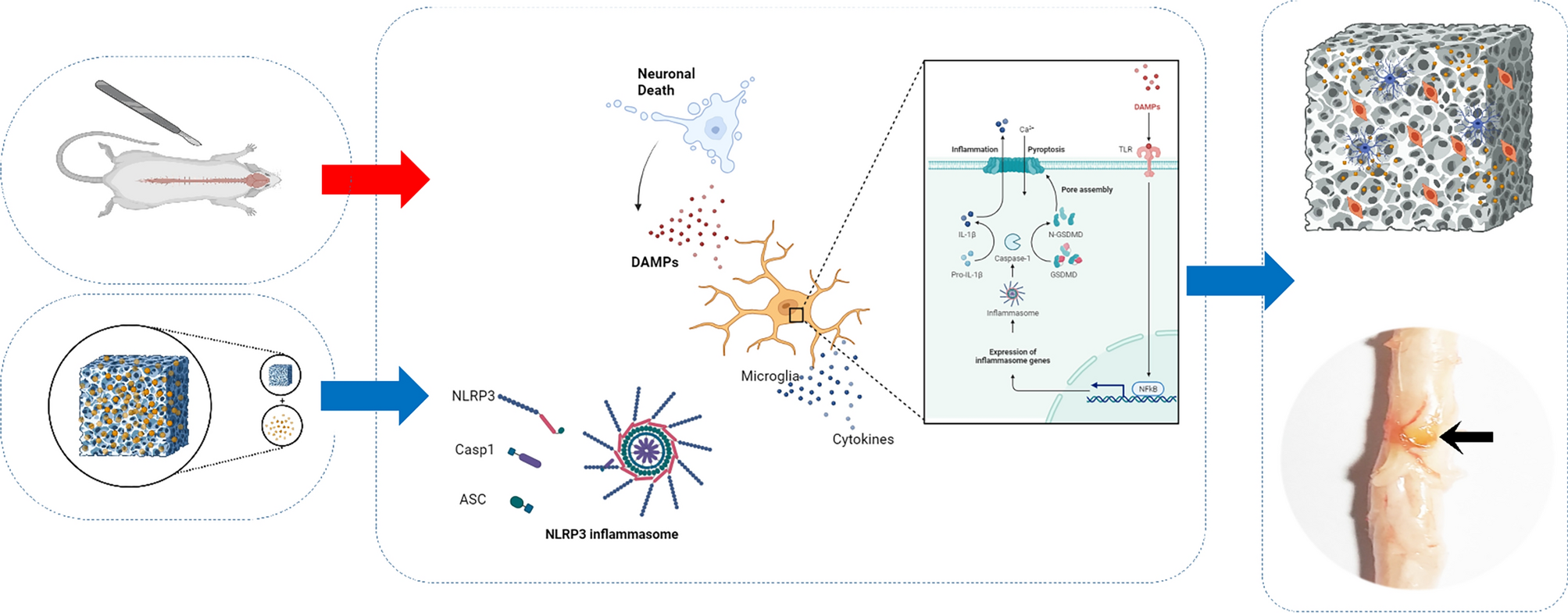
Xenograft models
A 200 µl cell suspension containing 2 × 10⁶ cells was subcutaneously injected into 5-week-old female BALB/c nude mice (Beijing Vital River Laboratory Animal Technology Co., Ltd.). After the tumors grew to approximately 0.5 mm3, 15 mice were randomly divided into the following 3 groups: the control group, in which mice were given 0.9% saline intragastrically, and the ibuprofen groups, in which mice were given 10 or 20 mg/kg ibuprofen. All groups were given solutions once every two days. The tumor volumes were monitored using a digital caliper. The xenograft tumor volume (mm3) was calculated as 0.5 × (shortest diameter)2 × (longest diameter). When some tumors had grown to about 2 mm3, the mice were killed and the size and weight of the xenograft tumors were measured.
Immunohistochemistry analysis and hematoxylin–Eosin (HE) staining
The xenografts tumor, normal thyroid tissues, and stomach were separated from mice and fixed in 4% paraformaldehyde buffer for HE staining or immunohistochemistry staining. The operation procedure of this animal experiment was carried out under the program approved by the Ethics Committee on Laboratory Animal Management of Zhengzhou University.
Statistical analysis
GraphPad Prime 9.0 was used for mapping and statistical analysis of the experimental data. Measurement data are expressed as the mean ± standard deviation (mean ± SD). Paired T-test and one-way ANOVA were used for statistical analysis. P < 0.05 indicated a statistical difference between the samples.


留言 (0)