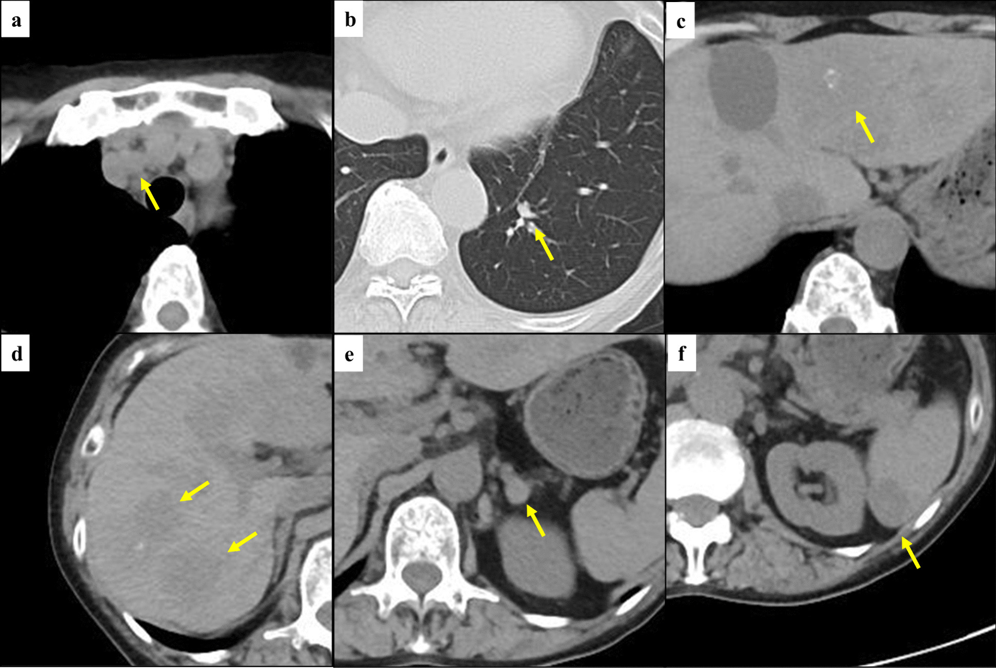
In this study, we described our experience with two patterns of reconstruction using the RGEV during PD and TP to prevent LSPH including gastric venous congestion. One pattern is RGEV–MCV anastomosis and the other is RGEV–LRV anastomosis. Both patients had short-term shunt patency and showed no signs of LSPH. There were no procedure-related morbidities during the postoperative follow-up.
SV ligation during PD or TP may result in the development of LSPH. LSPH is caused by insufficient splenic/gastric venous drainage and may induce splenomegaly, varices, and severe gastric bleeding/congestion. Variceal bleeding after LSPH is recurrent or massive in some patients, resulting in fatal hypovolemic shock. Splenomegaly causes pancytopenia, resulting in anemia, compromised status, and easy bleeding [13,14,15]. Regarding TP, Loos et al. [16] reported that the overall 90-day mortality after TP was 4.1%, and 7.4% in patients with gastric vein congestion and 2.8% in those without gastric vein congestion. The authors also reported that half of the patients who died after TP had gastric vein congestion. Mehrabi et al. [17] also described gastric vein congestion after TP, which led to gastric venous infarction and ischemia with subsequent gastric perforation and abdominal sepsis. These complications increase patient morbidity and mortality, and reconstruction of gastric venous drainage is useful to avoid gastric vein congestion. In addition to these postoperative complications, gastric vein congestion can increase difficulty controlling intraoperative hemostasis, which affects the surgery. For these reasons, it is important to prevent LSPH. We have experienced 86 cases of PD with PV resection in the 10 years from 2013 to 2022, of which the SV was sacrificed in 40 cases. LSPH occurred in eight cases, in which perigastric varices, gastric congestion, or splenomegaly occurred, among the cases with SV resection. Although LSPH is a rare complication, its frequency cannot be ignored.
Tanaka et al. [14] reported that the risk of LSPH after PD with portomesentericosplenic confluence resection could be stratified based on the number of preserved critical veins among the LGV, MCV, and superior right colic vein arcade. The authors reported that, in patients who underwent SV ligation during PD in whom none of the three critical veins were preserved and the SV was not reconstructed (n = 29), all patients developed LSPH. In patients with only one of the critical veins preserved and no SV reconstruction (n = 51), 12 of the 51 (24%) patients developed LSPH. In contrast, no patients with preservation of two or three of the critical veins (n = 8) developed LSPH. The authors also reported that in patients with no preserved critical veins who underwent successful SV reconstruction (n = 5), LSPH developed in three of the five patients. In addition, in patients with only one preserved critical vein and successful SV reconstruction (n = 10), none developed LSPH. Therefore, regarding the indications for venous reconstruction when SV ligation is performed during PD or TP, the number of preserved critical veins is helpful. In our cases, only one critical vein, the right colic vein arcade, was preserved in both case 1 and case 2. In case 2, gastric venous congestion was observed, which is considered a good indication for venous reconstruction. In addition, in case 1, considering the incidence of LSPH in the above study in patients with only one remaining critical vein, and the usefulness of venous reconstruction, we believe that venous reconstruction was appropriate.
Partial gastrectomy is an alternative to venous reconstruction. Nakao et al. [18] reported that distal gastrectomy may be a safe method with which to prevent gastric venous congestion and bleeding when combined with TP. However, extended resection of the stomach with pancreatectomy may lead to functional and structural dysfunction, resulting in worsening of the patient’s nutritional status [2].
Regarding venous reconstruction, there are many reports of the usefulness of reconstruction with anastomosis with the SV, such as SV–SMV anastomosis [1], SV–IMV anastomosis [1, 4,5,6], SV–renal vein anastomosis [1, 3, 7,8,9,10], and other anastomoses [1, 11, 12]. We have experienced a case in which venous reconstruction using the SV was performed during PD with SV ligation. In the case, we performed SV–IMV anastomosis, and there were no findings suggesting LSPH, such as splenomegaly, varices, and thrombocytosis. However, these anastomoses are complicated because the length of the resected SV is long owing to tumor invasion, and it is necessary to separate the SV from the pancreatic parenchyma [15]. In this situation, we suggest performing anastomosis with the RGEV.
To date, there have been few reports of anastomosis using gastric veins (Table 1). In our search of PubMed, we found five cases of reconstruction using a gastric vein during PD or TP [2, 18,19,20]. In four cases, reconstruction was performed using the LGV [2, 19, 20], and right gastric vein reconstruction was also performed in one of these cases [20]. In the other case, reconstruction was performed with the RGEV, as in our report [18]. In the previous case, emergency anastomosis between the RGEV and the left ovarian vein was performed because severe gastric vein congestion and bleeding occurred during TP. Hemostasis was achieved after the reconstruction.
Table 1 Previously reported cases of vascular reconstruction using a gastric vein to prevent left-sided portal hypertensionThe RGEV is useful for venous reconstruction for the following reasons. First, the RGEV has a certain distance from the SV and PV, which pancreas cancer often invades. In addition, because the RGEV is located in the greater omentum, this vein is mobilized easily by incising the omentum. Furthermore, side-to-side anastomosis for reconstruction is possible because the distance between the anastomosed vessels is short. However, high mobility of the anastomosis site may cause rupture of the anastomosis as a result of body movements. To prevent rupture of the anastomosis, we placed a small number of sutures to fix the anastomosis in the fatty tissue in case 2.
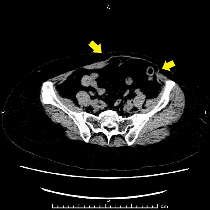
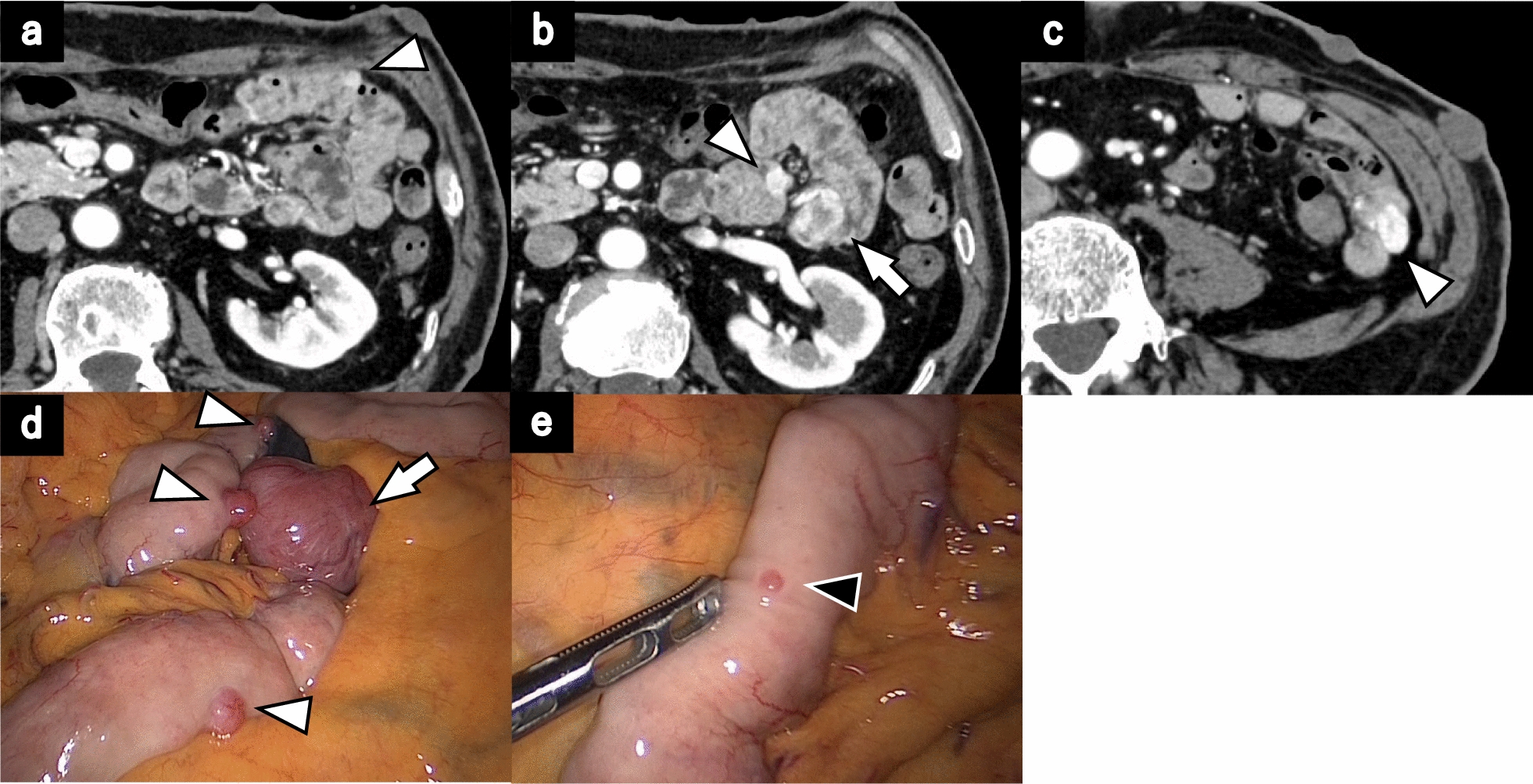
The following points are important during reconstruction with the RGEV. First, because the diameter of the vein is small, a surgical loupe is necessary for the procedure; however, a surgical microscope is unnecessary. In addition, the vessel walls are thin, so care must be taken for not to tear the vessel during anastomosis. Finally, for the venous reconstruction, we chose a large-diameter vein located close enough to be easily anastomosed. During PD, the blood vessels that can be anastomosed are limited because the pancreas overlies the retroperitoneum, and pancreatic-jejunal anastomosis is part of PD. In contrast, during TP, a relatively large number of blood vessels are options, such as the LRV or left gonadal vein, because the anterior layer of Gerota’s fascia is completely exposed, and the vein can be mobilized easily. In case1, because the site and size of the vessel were suitable, and anastomosis did not interfere with the gastrojejunostomy, we anastomosed the gastric side stump of the RGEV with the colonic side stump of the MCV side-to-side. In case 2, we chose the LRV because the anterior layer of Gerota’s fascia is completely exposed, and the LRV, which had a large diameter and was close to the RGEV, could be easily mobilized.

Comments (0)