Recombinant antibody production
D4a mAb (patent number WO2016091891A1) is specific to human AXL tyrosine kinase receptor since it does not bind to mouse Axl and other human receptors of the same family, TYRO3 or MER tyrosine kinase receptors. 13R4a mAb is specific of E. coli beta-galactosidase specific and this antibody is used as an isotype irrelevant control [9]. Interestingly, the synthetic library is build using 13R4a clone as template, meaning that there are only few differences between D4a and 13R4a exclusively located in the 6 CDR loops. These two mAbs have been isolated in the laboratory from a human synthetic library of scFv using phage display [10]. ScFv were reformatted as full human IgG1 with wild type (WT) Fc by cloning variable heavy and light chain in an expression plasmid. The L234A/L235A/P329G mutations were introduced in coding plasmid by targeted PCR-mutagenesis and validated by sequencing. 13R4a and D4a antibodies with wild type Fc and with the L234A/L235A/P329G mutation (LALAPG Fc) were produced in HEK293T cells by transient transfection with polyethylenimine. Antibodies were purified from supernatant using Protein-A agarose beads and dialyzed against PBS. Purity was verified on SDS-PAGE. Antibody binding was validated in vitro by enzyme linked immunosorbent assay and by fluorescent-activated cell sorting in different cell lines.
Radioimmunoconjugation
Antibodies (human IgG format) were functionalized with pSCN-Bn-deferoxamine in a non-site-specific manner before radiolabeling with 89Zr. Briefly, the antibody buffer was exchanged to chelexed PBS using Amicon® Ultra Centrifugal filters (30 kDa cut-off). pH was adjusted to 8.5–9.0 using 0.2 M chelexed Na2CO3, and a 15-fold excess of pSCN-Bn-deferoxamine was added to the solution (1.6–2.1 mg/mL, 500 µL). After incubation at 37 °C with gentle shaking for 60 min, excess pSCN-Bn-deferoxamine was removed using Amicon® Ultra Centrifugal filters as before. [89Zr] Zr-oxalate (Perkin Elmer) (30 MBq) was neutralized to pH 6.9–7.2 with 1 M chelexed Na2CO3 before addition of the deferoxamine-immunoconjugates and incubation at room temperature with gentle shaking for 1 h. Purity and radiolabeling efficiencies were determined using instant thin-layer chromatography with 0.1 M sodium citrate (pH 5.0) as mobile phase. Radiolabeling yield and radiochemical purity were routinely > 99%. No purification step was performed. Radioimmunoconjugates had a specific activity of 200 MBq/mg and were formulated in 0.9% NaCl for in vivo use. Analysis of the immunoconjugate was performed by Maldi-tof (Rapiflex, Bruker) to determine the number of DFO conjugated to mAbs. The number of DFO molecules is determined by dividing the difference in the m/s ratio of the peak of the whole antibody conjugated with DFO and unconjugated with the molecular weight of DFO (752 Da) (Additional file 1: Figure S1). Binding affinity of D4a mAbs conjugated with DFO was performed by flow cytometry using AXL expressing cell line. A binding affinity experiment was presented in Additional file 1: Figure S2.
Cell lines and mice
The AXL-expressing [11, 12] human MDA-MB-231 (triple negative breast cancer) and CFPAC-1 (pancreatic cancer) cell lines were from American Type Culture Collection. Immortalized human myoblasts (provided by Vincent Mouly, UMR-S 974) express low AXL level and were used to mimic healthy human tissues. Cancer cells were cultured in Dulbecco’s Modified Eagle’s Medium (DMEM) with 10% Fetal Bovine Serum (FBS, Eurobio), and myoblasts in skeletal muscle cell growth medium (C-23060, Promocell) with 20% FBS at 37 °C and 5% CO2. All animal experiments were performed in compliance with the European directive (2010/63/EU) and the INSERM standards for experimental animal studies (agreement E34-172-27). They were approved by the Institut de Recherche en Cancérologie de Montpellier (IRCM/INSERM U1194) and the Languedoc Roussillon region (CEEA LR France No. 36) ethics committees. Cells (5.106) in Matrigel (Corning) were injected subcutaneously in 6-week-old female athymic nude mice (Crl: Nu (NCr)-Foxn1nu, Charles River).
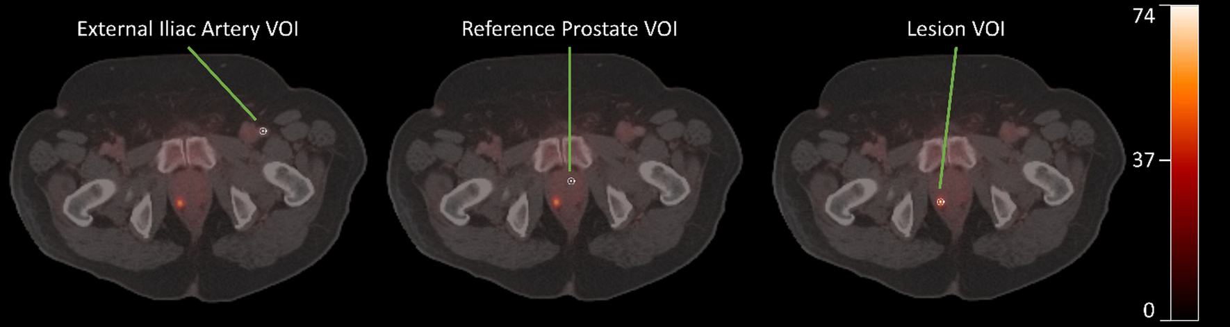
Imaging
Four weeks after cell xenografts, [89Zr] Zr-deferoxamine-labeled antibodies (50 µg, 200 MBq/mg) were injected in the tail vein and in vivo images were acquired with a Mediso NanoScan PET82S/CT80 system at 48, 72, and 96 h post-injection. Anesthesia was induced with 4% isoflurane in air followed by maintenance with 2% isoflurane in air. Images were acquired with Nuclide and processed with Interview™ FusionTM. Volumes of interests were manually drawn on fused PET-CT images using 3D Slicer. Results are expressed as percentage of the injected activity per cm3 (%IA/cm3).
Conventional biodistribution analysis
Biodistribution was assessed at 96 h post-injection after the last imaging time-point. After euthanasia, tumor, myoblast xenografts, and liver were excised, weighted, and activity counted with a gamma-counter (Hidex AMG) together with standards of the injected radiolabeled antibodies. Results were expressed as percentage of the injected activity per gram of tissue (%IA/g).


留言 (0)