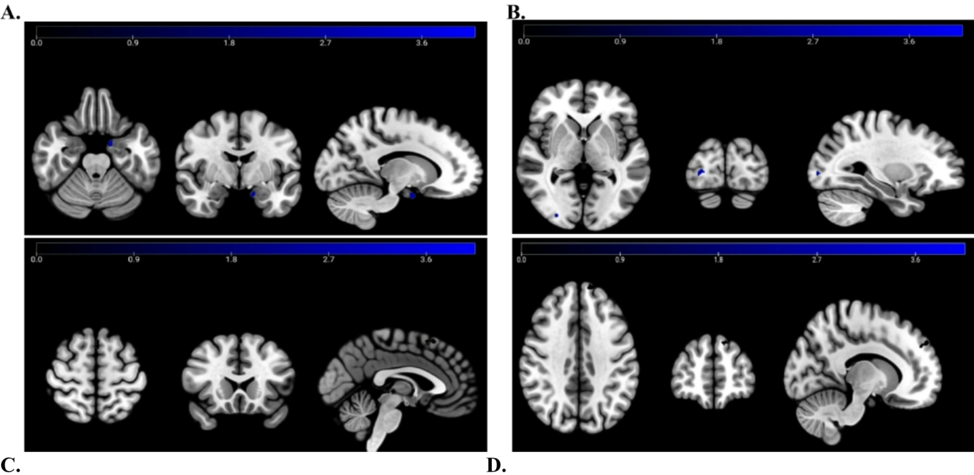
In this preliminary study, we applied FreeSurfer-based hippocampal subregion segmentation to examine volumetric differences among CM, EM and HC groups. No statistically significant differences in hippocampal subregion volumes were observed across the three groups. Nevertheless, exploratory analyses revealed trend-level alterations: (1) compared with HCs, patients with CM exhibited trend-level smaller volumes in the bilateral whole hippocampus, subiculum, CA1, and molecular layer; and (2) compared with the EM group, CM patients showed trend-level reductions in the bilateral whole hippocampus, molecular layer, CA1, as well as in the left subiculum, GC-ML-DG, and CA4. Overall, these analyses provide preliminary indications of trend-level volumetric alterations in hippocampal subregions among CM patients.
Similar to our findings, a previous study also reported no statistically significant differences in the left and right hippocampal volumes among CM, EM, and HC groups [28]. The lack of statistically significant differences in our study may be explained by several factors, including limited sample size, individual variability, and the resolution and sensitivity constraints of MRI. In addition, it has been demonstrated that a hippocampal subregion volume exceeding 300 mm³ offers the best test-retest reliability and stability [29], implying that smaller hippocampal volumes in our study might have lower measurement reliability. Indeed, some hippocampal subregions have been excluded or merged in previous studies [30,31,32]. Furthermore, the anatomical boundaries of small subregions such as CA3, GC-ML-DG, and the molecular layer are not well characterized [33], which may further compromise accuracy. Taken together, the absence of significant findings in our study may be explained by the combined influence of multiple factors. In this study, we reported the original results while acknowledging the need for future research with larger samples, optimized imaging protocols, and improved subregion segmentation strategies.
Despite the absence of statistically significant findings, trend-level variations in hippocampal subregion volumes were observed across the three groups. In general, patients with CM tended to show reduced hippocampal volumes compared with the other two groups, a pattern that is partly consistent with previous reports [10, 14]. As a structure involved in the perception and modulation of pain, the hippocampus is linked to susceptibility to chronic pain [34]. It has been demonstrated that patients with CM have abnormalities in both the structure and functional connectivity of the hippocampus [35, 36]. One possible explanation is that chronic pain-induced stress influences the hypothalamic-pituitary-adrenal axis, leading to elevated glucocorticoids, which may inhibit hippocampal neurogenesis and influence hippocampal remodeling, ultimately causing hippocampal volume reduction [37,38,39]. Additionally, neuroinflammatory mechanisms play a crucial role in the pathogenesis of CM, which can affect hippocampal neurogenesis and synaptic plasticity, and lead to memory impairment [40, 41]. Considering that memory dysfunction could be a symptom in CM patients [42], it is plausible to suggest that the observed trend-level reductions in hippocampal volumes might serve as a mediator between neuroinflammation and memory dysfunction in CM.
The hippocampus is a highly complex and heterogeneous brain structure, with each subregion serving distinct functional roles. Specifically, the CA1 plays a crucial role in emotion regulation and memory, and its volume reduction has been found to be correlated with memory scores [43]. Furthermore, this subregion forms a functional circuit with the medial prefrontal cortex, which is essential for encoding and retrieving episodic memories [44]. Besides the aforementioned memory issues in CM, co-occurring emotional problems are also common in CM patients and may exacerbate headaches [45]. Therefore, we tentatively suggest that the trend-level involvement of CA1 may be linked to both memory and emotional dysfunctions associated with CM. The CA1/subiculum is a major anatomical connection area for several cortical regions, including the hypothalamus, which is regarded as one of the primary migraine generators [20]. Altered connectivity between the hypothalamus and other brain regions, along with its correlation to the severity of headache, has been observed in patients with CM [46]. Consequently, the trend-level alterations observed in the subiculum, as the primary output region of the hippocampus, may suggest its potential involvement in the physiological mechanisms underlying migraine chronification through its connectivity with the hypothalamus. The molecular layer, composed of interneuron synaptic connections, plays a crucial role in hippocampal synaptic circuitry and the temporal processing of events [47]. Considering that chronic pain is closely associated with abnormal changes in synaptic connections [48], we assume that the observed decreased trend-level molecular layer volumes in CM patients may be associated with impaired synaptic transmission and related pain processing issues.
Moreover, it was found that CM patients exhibited a trend-level reduction in the volume of CA4/DG region compared to EM patients. The CA4/DG can induce neurogenesis and drive neuronal plasticity, with neurogenesis being considered one of the key factors in maintaining cognitive function [49]. Noorani et al. showed that the volume of the CA4/DG in patients with trigeminal neuralgia, a chronic pain disorder, was reduced and subsequently recovered after pain relief achieved through surgical interventions [50], suggesting the CA4/DG region serves as a biomarker for changes in neuronal plasticity and neurogenesis associated with pain. Accumulating evidence [42, 51, 52] indicates that cognitive deficits are common in CM, potentially linked to intracranial pathological changes resulting from recurrent migraine attacks [53]. Based on the above observations, we tentatively suggest that the trend-level reduced volume of CA4/DG in CM patients may be related to their long-standing chronic pain, which might hinder neurogenesis and could be linked to cognitive dysfunction.
It is worth noting that the trend-level changes in hippocampal subregions in CM patients were primarily concentrated in the left hemisphere. Similar reports of left hippocampus involvement have been described in prior CM studies using voxel-based morphometry [10] and resting-state functional connectivity [54]. Taken together, these observations suggest that the hippocampal abnormalities associated with migraine chronification may demonstrate a left-sided lateralization. Indeed, it has been recognized that the left hippocampus plays a predominant role in verbal memory, whereas the right hippocampus is more closely associated with visuospatial memory [55]. According to a clinical investigation, CM may primarily affect verbal memory [56], which is predominantly associated with the left hippocampus. Therefore, it is not surprising that trend-level alterations in CM were observed mainly in the left hippocampus. However, it should also be noted that the trend-level reduction in the CA1, molecular layer, and subiculum subregions in CM patients occurred in both hemispheres. This bilateral reduction cannot be conclusively explained by the current study design. One tentative interpretation is that, while CM has been reported to primarily affect verbal memory, it may also impact other functions related to the right hippocampus. The left-sided lateralization, together with the partial bilateral involvement observed, may be related to the multifaceted and widespread functional impact of migraine chronification. Nonetheless, as our study did not collect the relevant scales about memory as well as other cognitive functions, these interpretations remain speculative and require further validation in future research.
Interestingly, the present study showed no trend-level alterations in hippocampal subregion volumes between the EM and HC groups. As seen in Fig. 2, in the subregions exhibiting trend-level differences across the three groups, the EM and HC groups had comparable volumes, whereas the CM group appeared to show a trend-level reduction. One explanation could be that, during the EM phase of migraine (with a relatively low frequency of headache attacks), hippocampal subregions may undergo compensatory adaptive changes through neuroplastic mechanisms, which may help preserve subregion volumes at this stage. As the condition progresses to the CM phase, however, these adaptive changes may become maladaptive, contributing to the trend-level reductions in hippocampal subregion volumes observed in CM. Therefore, this observation may underscore the potential importance of early intervention in EM to reduce the risk of progression to CM in terms of hippocampal subregion alterations.
Our research had several limitations. First, one limitation of this study is the relatively small and unequal sample size, particularly for CM group, which is due to its lower prevalence compared to EM in the general population. The overall smaller sample size increases the risk of Type II errors, meaning that we may miss some hippocampal subregions that are potentially significant. This limitation should be considered when interpreting the findings, and future research with larger and more balanced samples is needed to confirm these results. Second, the participant groups were not perfectly sex- or age- matched, but rather balanced. Future research should focus on achieving a more optimal sex ratio and a closer age match between groups to enhance the robustness of the results. Third, as the participants were recruited from a single center, the generalizability of the results requires further validation using a multicenter dataset. Fourth, as previously mentioned, one study suggests that hippocampal subregion volumes exceeding 300 mm³ provide the best test-retest reliability and stability [29], meaning that caution should be taken when evaluating our measurements of smaller hippocampal subregions. Fifth, some patients had a history of using acute-phase therapeutic medications (e.g., non-steroidal anti-inflammatory drugs), but the specific types and dosages of these medications were not thoroughly recorded. This represents an important limitation that should be further explored and addressed in future research. Last, it is important to note that our study is only a cross-sectional study, and therefore it is not yet possible to infer any causal relationships. In the future, prospective longitudinal studies tracking the dynamic changes are needed to address this issue.
In conclusion, this study did not identify statistically significant volumetric differences in hippocampal subregions among the three groups. However, exploratory analyses revealed trend-level reductions in several subregions in CM patients, including CA1, CA4, molecular layer, GC-ML-DG, and subiculum. These preliminary observations may suggest potential—but unconfirmed—associations between hippocampal subregional characteristics and processes such as pain perception, emotion, memory, and cognition during migraine chronification. Future studies with larger, well-balanced samples and higher-resolution imaging are required to clarify these tentative findings.


Comments (0)