
Remember me
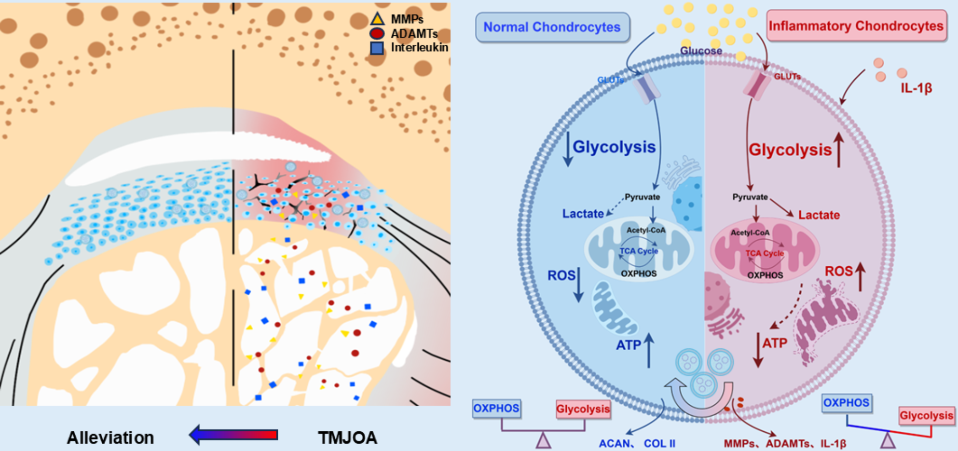


To explore gene expression alterations in the condyles of TMJOA mice, condyle tissues from both normal and TMJOA mice were collected for transcriptomic analysis. As Fig. 2A depicted, sequencing results demonstrated a significant upregulation of glycolysis-related genes, including HK3 (Hexokinase 3) and Pklr (Pyruvate kinase). In contrast, key genes related to oxidative phosphorylation (OXPHOS) and TCA cycle, including Cs (Citrate synthase), Mdh2 (Malate dehydrogenase), and Sdhc (Succinate dehydrogenase), were markedly downregulation.
Hexokinase (HK), including HK1, HK2, HK3, HK4, and HKDC1, play critical roles in glucose metabolism, catalyzing the initial step of glucose utilization [25]. The upregulation of HKs suggests enhanced glycolytic activity. Citrate synthase (Cs) initiates the tricarboxylic acid (TCA) cycle by catalyzing the condensation of oxaloacetate and acetyl-CoA [26]. Moreover, Mdh2, a crucial enzyme in the TCA cycle, contributes to energy production and stabilizes hypoxia-inducible factor 1 alpha (HIF-1α), linking metabolism to cellular responses to hypoxia [27]. Sdhc, serves as a pivotal enzyme bridging the TCA cycle and OXPHOS pathway, its downregulation further indicates disrupted energy metabolism in TMJOA cartilage [28].
Additionally, the expression of structural cartilage matrix genes, Col2a1 (collagen II) and acan (aggrecan) was significantly decreased, while IL-1β and matrix metalloproteinases 10 (MMP10) levels were markedly elevated in the TMJOA condylar cartilage. Kyoto Encyclopedia of Genes and Genomes (KEGG) analysis indicated that differentially expressed genes were primarily enriched in pathways related to “Oxidative phosphorylation”, “Citrate cycle (TCA cycle)”, “Glycolysis/Gluconeogenesis” and others (Fig. 2B). These alterations indicate a metabolic shift towards enhanced glycolytic activity in TMJOA condylar cartilage, contrasting with a decrease in TCA cycle and OXPHOS functionality compared to normal cartilage.
To validate the expression of key metabolic enzymes, qRT-PCR was performed, revealing significant upregulation of glycolysis-associated genes, including HK1, HK2, Lactate dehydrogenase (LDHA), and Pyruvate kinase (PKM2) in OA cartilage (Fig. 2C). The expression levels of Lacc1 and pro-inflammatory cytokines were also assessed. As shown in Fig. 2D, pro-inflammatory markers such as IL-1β, matrix metalloproteinases (including MMP9, MMP13) and tumor necrosis factor-α (TNF-α) were markedly elevated in OA cartilage. Previous researches indicated that IL-1β played a crucial role in OA by promoting the upregulation of matrix metalloproteinases (MMPs), which contributed to the irreversible breakdown of extracellular matrix (ECM) and progression of OA [1, 29]. In contrast, both qRT-PCR results (Fig. 2D) and immunofluorescence staining for Lacc1 (Fig. 2E) demonstrated a significant downregulation of Lacc1 in TMJOA cartilage, suggesting its potential anti-inflammatory role and its relationship with glycolysis in chondrocytes.
Furthermore, Fig. 2F illustrated a pronounced expression of PKM2, a key glycolytic enzyme, in TMJOA group. Previous studies have highlighted that the metabolism reprogramming played a vital role in knee OA, with inflamed chondrocytes shifting their metabolism towards glycolysis, resulting in LDHA accumulation and increased reactive oxygen species (ROS) generation [29]. Enzymes such like pyruvate kinase (PKM), lactate dehydrogenase (LDHA) and hexokinase (HK) are integral to regulating glucose metabolism, and IL-1β induction significantly elevated the expression of those genes in chondrocytes [29, 30]. Collectively, our findings indicate an imbalance between glycolysis and OXPHOS in TMJOA cartilage, with Lacc1 potentially acting as a metabolic link in the pathogenesis of TMJOA.
Fig. 2
Metabolic Dysregulation in TMJOA Cartilage. (A) Volcano plot of differentially expressed genes in TMJOA cartilage. (B) KEGG pathway enrichment analysis of metabolism-related signaling pathways. (C&D) qRT-PCR analysis of glycolytic enzymes, Lacc1, and inflammatory cytokines in TMJOA cartilage. (E&F) Immunofluorescence staining of Lacc1 and PKM2 in TMJOA cartilage. (n = 3, *p < 0.05, **p < 0.01, and ***p < 0.001)
Validation of the function of Lacc1 in RAW264.7 macrophagesTo elucidate the role of Lacc1 in RAW264.7 macrophages, we performed both siRNA-mediated knockdown and Lacc1 overexpression experiments. Initially, qRT-PCR was employed to identify the most effective siRNA for Lacc1 knockdown (Fig. S1A). The optimal siRNA was selected for subsequent analysis to evaluate its effect on macrophage polarization upon LPS treatment. As Fig. S1B depicted, siLacc1 treatment increased the expression of pro-inflammatory cytokines including TNF-α, IL-1β and inducible nitric oxide synthase (iNOS), while significantly reducing the expression level of Arginase-1 (Arg-1), a marker associated with anti-inflammatory M2 macrophages [31, 32], suggesting a shift toward a pro-inflammatory phenotype in siLacc1-treated macrophages. Subsequently, the expression of glycolysis related genes were also assessed by qRT-PCR. Fig. S1C illustrated that siLacc1 markedly elevated the expression of aconitate decarboxylase 1 (Acod1), PKM2, phosphofructokinase (PFKp) and HK1, which served important roles in the process of glycolysis [25, 33]. Furthermore, ATP level measured in Fig. S1D indicated that siLacc1 led to lower ATP production, regardless of LPS stimulation, compared to the siNC group.
Subsequently, Lacc1 overexpression plasmids were transfected into RAW264.7 macrophages to assess the effects of Lacc1 on inflammation and metabolism. The enhanced expression of Lacc1 in the transfected cells, compared to the negative control (NC) group, was confirmed through fluorescence microscopy, quantitative real-time PCR (qRT-PCR), and western blot analyses (Fig. S2A-S2C). Then qRT-PCR and Western blot analysis was conducted to detect the effects of Lacc1 overexpression on inflammatory responses of macrophages. As presented in Fig. S2D & S2E, Lacc1 expression was significantly elevated in the overexpression (OE) group. In contrast, the levels of IL-1β, IL-6, iNOS, MMP9, and MMP13 were substantially decreased in the OE group compared to the NC group.
In summary, our data indicate that Lacc1 plays a pivotal role in regulating both inflammation and glycolysis in RAW264.7 macrophages. Thus, RAW264.7 macrophages engineered to overexpress Lacc1 could represent a promising source for the development of extracellular vesicles aimed at regulating metabolism and suppressing inflammation.
Lacc1-Engineered EVs (OE-EVs) alleviated IL-1β induced inflammation in chondrocytesConstruction and characterization of OE-EVsIn this study, RAW264.7 macrophages were transfected with a plasmid for Lacc1 overexpression. Following transfection, Lacc1-engineered vesicles (OE-EVs) were isolated from the treated RAW264.7 macrophages using ultracentrifugation, while the vesicles from NC plasmid-treated cells were designated as NC-EVs.
The morphology of OE-EVs and NC-EVs was evaluated using transmission electron microscopy (TEM). As illustrated in Fig. 3A, both OE-EVs and NC-EVs exhibited typical exosomal morphology, characterized by a saucer-like or hemispherical shape with concave surfaces. Nanoparticle tracking analysis (NTA) (Fig. 3A) revealed average sizes of 126.1 nm for OE-EVs and 128.3 nm for NC-EVs, aligning with the expected EV size range (30–200 nm). The concentrations of OE-EVs and NC-EVs were quantified as 1.35 × 10¹⁰ particles/mL and 1.6 × 10¹⁰ particles/mL, respectively. The protein content of OE-EVs was determined to be 0.82 mg/mL, while NC-EVs had 0.66 mg/mL. Western blot analysis in Fig. 3C confirmed the presence of exosomal markers CD63, CD81, and TSG101 in both OE-EVs and NC-EVs, while the negative marker Calnexin was not detected. Additionally, Lacc1 expression was significantly higher in OE-EVs group than that in the NC-EVs group.
The uptake efficiency of OE-EVs by chondrocytes was assessed by labeling the vesicles with PKH26 dye [34]. As illustrated in Fig. 3B, both OE-EVs and NC-EVs were effectively internalized by chondrocytes, primarily localized in the cytoplasm. PKH26 was selected for EV labeling due to its high membrane affinity, strong fluorescence stability, and extensive use in EV research. PKH26 has been widely utilized to track EV uptake and biodistribution in various disease models, including osteoarthritis and rheumatoid arthritis [35]. Compared to other dyes, PKH26 offers low photobleaching, ensuring long-term visibility of labeled EVs in both in vitro and in vivo settings, making it a reliable tool for tracking EV-mediated cellular interactions. To assess biocompatibility, chondrocytes were treated with various concentrations of OE-EVs and NC-EVs for 1, 3, and 5 days, followed by cell viability assessment using the CCK-8 assay. As shown in Fig. S3A, chondrocytes treated with 15 µg/mL OE-EVs demonstrated the highest viability across all time points. Consequently, 15 µg/mL OE-EVs and NC-EVs were utilized in subsequent experiments. To further assess the protective effects of EVs on chondrocytes in an inflammatory environment, calcein-AM/PI live/dead cell staining was performed. Fig. S3B revealed more dead cells in the IL-1β-treated group while fewer dead cells were observed in the Con, NC-EVs, and OE-EVs groups, suggesting that both OE-EVs and NC-EVs had favorable biocompatibility and protective effects. These results confirm that OE-EVs and NC-EVs have favorable biocompatibility and are efficiently endocytosed by chondrocytes.
Fig. 3
OE-EVs attenuate IL-1β-induced inflammation in chondrocytes. (A) TEM and NTA characterization of NC-EVs and OE-EVs. (B) PKH26-labeled EVs internalized by chondrocytes. (C) Western blot analysis of exosomal markers and Lacc1 in NC-EVs and OE-EVs. (D) qRT-PCR of inflammatory and cartilage matrix markers. (E&G) Immunofluorescence staining of Aggrecan and MMP13. (F) IL-1β levels measured by ELISA. (H) Western blot of inflammation- and ECM-related markers. (n = 3, *p < 0.05, **p < 0.01, and ***p < 0.001)
OE-EVs alleviated IL-1β induced inflammation in chondrocytes in vitroDiminishing inflammation and promoting cartilage matrix synthesis are crucial for the effective treatment of TMJ osteoarthritis. To evaluate the therapeutic effects of OE-EVs on IL-1β induced inflammatory chondrocytes, we assessed the expression of key markers involved in cartilage matrix formation and the inflammatory cytokines. As shown in Fig. 3D, qRT-PCR results indicated a significant reduction in the expression of collagen II, aggrecan, and Sox9, key markers for cartilage matrix synthesis in IL-1β group. However, treatment with OE-EVs resulted in a significant upregulation of these markers, indicating that OE-EVs promote cartilage matrix synthesis under inflammatory conditions. Moreover, the expression of inflammatory cytokines (including IL-1β and IL-6), and matrix metalloproteinase (including MMP9 and MMP13) was markedly elevated following IL-1β induction, whereas OE-EVs treatment effectively downregulated these cytokines.
Western blot analysis corroborated these findings. As illustrated in Fig. 3H, the levels of IL-1β, IL-6, MMP3, and MMP9 were significantly enhanced with IL-1β stimulation. However, the OE-EVs treatment effectively counteracted the pro-inflammatory effects of IL-1β. The expression of Sox9 showed an opposite trend, further supporting the potential of OE-EVs in promoting cartilage matrix synthesis and chondrocyte anabolic metabolism.
To further assess the impact of OE-EVs on inflammatory cytokine secretion, ELISA was conducted. Figure 3F demonstrated that OE-EVs reduced the excessive secretion of IL-1β, further confirming their anti-inflammatory properties. In addition, immunofluorescence staining for aggrecan (Fig. 3E) and MMP13 (Fig. 3G) in chondrocytes showed that IL-1β stimulation decreased aggrecan expression while increasing MMP13 expression. Conversely, OE-EVs treatment counteracted these effects, resulting in higher aggrecan levels and lower MMP13 expression.
To further investigate the mechanism by which OE-EVs modulate the inflammatory response, we examined their effect on macrophages. RT-PCR and Western blot analyses of LPS-stimulated macrophages treated with OE-EVs revealed a significant downregulation of pro-inflammatory markers, including TNF-α, MMP9, MMP13, IL-1β, and iNOS, while the expression of Arg1 was markedly increased (Fig. S4). These findings indicate that OE-EVs primarily function by suppressing M1 macrophage polarization, thereby mitigating the inflammatory phenotype.
In articular cartilage, the extracellular matrix (ECM) is primarily composed of aggrecan and type II collagen. Aggrecan plays a pivotal role in maintaining cartilage phenotype and promoting chondrocyte proliferation, while type II collagen, encoded by the gene COL2A1 and synthesized by chondrocytes, is a major structural collagen [36,37,38]. Sox9, a transcription factor crucial for chondrogenesis, is expressed from the multifunctional mesenchymal precursor stage to the cell differentiation stage, and its inactivation hindered the development of cartilage [39]. Therefore, these cartilage-specific genes are essential for sustaining chondrocyte function and programming anabolic metabolism.
However, in the inflammatory environment of OA, pro-inflammatory cytokines (such as IL-1β and IL-6) and matrix-degrading enzymes (such as MMP3, MMP9, MMP13, and ADAMTS5) promote inflammation and catabolic metabolism, leading to ECM degradation and impaired chondrocyte function. IL-1β is significantly increased in synovial fluid of TMJOA patients and mediates matrix degradation via inducing expression of matrix degradative enzymes such as MMP3, MMP9, MMP13 and ADAMTS5 [39, 40]. Besides, elevated by inflammation, excessive production of reactive metabolites, including reactive oxygen (ROS) and reactive nitrogen species (RNS), contribute to mitochondrial dysfunction, matrix degradation, and cell damage in OA [9]. These factors create a vicious cycle that exacerbates cartilage degeneration.
Previous studies have demonstrated that MSC-derived exosomes can ameliorate inflammation in TMJOA by reducing IL-1β and iNOS expression [40]. Moreover, engineered EVs have been reported to deliver functional molecules, such as miRNAs, to repair the immune microenvironment in OA joints, thereby alleviating OA progression [41]. Recent research highlights the crucial role of synovial macrophages in OA pathogenesis, as they secrete pro-inflammatory cytokines that interact with chondrocytes and exacerbate cartilage degradation [14]. Additionally, macrophage-derived EVs have been implicated in cartilage catabolism and synovial inflammation, further underscoring their contribution to OA progression [42]. In line with these findings, our results demonstrate that OE-EVs not only protect cartilage ECM homeostasis by enhancing matrix synthesis and inhibiting degradation but also mitigate chondrocyte inflammation in vitro. These findings strongly support the potential of OE-EVs as a novel metabolic and immunomodulatory therapy for OA, paving the way for future investigations into their role in regulating energy metabolism.
OE-EVs reprogrammed chondrocytes’ energy metabolism by alleviating mitochondria dysfunctionEnergy metabolism is a crucial mediator of cellular function and is often altered during disease states, particularly in response to inflammatory stimuli in chondrocytes. Previous studies have demonstrated the therapeutic effects of OE-EVs on chondrocytes in vitro. To further investigate the metabolic changes in IL-1β-induced inflammatory chondrocytes, gene and protein expression related to key enzymes involved glycolysis were analyzed by qRT-PCR and Western blotting. As shown in Fig. 4A and B, IL-1β treatment resulted in a significant increase in the expression of HK1, HK2, LDHA, PKM2 and hypoxia-inducible factor 1-alpha (Hif-1α), indicating enhanced glycolysis activity in OA chondrocytes. Notably, treatment with OE-EVs effectively counteracted these changes.
Mitochondria play a central role in ATP synthesis and cellular metabolism. Inflammation-induced mitochondrial dysfunction is characterized by impaired ATP production. To assess the impact of OE-EVs on mitochondria dysfunction induced by inflammation, we performed cell ATP assay. As illustrated in Fig. 4C, IL-1β significantly reduced ATP level in chondrocytes, while OE-EVs treatment effectively restored the ATP level.
Moreover, we measured the extracellular lactate levels in the supernatants of various groups. As depicted in Fig. 4D, lactate concentration was markedly elevated in the IL-1β group, whereas OE-EVs treatment led to a significant reduction. Additionally, mitochondrial dysfunction is characterized by an aberrant change in the mitochondrial membrane potential (ΔΨm). JC-1 staining (Fig. 4E) revealed that IL-1β-treated chondrocytes exhibited increased green fluorescence from JC-1 monomers (indicating ΔΨm loss), whereas OE-EVs treatment resulted in prominent red fluorescence from JC-1 aggregates, suggesting ΔΨm restoration.
Excessive ROS production contributes to oxidative stress and mitochondrial dysfunction, exacerbating cellular damage. DCFH-DA staining (Fig. S5) demonstrated a significant increase in intracellular ROS levels in the IL-1β group, which were effectively reduced by OE-EVs treatment. We also evaluated mitochondrial ROS levels using MitoSOX staining and examined mitochondrial morphology with MitoTracker Green staining. As shown in Fig. 4F, inflammatory chondrocytes exposed to IL-1β exhibited impaired mitochondrial morphology, including mitochondrial deformation, swelling. Conversely, OE-EVs treatment mitigated these detrimental effects. Mitochondrial ROS (mtROS) level significantly increased in IL-1β group, implying an escalation in mitochondrial oxidative stress damage, however, remarkably inhibited with OE-EVs treatment. As shown in Fig. 4G, the IL-1β group displayed severely damaged mitochondrial structures, such as reduced crista, disrupted membranes, and a swollen phenotype, compared to Control group. In contrast, chondrocytes treated with OE-EVs maintained relatively normal mitochondrial morphology including displaying elongated, rod-shaped structures with improved cristae and membrane integrity compared to both IL-1β and NC-EVs groups.
In summary, our results indicate that OE-EVs reprogrammed glycolytic metabolism in OA chondrocytes by inhibiting aerobic glycolysis and alleviating oxidative stress, while simultaneously improving mitochondrial morphology and function. Our findings indicate that Lacc1-enriched EVs regulate both chondrocyte metabolism and inflammation; however, whether Lacc1 itself plays a direct role in chondrocyte metabolic homeostasis remains unclear. Given that EV cargo exerts biological effects through intercellular communication, it is possible that the observed metabolic changes are mediated via multiple signaling pathways rather than direct intracellular Lacc1 activity. Future studies involving direct overexpression or knockdown of Lacc1 in chondrocytes will be needed to determine its potential cell-autonomous function. Clarifying this mechanism will be essential for fully elucidating the therapeutic potential of Lacc1-based EV interventions in TMJOA.
Fig. 4
OE-EVs reprogram metabolism in chondrocytes. (A&B) qRT-PCR and Western blot analysis of glycolytic enzyme expression. (C&D) ATP production and lactate levels in chondrocytes. (E) JC-1 staining of mitochondrial membrane potential (red: JC-1 aggregates, green: JC-1 monomers). (F) Mitochondrial ROS analysis using MitoSOX and Mitotracker staining. (G) TEM images of mitochondria in different groups. (n = 3, *p < 0.05, **p < 0.01, and ***p < 0.001)
RNA-Seq analysis of the impact of OE-EVs on glycolysis and inflammation in OA chondrocytesTo further explore the underlying mechanism behind the therapeutic effects of OE-EVs, transcriptomic analysis was performed. A heatmap (Fig. 5A) depicting differentially expressed genes (DEGs) in IL-1β and OE-EVs-treated groups revealed significant alterations in the expression of key regulators of glycolytic and inflammatory. Notably, in the IL-1β group, the expression of glycolytic genes, including HK2, Pfkfb3, and Eno3, was significantly upregulated, suggesting enhanced glycolytic metabolism (Fig. 5A). In contrast, OE-EVs treatment markedly downregulated the expression of these genes (Fig. 5A), indicating a potential role of OE-EVs in glycolytic reprogramming in chondrocytes.
Additionally, inflammatory genes, such as Cxcl5, Ccl2, Nfkbia, Tnfaip3, and IL6, were significantly downregulated in the OE-EVs group compared to the IL-1β group. This downregulation further supports the anti-inflammatory effects of OE-EVs in OA chondrocytes. Moreover, the expression of genes involved in ECM degradation, including MMP3 and Adamts18, was significantly reduced in the OE-EVs-treated group, suggesting that OE-EVs may play a role in restraining ECM breakdown in OA. In addition, the expression of JAK2, a key component of the JAK-STAT signaling pathway, was notably decreased following OE-EVs treatment, further suggesting that OE-EVs may modulate glycolysis and inflammation, while suppressing ECM degradation and promoting proteoglycan synthesis in OA chondrocytes.
To gain further insights into the biological processes underpinning these changes, Gene Ontology (GO) enrichment analysis was performed. As shown in Fig. 5C, the most significantly enriched GO terms included processes related to glycolytic metabolism, carbohydrate metabolism, ATP generation from ADP, extracellular matrix organization, and glycosaminoglycan binding. These findings highlight the potential of OE-EVs in regulating metabolic processes and maintaining ECM homeostasis in OA.
In support of these results, Kyoto Encyclopedia of Genes and Genomes (KEGG) pathway analysis (Fig. 5B) identified several enriched pathways, including HIF-1 signaling, glycolysis/gluconeogenesis, TNF signaling, and JAK-STAT signaling. These pathways are consistent with OE-EVs’ role in modulating glycolytic metabolism and inflammation. In addition, Gene Set Enrichment Analysis (GSEA) revealed notable variations in JAK-STAT signaling pathway and extracellular matrix (Fig. 5D), TNF signaling pathway and cell adhesion (Fig. S6). Compared to the IL-1β group, the enrichment of genes associated with extracellular matrix and cell adhesion was significantly increased in the OE-EVs group, meanwhile the genes relating to JAK-STAT and TNF signaling pathway displayed a significant decrease in the OE-EVs group. These results suggest that OE-EVs not only promote ECM synthesis but also inhibit the inflammatory response, likely through modulation of the JAK-STAT signaling pathway.
Previous studies have shown that pro-inflammatory cytokines (such as IL-1β, IL-6) and matrix metalloproteinase (MMP) activate the Janus Kinase/Signal Transducers and Activators of Transcription (JAK/STAT) signaling pathway, contributing to inflammation and cartilage degradation in OA. Inhibition of the JAK-STAT pathway has been shown to alleviate chondrocyte inflammation and protect against cartilage degeneration [43, 44]. To further verify the correlation between anti-inflammation effects of OE-EVs with regulating JAK-STAT signaling, we performed western blot analysis. As shown in Fig. 5E and F, the levels of phosphorylated JAK2 (p-JAK2) and STAT3 (p-STAT3), markers of JAK-STAT activation, as well as the ratios of p-JAK2/JAK2 and p-STAT3/STAT3, were significantly reduced in the OE-EVs group compared to the IL-1β group. These results indicate that OE-EVs exert inhibitory effects on the JAK-STAT pathway, thereby suppressing inflammation in OA chondrocytes.
In summary, our findings suggest that OE-EVs play an active role in regulating inflammation microenvironment of TMJOA. By reprogramming glycolysis, inhibiting key inflammatory pathways (including JAK-STAT, TNF, and HIF-1 signaling), and promoting ECM synthesis, OE-EVs help create a favorable microenvironment for chondrocyte function and protect against cartilage degradation.
Fig. 5
Transcriptomic and signaling pathway analysis of IL-1β- and OE-EV-treated chondrocytes. (A) Heatmap of differentially expressed genes. (B&C) KEGG and GO enrichment analysis. (D) GSEA analysis of key pathways. (E&F) Western blot and semi-quantification of JAK-STAT signaling. (n = 3, *p < 0.05 and **p < 0.01)
OE-EVs alleviate inflammation and promote cartilage repair in TMJOA in vivoBuilding upon the promising in vitro results, the therapeutic effects of OE-EVs were further assessed in vivo using a surgical induced mouse model of TMJOA [45, 46]. As illustrated in Fig. 6A, treatments began two weeks after unilateral discectomy to allow for TMJOA development. Mice in TMJOA group received intra-articular injections of PBS, while the NC-EVs and OE-EVs groups received NC-EVs and OE-EVs, respectively. Four weeks post-treatment, TMJ tissues were harvested for analysis.
To examine the retention ability of EVs in vivo, PKH26 labeled OE-EVs and NC-EVs were injected into the TMJ, and fluorescence signals were tracked using an in vivo imaging system (Berthold, Germany). Fig. 6B showed that both OE-EVs and NC-EVs were retained in the tissue for over 72 h. For assurance of the sustained effects of OE-EVs, intra-articular injection was administered twice weekly. TMJs were harvested after four weeks of treatment.
Since inflammation in TMJOA also leads to subchondral bone deterioration, we evaluate the therapeutic effects of OE-EVs on subchondral bone remodeling using Micro-CT and parametric analysis. The Micro-CT reconstruction images of condyle in Fig. 6C showed severe condylar cartilage and subchondral bone destruction including irregular trabecular structures and typical subchondral bone loss in the TMJOA group. In contrast, OE-EVs treatment notably preserved subchondral bone integrity, presenting a more regular trabecular pattern compared to the TMJOA and NC-EVs groups.
Subsequently, Bone structural parameters—including the ratio of bone volume to tissue volume (BV/TV), trabecular thickness (Tb.Th), trabecular number (Tb.N) and trabecular separation (Tb.Sp) were also examined. As shown in Fig. 6D, the TMJOA group had significantly lower BV/TV, Tb.Th, and Tb.N, along with increased Tb.Sp, indicating substantial bone structure deformation. OE-EVs treatment significantly improved BV/TV, Tb.Th, and Tb.N while reducing Tb.Sp, suggesting an effective amelioration of bone loss. No significant differences were observed between the TMJOA and NC-EVs groups. Thus, these results suggested that OE-EVs treatment effectively alleviated subchondral bone erosion caused by TMJOA and protected condylar subchondral bone structure.
OE-EVs exhibit excellent biocompatibility and do not cause systemic toxicity. To further assess the safety of OE-EVs in vivo, we performed H&E staining of major organs, including the heart, liver, spleen, lungs, and kidneys. The histological analysis (Fig. S7) revealed no detectable pathological abnormalities across all groups, indicating that OE-EV treatment does not induce observable toxicity or tissue damage. These findings, together with our in vitro CCK-8 and live/dead staining assays (Fig. S3), confirm that OE-EVs are well-tolerated and biocompatible, supporting their potential for clinical application.
Histological analysis was further conducted to evaluate changes in articular cartilage and subchondral bone. H&E staining (Fig. 6E) revealed characteristic TMJOA pathology in the TMJOA group, including disorganized cellular layers, a rough surface, inflammatory cells infiltration, and severe subchondral bone damage, whereas the OE-EVs group exhibited reduced TMJOA related histological alterations compared to the TMJOA and NC-EVs groups. Moreover, the Safranin O-fast green staining (Fig. 6F) was performed to assess the glycosaminoglycan (GAG) level deposited in cartilage, with TMJOA cartilage showing diminished and irregular Safranin O+ matrix compared to the Con group. Notably, OE-EVs treatment resulted in a uniform and abundant distribution of the safranin O+, suggesting enhanced GAG synthesis. Additionally, TB staining (Fig. 6G) revealed marked cartilage loss in the TMJOA group, while OE-EVs treatment preserved cartilage thickness, indicating effective protection against cartilage matrix degradation. Semi-quantitative analysis of toluidine blue-stained cartilage area in Fig. S8 showed a 1.55-fold increase in the OE-EV group compared to the TMJOA group, indicating enhanced proteoglycan accumulation. Previous study reported that EVs or engineered EVs restore TMJ condylar structure with amelioration in cell arrangement, cartilage thickness, cellularity and matrix synthesis and subchondral bone remodeling [47, 48]. Consistent with curative effects as previous work reported, OE-EVs treatment exhibited remarkable therapeutic effects in restoration of the TMJ condylar structure, including improvements in morphology and functional ingredients.
Fig. 6
OE-EVs promote cartilage repair and suppress inflammation in TMJOA in vivo. (A) Schematic of OE-EV treatment in a TMJ-OA mouse model. (B) In vivo fluorescence imaging of TMJ-injected OE-EVs and NC-EVs. (C&D) Micro-CT images and bone structural parameters. (E-G) Histological staining (H&E, Safranin O-Fast Green, Toluidine Blue). (n = 6, *p < 0.05, and ***p < 0.001)
Collagen II and Aggrecan are essential for chondrogenesis, and their expression levels were evaluated by immunohistochemistry and immunofluorescence staining, respectively. Collagen II (Fig. 7A) and Aggrecan (Fig. 7C and F) expression was significantly suppressed in TMJOA group compared to the Con group. While OE-EVs markedly enhanced their expression, indicating that OE-EVs treatments effectively promote cartilage matrix regeneration and anabolic activity.
Moreover, inflammatory cytokines (such as IL-1β) and matrix metalloproteinases (MMP3, MMP9, MMP13) played pivotal roles in cartilage matrix degradation. Herein, the impact of OE-EVs on alleviating cartilage inflammation was investigated by immunohistochemical staining for IL-β (Fig. 7B) and immunofluorescence staining for MMP13 (Fig. 7D and G) and MMP3 (Fig. 7E and H). As the results showed, the expression of these proteins was significantly elevated in the TMJOA group; however, OE-EVs treatment effectively reduced IL-1β, MMP3 and MMP13 levels, demonstrating that OE-EVs significantly suppress catabolic and pro-inflammatory factors. As Fig. 7I showed, histological analysis was conducted using the OA Research Society International (OARSI) scoring system. The OE-EVs group displayed a significantly lower score compared to the TMJOA group, indicating a substantial therapeutic effect in mitigating OA-related damage.
We also conducted immunofluorescence staining of iNOS and Arg1 to verify the anti-inflammatory effects of OE-EVs in vivo. As Fig. S9 showed, these results demonstrated that in the OE-EV-treated group, Arg1 expression was upregulated, whereas iNOS expression was significantly reduced. This suggests that OE-EVs may mitigate TMJOA inflammation by promoting macrophage polarization toward an anti-inflammatory M2 phenotype, further supporting their therapeutic potential in modulating the joint microenvironment.
Based on the above results, OE-EVs exhibited favorable therapeutic potential by attenuating inflammation, enhancing cartilage regeneration, rebalancing cartilage anabolism and catabolism, preventing cartilage matrix degradation, and remodeling subchondral bone. Overall, OE-EVs successfully promoted cartilage repair and alleviated TMJOA inflammation progression in vivo.
While our 4-week data demonstrate the capacity of OE-EVs to mitigate early-stage TMJOA inflammation, chronic disease modification requires evaluation over extended periods (≥ 8 weeks). Future studies will optimize dosing schedules and incorporate longitudinal imaging (e.g., in vivo micro-CT) to track OA progression dynamically. This is critical for clinical translation, as TMJOA therapies must demonstrate durability beyond acute symptom relief.
While our TMJOA animal model offers valuable insights into disease mechanisms, it may not fully replicate the complexity of human TMJOA. Notably, anatomical and physiological differences exist between species, and animal models often simul
Comments (0)