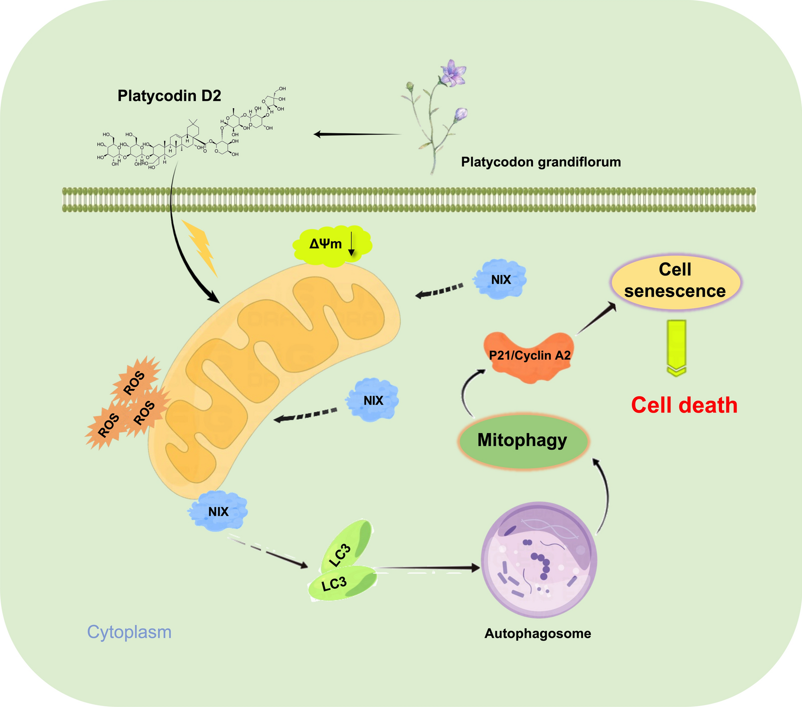
Cells and reagents
The liver cancer cell lines, Huh-7, MHCC97H, HCCLM3, HepG-2, and SK-Hep1, Huh-6, and normal liver cell lines, THLE-2 and L02, were purchased from the Guangzhou Saiku Biotechnology Co. (Guangzhou, China). PD2 (cat. HY-N4087), 3-methyladenine (3-MA) (cat. HY-19312), chloroquine (CQ) (cat. HY-17589A), and rapamycin (cat. HY-10219) were purchased from MedChemExpress (MCE, USA).
We purchased the control siRNA, NIX, and P21 siRNAs from RiboBio (China). Si-NIX (cat. SIGS0005709-1) and si-P21 (cat. SIGS0001992-1) were used in this study. We transfected 30 nM siRNA into cells using the transfection reagent in the kit.
Human cyclin A2 cDNA was purchased from the Public Protein/Plasmid Library, (Jiang su, China) and cloned into the pCDNA 3.1 plasmid. Following the manufacturer’s protocol, the cyclin A2 plasmid and corresponding empty vector were transfected into HCC cells using Advanced DNA/RNA Transfection Reagent (ZETA life, USA).
Five- to six-week-old female BALB/c nude mice were used in this study. Mice were fed in a specific pathogen-free environment at the Animal Experimental Center of the Changchun University of Chinese Medicine. Their food was sterilized with cobalt-60 radiation, and their ultrapure water was sterilized by autoclaving. This study was approved by the Institutional Animal Care and Use Committee of Changchun University of Chinese Medicine (Approval No. 2022062).
Cell viability assay
HCC cells were cultured in DMEM medium supplemented with 10% FBS and 1% penicillin/streptomycin dual antibody and incubated in an incubator at 37 ℃ and 5% CO2. HCC cells were passaged into 96-well plates at a rate of 5 × 10⁴/well. After 24 h, Huh-7, MHCC97H, HCCLM3, HepG-2, SK-Hep1, and Huh-6 cells and normal liver cells, THLE-2 and L02, were treated with different doses of PD2 from 1 to 100 μM. 5-FU was used as a positive control. After 48 h, the culture medium was replaced with 100 μL of medium containing 10 μL of the CCK-8 reagent (Cat#C0038, Beyotime Biotechnology). After 2 h of incubation at 37℃ and 5% CO2, the absorbance value was measured at 450 nm.
Detection of apoptosis rate
HCC cells were passaged into six-well plates (2 × 10⁵/well) and cultured in an incubator for 24 h. Double-free DMEM was added, and then 10 μM of PD2 was added to each well. After 48 h of incubation, the culture medium was removed and washed with PBS. Cells were digested with trypsin and centrifuged at 500 g for 5 min. The cell precipitate was washed with PB and resuspended in 1 × binding buffer. Subsequently, 5 μL of FITC and 5 μL of PI were added. After gentle blowing, cells were stained in the dark at room temperature for 20 min, and analyzed using a Beckman Coulter flow cytometer (CytoFLEX A00-1–1102, USA).
Cell cycle detection
HCC cells were seeded in six-well plates at a density of 2 × 10⁵ cells/well. After 24 h of culture in the incubator, double-free DMEM was added. Thereafter, 10 μM of PD2 was added. After 48 h of culture in the incubator, PBS or the drug was removed, and PBS was washed. Cells were digested with trypsin, centrifuged for 5 min (500 g), and washed with PBS. Precooled 70% ethanol was added to the samples, and the samples were placed in a refrigerator at 4 °C for 2 h. Then, the samples were centrifuged for 5 min (300 g), and PBS was added to re-suspend the cell precipitate. After centrifugation at 300 g for 5 min, the supernatant was removed, and PBS (0.5 mL) was added. Then, 5 μL of cell cycle solution was added, and the mixture was fully shaken and mixed with a vortex oscillator in the dark. The mixture was placed in an incubator for 15 min and analyzed using a Beckman Coulter flow cytometer (CytoFLEX A00-1-1102, USA).
Autophagy change detection
Cells were grown in six-well plates and treated with PD2 for 24 h. Next, cells were transfected with the plasmid EGFP-LC3. After 48 h, cells were fixed with 4% paraformaldehyde for 15 min, and changes in the green fluorescence of LC3 were observed under a confocal microscope (CARL ZEISS LSM980, Germany).
The cells were grown in six-well plates and treated with PD2 for 24 h. Next, the cells were infected with mRFP-GFP-LC3 double labeled adenovirus (Cat#HB-AP2100001, Hanbio Biotechnology, China) at a dose of 20 MOI. After 48 h, the cells were fixed with 4% paraformaldehyde for 15 min, and changes in the green fluorescence of LC3 were observed under a confocal microscope (CARL ZEISS LSM980, Germany).
The cells were grown in six-well plates and treated with PD2 for 48 h. The cells were collected and centrifuged, and then subjected to fixation, dehydration, embedding, slicing, and staining. Autolysosome formation was observed using a transmission electron microscope (JEOL 1400, Japan).
Detection of mitochondrial membrane potential
The qualitative and quantitative changes in mitochondrial membrane potential can be detected using JC-1 staining. HCC cells were passaged into 12-well plates at 2 × 10⁵/well. After 24 h, 10 μM PD2 was added to each well. After 48 h, the cells were stained with JC-1 staining solution. After 15 min of staining, cells were washed three times and then observed using a fluorescence microscope.
HCC cells were passaged into 96-well plates at 5 × 10⁴/well and treated with 10 μM PD2 24 h later. After 48 h, the culture medium was discarded and replaced with 100 μL of JC-1 staining solution. Absorbance was measured at 435 and 585 nm after incubation at 37 ℃ for 2 h.
The changes in mitochondrial membrane potential were measured by TMRM staining. Hepatoma cells were passaged into 12-well plates at 2 × 10⁵/well. After 24 h, 10 μM of PD2 was added to HCC cells. After 48 h, the cells were stained with TMRM staining solution, washed three times after 30 min of staining, and then observed using a fluorescence microscope.
ROS detection
HCC cells were passaged into 12-well plates at 2 × 10⁵/well. After 24 h, 10 μM PD2 was added to each well. After 48 h, the cells were stained with a DHR123 staining solution (Sigma, USA). After 30 min of staining, cells were washed once and transferred to flow tubes for detection and analysis with a flow cytometer (CytoFLEX A00-1-1102, USA).
Transcriptomic analysis
HCCLM3 cells were seeded in 6-well plates 12 h before experiments, then treated with Lico A (30 μM) for 48 h. The cells were collected for transcriptomic analysis. Total RNA extraction was performed on cells treated with Lico A. The extracted total RNA was then subjected to a sample quality check, and samples passing the check were subjected to PCR to obtain cDNA libraries. RNA concentration and purity was measured using NanoDrop 2000 (Thermo Fisher Scientific, Wilmington, DE). RNA integrity was assessed using the RNA Nano 6000 Assay Kit of the Agilent Bioanalyzer 2100 system (Agilent Technologies, CA, USA). The concentration is ≥ 25 (ng/μL), while the OD260/280 is 1.7–2.5 and the OD260/230 is 0.5–2.5. Quality-checked libraries were sequenced in PE150 mode with the Illumina NovaSeq6000 sequencing platform. Transcriptomic analysis was performed using the assay analysis service provided by Biomarker Technologies, Inc.
Western blotting
The cells were passaged to six-well plates at 2 × 10⁵ cells/well, and cultured in 5% CO2 and 37 °C in a cell incubator for 24 h. Next, 1 mL of double-free DMEM was added, followed by PBS, 10 μM PD2. After 48 h, the cells were collected and centrifuged to remove the supernatant, and total cell protein was extracted using a protein extraction kit (Cat# DE101-01, TransGen Biotech, China). Subsequently, the protein concentration was determined using a BCA protein assay kit (P1011, Beyotime Biotechnology, China), and the absorbance was read using a microplate reader. A standard curve was constructed, and the protein concentration of each sample was calculated. The sample was quantified with the minimum protein sample mass. Then, the sample protein was fully denatured, and 30 μg of total protein from each sample was loaded onto SDS-PAGE and transferred onto PVDF membranes. After 2 h of incubation in the blocking solution, the primary antibodies (1:1000) were incubated overnight at 4 °C. The membranes were then incubated with goat anti-rabbit or goat anti-mouse secondary antibodies for 40 min and washed three times with TBST for 10 min each. ECL immunoblotting chemiluminescence solution was used for dark color development. An ultrasensitive multifunctional imager was used for exposure (BIO-RAD ChemiDoc Imaging System 733BR3967, USA) (Additional file 1).
Co-localization observation assay
Cells in the logarithmic growth phase were seeded at 1 × 10⁵ cells/well in a 12-well cell culture plate pre-coated with cell slides and cultured at 37 °C and 5% CO2. Next, cells were transfected with the plasmid EGFP-LC3 for 24 h. Subsequently, adding 10 μM of PD2 for 48 h, samples were fixed with 4% paraformaldehyde for 30 min. After washing, the membranes were blocked with a blocking solution for 30 min. After discarding the blocking solution, the primary antibody solution was added to the 12-well plate, and the plate was gently shaken at 4 °C overnight. The primary antibody solution was removed, and cells were washed with PBS. Subsequently, the corresponding secondary antibody solution was added to meet different immunofluorescence co-localization requirements. After cleaning with PBS, the climbing piece was removed and attached to a slide. A nail polish seal was used to avoid drying, and photographs were taken under an inverted fluorescence microscope. Analysis of fluorescence intensity using ZEN 2.3 software.
β-galactosidase staining
HCC cells were passaged into 12-well plates at 2 × 10⁵/ well, and after 24 h, 10 μM PD2 was added to HCC cells. After 48 h, the cells were fixed for 15 min and then stained with a senescence-associated β-galactosidase staining kit (Beyotime, China). The cells were incubated at 37 ℃ overnight and observed using a microscope.
In vivo anti-tumor assays
SPF female BALB/c nude mice were housed in a sterile environment. SPF sterile mouse food and autoclaved water were fed for one week of adaptive feeding.
HCCLM3 cells in the mid-log phase were selected and digested with 0.25% trypsin. After centrifugation at 500 g for 10 min, cells were washed with PBS, mixed with double-free culture medium, and counted. Then, cells were adjusted to 5 × 10⁷ cells/mL concentration. A 100 μL cell suspension was injected into the right limb of each nude mouse. Seven-ten days after tumor implantation, mice were randomly divided into seven groups: PD2 (5, and 10 mg/kg), 5-FU (10 mg/kg, as positive control), siNIX + 10 mg/kg PD2, siP21 + 10 mg/kg PD2, cyclin A2 + 10 mg/kg PD2, and PBS (control). The drug was administered via intratumoral injection once every three days for four consecutive doses. Body weight change and tumor size of nude mice were recorded. Finally, histopathological tests were performed.
Statistical analysis
Data are expressed as mean ± standard error. GraphPad Prism software (version 8.0) was used for statistical analysis. T-test and one-way analysis of variance (ANOVA) were used for comparing groups. When P < 0.05 in ANOVA, the Student–Newman–KeuLs test was used for multiple comparisons. P < 0.05 was considered statistically significant.


Comments (0)