In this [15O]H2O-PET study, we demonstrated that acoustic stimulation of patients with SSD evoked more symmetric patterns of activation in the auditory cortices than those of a matched group of subjects with normal hearing on both sides (Figs. 2, 3 and 4). Moreover, we showed that the use of a CI reversed these changes towards more normal patterns. When patients with SSD used a CI on their deaf ear, they developed a more asymmetric activity of the auditory cortices to an unchanged acoustic stimulation of their normal hearing ear. The patterns with CI became more similar to those of the matched HCS. These findings support the view that CI in SSD affects the entire auditory processing permanently. Regaining hearing on the deafened side induced higher neuroplastic changes, including processing by the contralateral hearing ear. It may be assumed that at least part of the salutary effects of CI on hearing and on tinnitus in SSD is related to such changes.
It has been shown in many clinical studies that CI in SSD can improve speech understanding in noise, the localization of sound, and that it can suppress tinnitus (e.g., [17,18,19,20,21,22,23,24,25]). Imaging studies of neuronal plasticity induced by CI are more difficult to obtain than auditory studies. Even though ongoing developments on CI devices may improve compatibility with MRI examinations [2], artifacts caused by the case and the magnet will remain, affecting in particular the area of the temporal cortex. Examining the auditory cortex as the primary region of interest by fMRI may therefore continue to be problematic.
Our functional imaging study investigating neuroplastic changes after CI in SSD was performed by [15O]H2O-PET. The [15O]H2O-PET examined changes of the rCBF in the auditory cortices during acoustic stimulation compared to a baseline recording without stimulation. Compared to other imaging techniques used to examine the effects of SSD such as fMRI [2,3,4,5,6,7,8,9,10,11], EEG [12, 13], or MEG [14], [15O]H2O-PET can only examine relatively slow changes in minutes with relatively limited resolution of 4–7 mm FWHM (full width at half maximum). Only rCBF and not direct neuronal activity is measured. This together with the limited resolution and partial volume effects leads to images representing not only the AC enriched during acoustic stimulation, but also surrounding structures. This limitation seems acceptable since conditions were similar for all participants and the focus was on changes. Within these limitations, we found a reduced hemispheric asymmetry of the auditory cortices in unilateral deafness. However, it must be assumed that other brain areas also involved in neuroplastic changes due to SSD were not visualized with the [15O]H2O-PET technique, possibly also due to the small sample size of our patient group.
A recent [15O]H2O-PET study [32] investigated cortical plasticity after cochlear implantation in asymmetric hearing loss (AHL) with similarities to ours. The size of the study was similar with an inclusion of ten patients with unilateral CI after postlingual AHL and ten controls with normal hearing. In contrast to our study, the patients were examined once only with four different conditions of acoustic stimulation (baseline, bilateral, right, left). Results of this [15O]H2O-PET study after CI implantation [32] were compared to those of a previous study of the same research group [8], which examined acoustic stimulation of the normal hearing ear in SSD patients with fMRI. A normalization of interhemispheric asymmetry was demonstrated. We confirm this finding with our prospectively collected data within the same patient group before and after CI. Karoui et al. investigated also stimulation by CI and found a tendency of contralateral predominance, but without statistical significance [32]. The AI of all test conditions was not significantly different in comparison to the control group [32]. Notably, contralateral dominance during stimulation of the non-implanted ear correlated with performance of sound localization [32].
Even though laterality of the postlingual SSD seems to have an influence on the patterns of adaptive changes [27, 35,36,37,38,39,40,41], our sample of four right- and four left-sided SSD was too small to investigate laterality differences. Nevertheless, the four patients with a left-sided CI had remarkable improvements of their AI in comparison to the four right-sided patients. It seems possible that they benefited more from their CI. Laterality differences may be attributed to the distinct lateralization asymmetries in normal binaural auditory central processing. Larger samples would be needed to investigate this further.
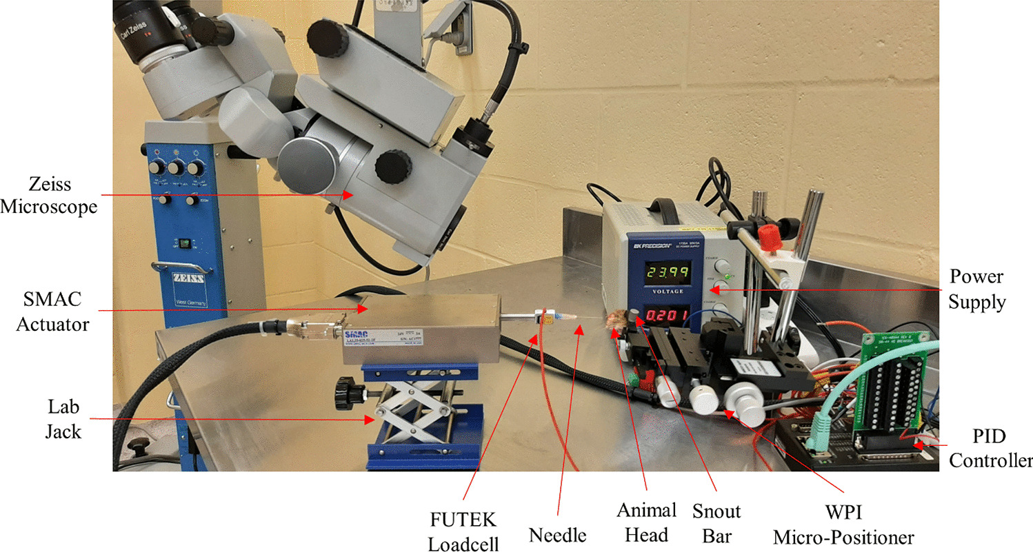
A major strength of our prospective study was the direct visualization of neuroplastic changes with [15O]H2O-PET before and after CI in the same subjects. These imaging methods were not influenced by the implantation of a magnet. Since the CI was not stimulated during examination, artifacts such as described, for example, in the CEAP responses were avoided [26].
There are several limitations to our study. The sample size was too small to allow for statements about right-left differences. Moreover, the use of radioactive tracers in [15O]H2O-PET examinations limited the number of examinations per subject. For this reason, our study design focused on postoperative measurements of the normal hearing ear only, and not on CI stimulation. In addition, the [15O]H2O tracer with a half-life of 2 min must be produced on the examination site, which causes waiting periods of 10 min between scans. Consequently, the total examination time was approximately 1.5 h in the scanner. Patients were reminded of the next examination before a scan, but fatigue with effects on the results of the cortical activities cannot be excluded. Unfortunately, additional unforeseen and prolonged technical problems with the onsite radionuclide production by a cyclotron necessitated deviation from the protocol and a wider time range of postoperative examinations than initially planned.
In summary, our [15O]H2O-PET study showed more symmetrical rCBF in the AC when the healthy ear was stimulated in SSD than in HCS. Secondly, we were able to show a reversion of these neuroplastic changes to more normal patterns in SSD patients after the regular use of a CI. This objective finding complements the well-established positive subjective effects of CI in SSD. Subjective benefits include improved speech understanding in noise, improved sound localization, reduced tinnitus perception, better general hearing ability, and improved quality of life (e.g., [17,18,19,20,21,22,23,24,25, 32]). In view of the fact that CI in SSD is still not reimbursed in several countries [2], further studies on neuroplastic changes caused by CI in SSD with larger numbers of cases are desirable, such as with PET-CT, EEG, or with other techniques. Such studies could also help to better predict the outcome of CI in SSD and determine the period of deafness in which a reversion of neuroplastic changes by CI can still be expected.


Comments (0)