
記住我


This study was approved by the State and Institutional Animal Care Committee (Landesuntersuchungsamt Rheinland-Pfalz, Koblenz, Germany; approval no. G 16-1-042). The study is a prospective, randomized trial and was conducted from May 2019 to April 2020. The study followed the ARRIVE guidelines and involved 45 juvenile, male pigs (Sus scrofa domestica; mean weight 30 ± 3 kg; age 12–16 weeks) from a local breeder.
The experimental setup was based on previously conducted resuscitation studies [3, 13]. After intramuscular injection of ketamine (Hameln Pharmaceuticals GmbH, Hameln, Germany; 1.5 mg/kg BW), azaperone (Lilly Deutschland GmbH, Bad Homburg, Germany; 2.5 mg/kg BW), and midazolam (Hameln Pharmaceuticals GmbH, Hameln, Germany; 0.3 mg/kg BW), the sedated animals were transported to the laboratory.
An IV access was established at the ear. Anesthesia was then induced by a bolus injection of fentanyl (Janssen-Cilag, Neuss, Germany; 4 µg/kg BW) and propofol (Fresenius Kabi, Bad Homburg, Germany; 2 mg/kg BW), and a single dose of atracurium (HEXAL AG, Holzkirchen, Germany; 0.5 mg/kg BW) was administered to facilitate endotracheal intubation. The animals were ventilated in volume-controlled mode [tidal volume 6 ml/kg BW, positive end-expiratory pressure of 5 mbar, inspired fraction of oxygen (FiO2) of 0.4, inspiration to expiration ratio (I:E) of 1:2 and variable respiratory rate to achieve an end-tidal arterial pressure of carbon dioxide (PaCO2) below 6 kPa using an intensive care respirator (Engstroem care station, GE healthcare, Munich, Germany)]. Peripheral oxygen saturation was measured continuously with a sensor clipped to the ear (Radical 7, Masimo Corp., Irvine, CA, USA). General anesthesia was maintained using continuous infusions of fentanyl (0.1–0.2 mg/kg BW/h) and propofol (8–12 mg/kg BW/h). The animals received intravenous infusions of an isotonic electrolyte solution (Sterofundin®, B. Braun Melsungen AG, Melsungen, Germany) initially as a bolus of 30 ml/kg BW and then continuously with 5 ml/kg BW/h.
Arterial and central venous access was established using ultrasound guidance as previously described [13], and a pulse contour cardiac output system (PiCCO, Pulsion Medical Systems, Munich, Germany), a pulmonary artery catheter (PA Katheter Swan Ganz, Edwards Lifesciences Corporation, Irvine, CA, US), and a fibrillation catheter (VascoStim B 2/5F, Vascomed GmbH, Binzen, Germany) were inserted.
Trial protocol and data collectionAfter the instrumentation, a half-hour consolidation phase followed, during which a set of six chemically inert gases with varying rates of transpulmonary elimination (sulfur hexafluoride, krypton, desflurane, enflurane, diethyl ether, and acetone) mixed in safe quantities with saline solution were injected intravenously. After allowing 30 min for stabilization and reaching a steady state, MIGET measurement (MIGET, MMIMS-System, Oscillogy LLC, Folsom, US) was conducted at the measurement time point baseline healthy (BLH). In this procedure, 10 ml of arterial and pulmonary arterial blood was drawn after heparinization of the blood, and the pulmonary elimination of inert gases was analyzed by mass spectrometry. Differences in transpulmonary elimination constants for those gases allow for a mathematical estimate toward ventilation/perfusion (V/Q) ratios and shunt measurement.
Arterial and mixed venous blood gases were obtained (radiometer, ABL90flex, Copenhagen, Denmark) at time point BLH. The hemodynamic parameters were recorded continuously using Datex Ohmeda S5 monitor (GE Healthcare, Munich, Germany) as well as the ventilation parameters using the internal software of the intensive care respirator.
The measurement time points after BLH are designated as follows: T4 denotes 4 h after ROSC, T20 denotes 20 h after ROSC, etc.
InterventionAfter BLH, the animals were randomized into one of three intervention groups by pulling sealed envelopes (Table 1). Ventricular fibrillation was induced via a venous fibrillation catheter, as previously described [13]. The sham group is the control group in which no cardiac arrest was induced, and accordingly, no ventilation intervention was performed.
Table 1 Group design and intervention parameters of the conducted experimentsAfter monitor-confirmed ventricular fibrillation, ventilation was disconnected, and the animal was left untreated for 8 min. Eight minutes after induction of cardiac arrest, basic life support (BLS) was initiated: the animal was mechanically resuscitated with the assigned ventilation mode (Table 1). Thoracic compressions were administered using a mechanical chest compression device (LUCAS 2, Physio-Control, Redmond, US). Thoracic compressions were performed at a fixed frequency of 100 compressions per minute and a compression depth of 5 cm. After 8 min of BLS, an arterial and mixed venous blood gas analysis was taken, and blood samples were collected for MIGET. Immediately afterward, advanced life support (ALS) was performed, adapted to the ALS algorithm of the ERC [1]. If ventricular fibrillation or flutter was detected, immediate defibrillation was performed (200 Joule; Zoll R Series Monitor Defibrillator, Zoll Medical Deutschland GmbH, Cologne, Germany), resuscitation continued, and medications were administered intravenously according to the following schedule: adrenaline (1 mg; Sanofi-Aventis GmbH, Frankfurt a.M., Germany), vasopressin (0.5 U/kg BW; Pfizer Inc., New York City, US) after the first, third, sixth and ninth defibrillation, and amiodarone [150 mg; Hikma Pharma GmbH, Martinsried, (DE)] after the third and sixth defibrillation. After 8 min of ALS, arterial and mixed venous blood gas analysis, and MIGET measurement was performed (Fig. 1).
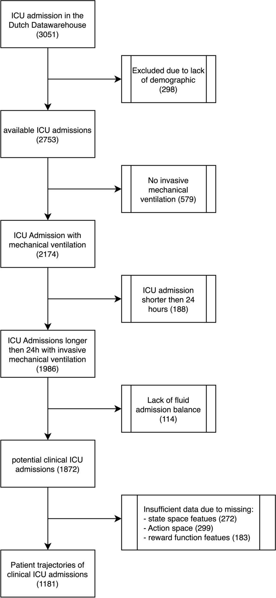
Fig. 1
This figure was created using the graphics software draw.io
Flowchart of the intervention groups during the peri-resuscitation phase. If no ROSC was achieved, this led to exclusion from further data analysis. ROSC return of spontaneous circulation, IPPV intermittent positive pressure ventilation, ULTVV ultra-low tidal volume ventilation, Tx timepoint (in hours) after ROSC, MIGET multiple inert gas elimination technique.
If return of spontaneous circulation (ROSC) was not achieved after ten ALS cycles, resuscitation was terminated, and the experiment was ended. If ROSC was achieved during a rhythm analysis, resuscitation was terminated, and the post-resuscitation phase began.
Post-resuscitation phaseAnimals which achieved ROSC were returned to standard ventilation, as described at baseline, and were monitored for 20 h with the aim of maintaining peripheral oxygen saturation above 93%. If necessary, the invasiveness of ventilation was adjusted according to the ARDS-network specifications [14], while mean arterial blood pressure was maintained above 60 mmHg using norepinephrine administration and volume boluses. Upon return of spontaneous circulation, the animals received a dose of 30 ml/kg BW of the electrolyte solution over a 2-h period, followed by an hourly rate of 2.5 ml/kg BW.
MIGET was taken again after 6 and after 20 h after ROSC. Blood gas analyses were performed repeatedly, and ventilation parameters were continuously recorded.
To prevent painful pressure sores, the animal was repositioned every 3–4 h between supine position, right and left side during the observation period.
The experiment was concluded by administering 40 mmol potassium chloride via the pulmonary artery catheter after inducing deeper general anesthesia with 200 mg propofol.
Sample collectionPostmortem, the lungs were dissected free under preserved ventilation. Before the lungs were collected in their entirety, the trachea was clamped at the end of inspiration to minimize the risk of developing postmortem artificial atelectasis. Lung tissue samples were collected from the dorsal and ventral regions of the peripheral upper and lower lobes of the lung and fixed with 4% formalin. The samples were processed by the tissue bank at the University Medical Center Mainz in Germany, where they were paraffinized, sliced into 2-µm-thick sections, and then stained with hematoxylin–eosin (HE).
Lung damage scoring systemUsing an Olympus microscope (CX43RF, Olympus Cooperation, Tokyo, Japan) and CellSens software (Olympus cellSens Entry, Version 2.1, Olympus Corporation, Tokyo, Japan), the lung tissue samples were examined and assessed in a blinded fashion by a trained investigator using the established diffuse alveolar damage (DAD) scoring system [15], containing seven sub-items: alveolar edema, interstitial edema, hemorrhage, inflammation, epithelial damage, microatelectasis and overdistension. The DAD scoring system is the recommended examination tool of the expert consensus recommendations of the American Thoracic Society for simulating lung injury in animal models [16].
Exclusion criteriaOnly animals that showed no obvious signs of illness (e.g., normal eating and drinking behavior in the days before the experiment, no apparent injuries or inflammations) were included. This was ensured prior to sedation and transportation.
For animals in the intervention groups, only those who achieved sustained ROSC within the aforementioned study protocol were included in the data analysis.
Statistical analysisStatistical analyses were performed using one-way ANOVA with post-hoc Bonferroni correction [17], if the prerequisites for the use of ANOVA were given [18, 19]. Effects over time were analyzed using repeated measures ANOVA; within-subject effects were assessed using the Greenhouse–Geisser correction in case of non-sphericity. For single measurements, if normal distribution was not given, Kruskal–Wallis test [20] was used. Statistical evaluation was performed using IBM SPSS Statistics (IBM SPSS Statistics for Windows, Version 20. IBM Corporation, Armonk, NY, USA). The data are presented as mean (standard deviation). A significance level of 0.05 was set.
留言 (0)