Participants
Patients received transepithelial CXL between September 2021 and January 2023 and were selected from the Fudan University-affiliated Eye and ENT hospital. The inclusion criterion was clear diagnosis of spontaneous keratoconus, pediatric keratoconus, increase of maximum keratometry by at least 1 Diopter (D) or increase of pachymetry by at least 20 microns in a year,
preoperative topography (examined with a Pentacam produced by Oculus Optikgeräte, Wetzlar, Germany) results indication of the thinnest corneal thickness > 380 um, and age ranges between 18–35 years old. Carriers of systemic diseases, autoimmune diseases, or psychological diseases were excluded. The evaluation of lens comfort was conducted by an experienced ophthalmologist, observed via a slit-lamp, and postoperative corneal morphologies were recorded for each individual.
In this prospective study, 60 participants (60 eyes) were recruited, with an average age of 25.42 ± 5.47 years. The male:female ratio was 39:2. The patients were divided into groups A and B based on the random number table method, and each group contained 30 patients. There were no significant differences between the two groups (P > 0.05) as indicated in Table 1.
Table 1 Participants’ basic information for two groups (Mean ± SD, N = 60)Study protocol
After a routine preoperative examination, each surgery was performed by the same experienced senior physician.
The surgical procedure was the same as that used in our previous study [13]. The cornea was infiltrated with Sufficient Paracel (Avedro, Waltham, MA, USA) which includes 0.25% riboflavin-5-phosphate, NaCl, 1.2% hydroxypropyl methylcellulose (HPMC), 0.01% benzalkonium chloride, sodium edetate and trometamol for 14 min, then exposed to UV light with a wavelength of 370 nm and intensity of 45 mW/cm2 (Avedro, Waltham, MA, USA). The exposure time was 5 min 20 s for pulse irradiation with 1 s bright and 1 s dark, and the total energy was 7.2 J/cm2.
Once the surgical procedure was completed, both groups were immediately fixed with AcuVue Oasys SiH contact lenses (Johnson&Johnson, ACUVUE, USA). In terms of postoperative medication use, each patient was provided with Clopito (Santen, Japan) and Flumei drops (Santen, Japan) after surgery, accompanied with a follow up period of 7 days.
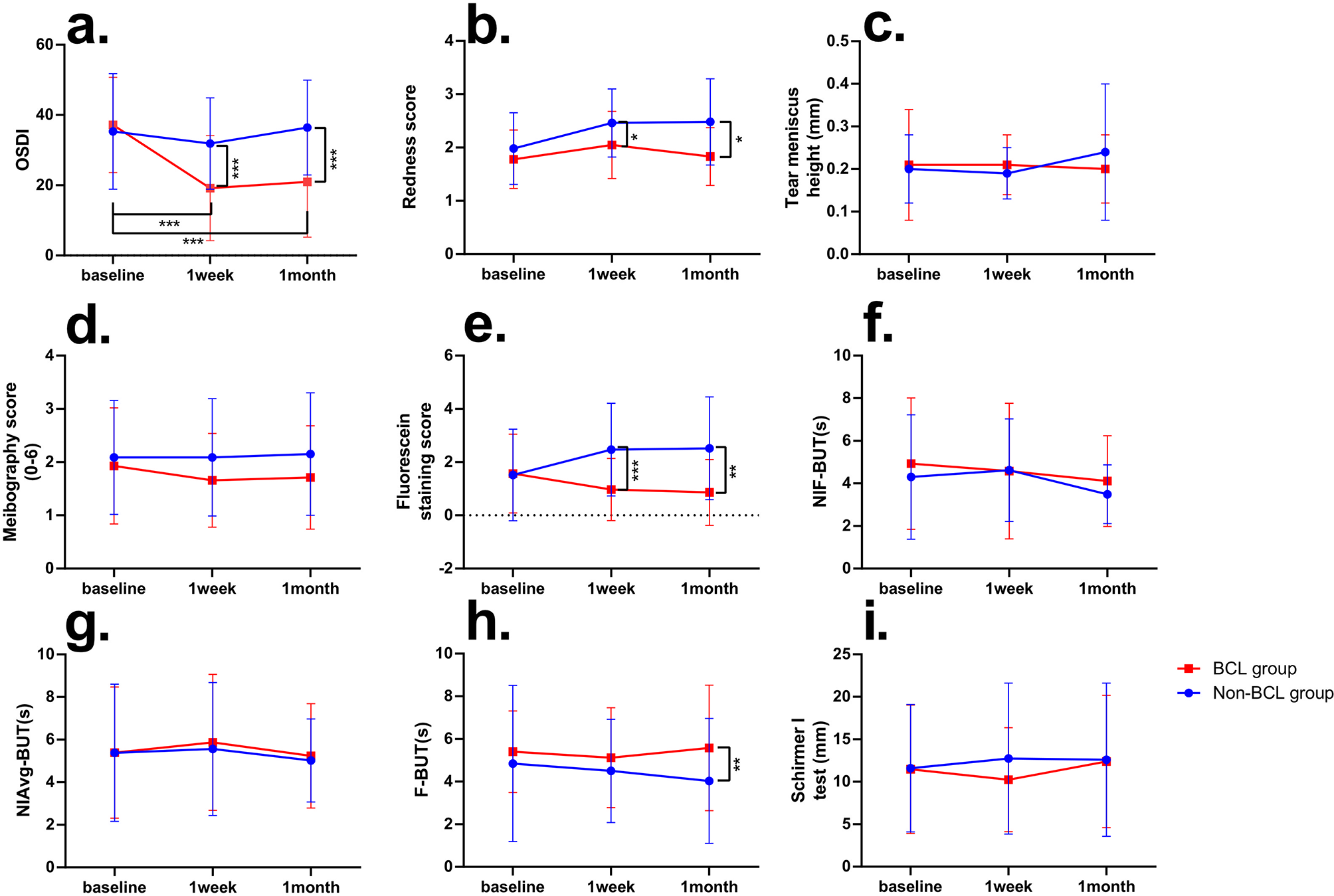
On the day after the operation, the researcher evaluated the lens condition of each patient using a slit-lamp microscope, including the central position of the lens, any horizontal or vertical lens displacement, degree of lens movement during natural blinking in the original ocular position and upward gaze position, and wettability of the lens. Patients in Group A underwent lens removal at postoperative day 7, whereas those in Group B underwent lens removal after 3 days.
Postoperative observationSubjective symptoms of the operative eye
Postoperative comfortability
A standard self-administered questionnaire was adopted for both groups to record the postoperative comfort of the ocular area, which was administered on postoperative days 1, 3, 5, and 7. The questionnaire assessed 10 symptoms, including photophobia, tearing, burning, pain, foreign body sensation, blurred vision, difficulty opening the eyes, dry eyes, edema, and sting. Each feature was scored from 0–3, where a score of 0 was considered asymptomatic, and scores of 1, 2, and 3 were rated as mild, moderate, and severe, respectively. The participants received a tutorial regarding the questionnaire, and the completed questionnaires were collected on the day of the re-examination.
Severity of postoperative ocular pain
The visual analogue scale (VAS) is used to assess the severity of postoperative ocular pain. The VAS for pain is composed of a 10 cm straight line, which is marked with 0 at one end and 10 at the other. A score of 0 represents “no pain,” score 1–3 represents “mild pain,” which does not affect sleeping; score 4–6 is defined as “moderate pain,” which affects night sleep; score 7–10 is regarded as “severe pain,” which severely affects sleeping [14]. The participants were asked to rate their scores based on their subjective pain sensation.
Objective signs of the operative eye
1)
A single blinded test was applied for corneal edema assessment, where one senior ophthalmologist masked the allocation of the participant’s group while examining the ocular condition of each participant using slit-lamp microscopy. The resulting ocular status was recorded using a score range between 0–3. A higher score represents worse symptoms, where 0 implies a symptom-free cornea and 3 indicates the most edematous cornea. In addition, conjunctival congestion was examined by the same experienced doctor via slit-lamp using the same single blinding method and the ophthalmologic status was recorded. Scoring ranged 0–3, and the higher the score, the worse the conjunctival congestion.
2)
Corneal contact lens conditions were also recorded at each examination.
Statistical analysis
The database was established in Excel using SPSS 20.0 statistical software. All the data were tested for homogeneity of variance and sphericity. Further, analysis of variance (ANOVA) and chi-square tests were used for comparison of preoperative basic condition in the two groups, and repeated measures analysis of variance (rANOVA) and one-way ANOVA were used for postoperative comparison between groups; P < 0.05 was considered statistically significant. Lens displacements from the center were analyzed using the mean standard deviation.


留言 (0)