Study design
This nonrandomized open-label controlled study enrolled patients who underwent small-incision lenticule extraction (SMILE) and femtosecond laser-assisted in situ keratomileusis (FS-LASIK) procedures between July 2021 and April 2022 at the Department of Ophthalmology, Peking Union Medical College Hospital (PUMCH). The participants were sequentially allocated to MCG and control groups. The study adhered to the tenets of the Declaration of Helsinki and was supervised by the institutional review board at PUMCH. Signed informed consent was obtained from all patients.
Inclusion criteria were as follows: (1) age between 18 and 40 years; (2) stable refractive error (≤ 0.5 D change of refractive error) in the past 1 year; (3) spherical equivalent (SE) between -2.50 D and -20.00 D; (4) astigmatism up to -5.00 D; (5) logarithm of the minimal angle of resolution (LogMAR) of BCVA of 0.1 or better; (6) clear crystalline lens.
Exclusion criteria included current or history of severe ophthalmic diseases including corneal diseases, cataracts, glaucoma, retinal detachment, neuro-ophthalmic diseases, trauma, ocular surgery, and diagnosed autoimmune diseases. Patients with severe preoperative dry eye, defined as prominent desiccation before surgery, a fluorescein TBUT < 2 s, and/or corneal epithelial defects in 2 quadrants or more and/or fluorescein staining ≥ 30, were excluded from the study [15, 16].
All participants underwent complete preoperative ophthalmic examinations, including uncorrected visual acuity (UCVA), best corrected visual acuity (BCVA), manifest and cycloplegic refraction by autorefractometery (RM-800, Topcon, Japan), standard slit-lamp biomicroscopy and funduscopic examinations, gonioscopy, intraocular pressure (IOP) by a non-contact tonometer (Canon, Japan), and corneal topography (Canon, Japan). The participants underwent routine follow-ups at 1 day, 1 week, and 1 month postoperatively, with examinations including BCVA, manifest refraction, standard slit-lamp biomicroscopy, and IOP measurement. OSDI, DED evaluation, and HOA examinations were performed at the preoperative examination and each follow-up session. Participants unable to complete preoperative examinations or more than 1 follow-up examinations were excluded from the analyses.
Surgical procedures
Surgical procedures were performed by an experienced surgeon (YL). SMILE was performed under topical anesthesia, with the VisuMax 500 kHz femtosecond laser (Carl Zeiss Meditec, Germany). Cap thickness was set at 110–120 μm, cap diameter at 7.0–7.5 mm, and lenticules diameter at 6.0–6.5 mm, with a transition zone of 0.1 mm. A 2 mm side cut incision was made at the 10 o’clock position of the cornea. Cut energy was set at 135 nJ. The stromal lenticules were removed using forceps.
FS-LASIK was performed using the VisuMax 500 kHz femtosecond laser (Carl Zeiss Meditec, Germany) for flap creation, and the Schwind Amaris 179 excimer laser (Schwind Eye-Tech-Solutions, Germany) for refractive correction. Flap thickness was set at 90 or 100 μm, flap diameter at 8.5 mm, and hinged at the 12 o’clock position of the cornea. The side cut angle was at 120 degrees.
The prescriptions for postoperative management were as follows: tobramycin dexamethasone eye drops (s.a. Alcon-Couvreur n.v.), four times a day for two weeks, deproteinized calf blood extract eye gel (Xingqi pharmaceuticals), once for four weeks, and sodium hyaluronate eye drops, four times a day for four weeks.
Moisture chamber goggle
The moisture chamber goggle consists of two moisture-retaining chambers and supporting structures. The chambers have rubber adapters to better fit the frame of the user, providing better sealing and a more comfortable wearing experience. At each junction of the chamber and the temples, a tank connected to the chamber, filled with water-absorbing material, was designed to provide moisture and maintain humidity levels in the chambers. The participants were asked to fill the tanks each time before use. The participants were instructed to use MCG for at least half of the day according to instruction postoperatively for 1 month.
DED evaluation
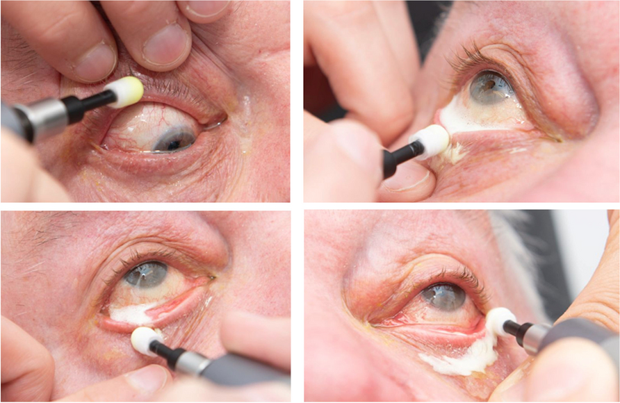
DED-1L Dry Eye Analyzer (Kanghuaruiming Science Technology, China) is a comprehensive dry eye diagnostic system that performs non-invasive tear film break-up time (NIBUT), tear meniscus height (TMH), lipid layer interferometry, and congestion assessment [17]. NIBUT and TMH measurement was performed under infrared light, while lipid layer interferometry and congestion examination was conducted under natural white illumination. NIBUT is automatically measured as the duration from the last complete blink to the first discontinuity in Placido ring reflections under infrared illumination. TMH was calculated as the average of three measurements at central, medial, and lateral paracentral locations. The congestion levels in the conjunctival and perilimbal regions were automatically measured under natural light, and an average congestion score was calculated.
The LipiView Ocular Surface Interferometer (TearScience, United States) measures the absolute thickness of the lipid layer using interferometric images of the tear film. During the examination, the participants were asked to maintain still and blink normally. The camera was adjusted to focus on the tear film plane and captured a 25-s video with a clear interferometric image of the tear film [18]. Lipid layer thickness (LLT) is measured in interferometric color units (ICU). LipiView automatically calculates the average (AvgICU), maximal (MaxICU), and minimal (MinICU) measurements of LLT. The homogeneity in lipid layer distribution is reflected by \(DevICU= \sqrt^+^}\).
To avoid introducing bias from diurnal variations, objective DED evalutions were performed in the mornings. The participants were asked to complete the OSDI questionnaire at preoperative examination and 1-month follow-up.
HOA evaluation
The iTrace aberrometer (Tracey Technologies, United States) was used to measure ocular HOAs following 10 min of dark adaptation without pharmacological pupil dilatation before the operation and at the follow-ups. The root means square values of total HOAs, spherical aberration, secondary astigmatism, coma aberration, and trefoil aberration were recorded.
Statistical analysis
Statistical analyses were performed with R (version 4.2.3) and RStudio (2023.03.1 + 446). BCVA and UCVA were converted to logMAR visual acuity. Continuous variables were presented as mean ± standard deviation (SD) under normal distribution or median with interquartile range (IQR) under non-normal distribution. Independent student’s t-test was used for comparisons of parameters between control and MCG groups. Paired student’s t-test was used for comparisons between preoperative and postoperative parameters within a group. A p-value of less than 0.05 was considered statistically significant.


Comments (0)