Preparation of nanoparticles
In this study, nanoparticles were made using the Diffusion Emulsification-So technique. A 1:1 mixture of benzyl alcohol and water was placed on the heater-stirrer at a temperature of 55 degrees Celsius, and It was allowed to mix well and saturate each other for 11 min. Then it was kept still for 21 min until the saturated phases were separated from each other (the organic phase saturated with water and the aqueous phase saturated with organic compound), and two phases were produced. Thus the lower phase containing benzyl alcohol saturated with water and the upper phase containing water saturated with benzyl alcohol was obtained. Compritol and oleic acid are melted together at a temperature of 55–65 degrees using indirect heat mixed, and then at the same temperature, the first part of the surfactant formula that includes lecithin or a 1:1 mixture of Span 20 and tween 80 was added to it. After mixing and becoming uniform, the desired amount of iodine (1% concentration). weight/weight) was added, and after complete mixing, the required amount of benzyl alcohol saturated with water was added to it and mixed well.
On the other hand, to prepare the aqueous phase, mix 1:1 propylene glycol and polyethylene glycol 300 in the amount of 3% of the total formula, with the second part of the surfactant-containing labrafil at a temperature of 55 degrees on bain-marie It was mixed. After mixing, the required amount of water saturated with benzyl alcohol was added and well mixed. The aqueous phase was added dropwise to the lipid phase. In contrast, the phase mentioned under the device, The high-speed homogenizer (HSH), was mixed well at 12,000 rpm for three minutes until the emulsion was formed. It was then placed at four °C for 31 min and then for 5 min. It was sonicated at room temperature. 18.75 mg of chitosan was dissolved in an aqueous solution (v/v) of 0.5% acetic acid. Then The resulting solution was diluted to a concentration of 2.5 ml/mg, and finally, its pH was adjusted with sodium hydroxide. to 5. The final solution was cooled to 4°C for the next steps. The emulsion formed in the previous step with 30 ml of 4 °C solution consisting of ml/mg 2.5 Chitosan was diluted to 60 ml and mixed and homogenized for 30 min at the same speed to form NLC particles. Several formulations were made in the form of pre-formulation, and it was observed that the considered range did not have the desired efficiency. Finally, A final formulation was obtained.
Characteristics of nanoparticlesParticle size
The formulation of solid lipid nanoparticles without iodine had a particle size of 133 ± 8.5 nm, and after loading iodine, this size increased to 168 ± 7.2, which is a significant increase. (P = 0.028). On the other hand, Poly Dispersity Index (PDI) parameter obtained is equal to 0.24, which indicates the homogeneity of the particle size in the aqueous suspension of nanoparticles, but still the size of Particles below 200 nm—considering that properties such as stability, and membrane permeability are affected by particle size. Therefore, this feature is one of the most essential features of nanoparticles. The size of the particles below 200 nm obtained in this study has increased the Brownian motions and the resulting suspension's stability. On the other hand, considering that Lipid nanoparticles are responsible for the passage of iodine through the cornea, particle size is very influential in this feature. These particles, having a size of 100–200 nm, can penetrate the membrane; therefore, the nanoparticles prepared in this study help the penetration of iodine into the eye.
Particle morphology
The images prepared by the AFM method show spherical particles with high homogeneity, proving the formation of lipid nanoparticles. The particle size obtained by the AFM method confirms the particle size results obtained by the optical diffraction method. Also, the pictures taken after three months of storage show the stability of the nanoparticles and the non-integration of the particles into each other. Percentage of iodine loaded in nanoparticles One of the most critical factors is Entrapment Efficacy (EE%), which expresses the percentage of the drug It is loaded with nanoparticles. The EE% value of iodine in iodine nanoparticles is 81.3 ± 2.7%. Considering the lipophilic nature of iodine, this percentage seems reasonable.
Iodine released test from lipid nanoparticles
The obtained results indicate that iodine was continuously released during the first ten hours, and after 4 h, about 21% was released. Therefore, lipid nanoparticles have acted as a reservoir for the iodine Act, continuously released it, and made it available to the environment. Considering that the retention time of nanoparticles on the eye's surface is also limited, the nanoparticles prepared in this study can provide about 21% of iodine to the eye during the first 4 h.
Animal clinical trial
In the present study, 15 New Zealand rabbits weighing 2 to 2.5 kg were studied. The rabbits did not differ regarding breed, sex, age, or physical health, and they did not have any eye abnormalities or corneal ulcers. They were kept under the same conditions and received the same proper nutrition. In this study, rabbits were randomly divided into groups of 5 (I, II, and III). After infecting the eyes of rabbits with Staphylococcus aureus bacteria and inducing corneal ulcers, the selected formulation of iodine lipid nanoparticle was used for group III rabbits, and iodine solution (1.25%) without nanoparticle structure was used as eye drops for group II rabbits. Group I was considered the control group. Nanostructured lipid carriers (NLC) formulations without drugs were used for Group I. The sample number was determined using the G*Power program. The minimal number of rabbits needed for the research was determined to be 13 based on the variables, the effect size of 0.35 with a confidence factor of 95%, the test power of 80%, and a related study by Brozou et al.
In this study, Staphylococcus aureus (ATCC 25923) was used. In the antibiogram, this bacterium was resistant to penicillin and ampicillin antibiotics but was sensitive to gentamicin, tetracycline, clindamycin, and erythromycin antibiotics.
Ketamine (30 mg/kg) and xylazine hydrochloride (10 mg/kg) subcutaneous injections were part of the anesthesia procedure, and tetracaine eye drops (0.5%) were used for local anesthesia of the eyes. A mydriasis procedure was also carried out using tropicamide (1%) droplets. A 27 gauge needle with a 1 ml syringe was introduced into the corneal stroma's middle depth and halted at the border of the 2 mm optical zone to cause an ulcer. Next, 100 microorganisms of Staphylococcus aureus were infused into 0.02 ml of physiological serum. After 24 h of injection using the score of Johnson et al. and Sanders et al.(Table 1), rabbits suitable for further study regarding the ulcer were isolated. Accordingly, rabbits with a score greater than two and less than 5, and a positive culture of Staphylococcus aureus from corneal secretions and necrotic tissue, were selected.
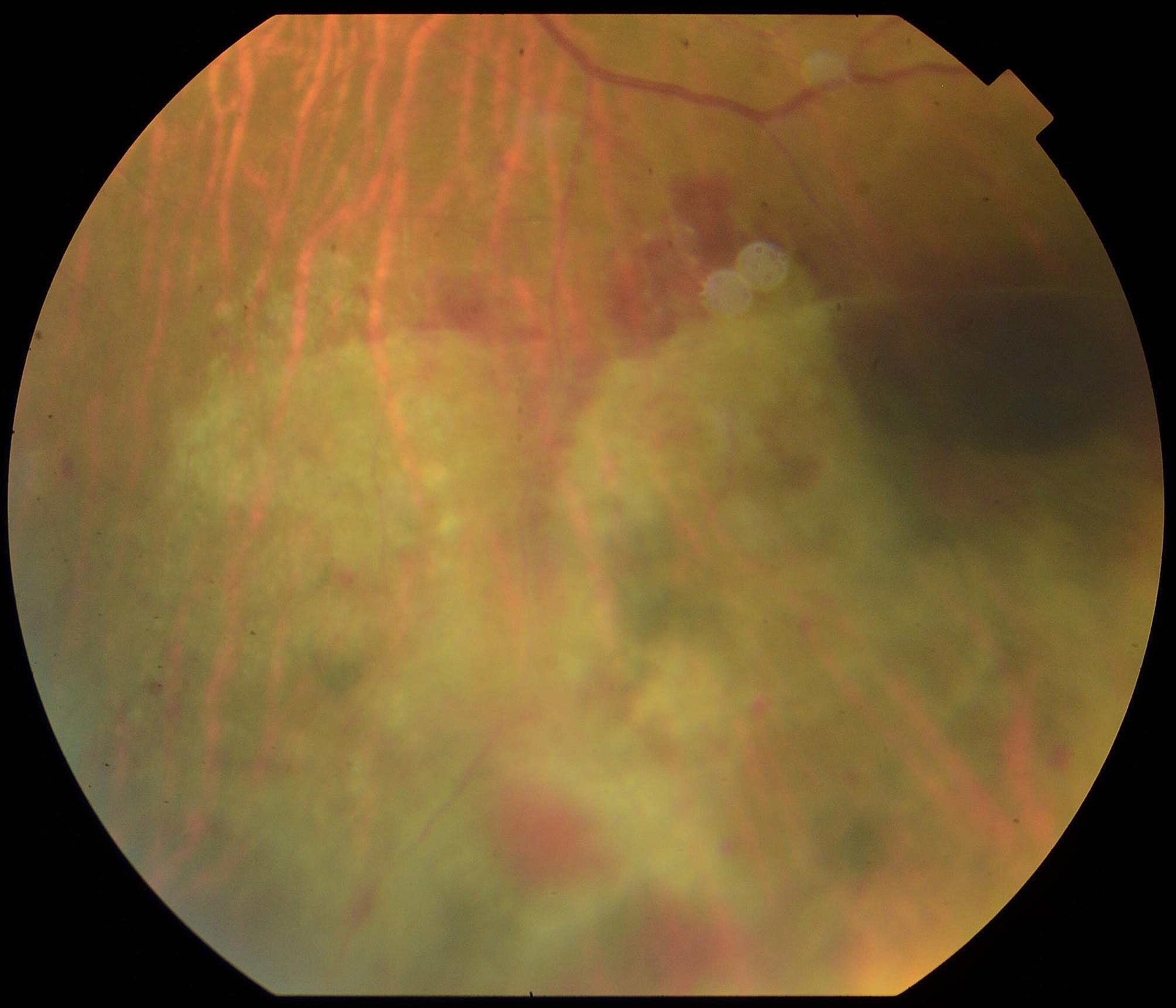
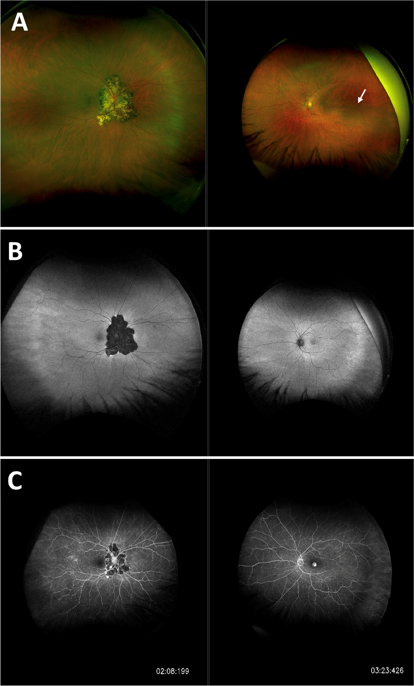
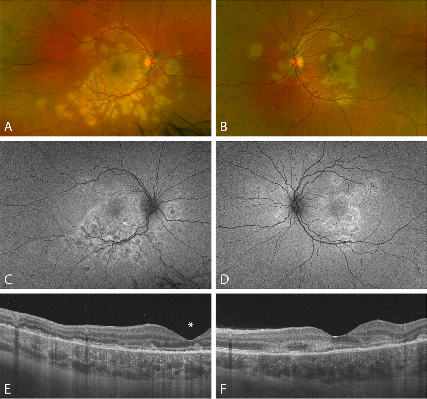
Table 1 Johnson et al. and Sanders et al. scores include the followingThe group with corneal ulcers applied drops five times daily. According to the described procedure, the rabbits' eyes were examined daily for up to 14 days using a slit light microscope and an indirect ophthalmoscope. Imaging was used to capture the alterations. To compare the effectiveness of the treatment, cultures were also taken from the corneal ulcer on the first, second, fifth, and fourteenth days.
SPSS17 software was used to analyze the data. The significance level was considered less than 0.05.


留言 (0)