
Remember me


Paecilomyces is a ubiquitous filamentous fungus commonly found in soil and air [1]. Paecilomyces rarely causes disease in humans; however, there have been increasing cases of infection in recent years [2]. Infection gives rise to a variety of clinical presentations, including but not limited to, keratitis, endophthalmitis, and cutaneous infections [2]. Established risk factors for infectious keratitis include contact lens wear [2], trauma to ocular surface [3], and chronic topical corticosteroid use [3]. The outcomes of fungal keratitis vary, and treatment is usually initiated with topical antifungal drugs [4].
The first line of treatment is topical natamycin [4]. The Mycotic Ulcer Treatment Trial (MUTT I) demonstrated the efficacy of natamycin over voriconazole in preventing corneal perforation and penetrating keratoplasty in smear or culture-proven fungal keratitis [4]. Intracameral amphotericin B has also been reported as an effective treatment in deep fungal corneal ulcers [5]. Despite consistent topical and intracameral antifungal therapy, treatment may fail. Oral posaconazole has demonstrated efficacy in treating resistant cases [6]. Even with these available treatments, infection with Paecilomyces has been associated with poor outcomes: 60% of the eyes required keratoplasty and 19%–35% resulted in enucleation of the eye [7, 8]. In this case series, we report the clinical progression of three patients with Paecilomyces keratitis, using in vivo confocal microscopy and histopathology to substantiate our clinical and microbiologic findings.
Patients and methodsThis retrospective case series examined the risk factors, clinical course, and characteristics of three patients with Paecilomyces keratitis presenting concurrently within three months in the same location. Data collected retrospectively included patient demographics, preexisting ocular conditions, exam findings, presenting symptoms, and treatment course (Table 1).
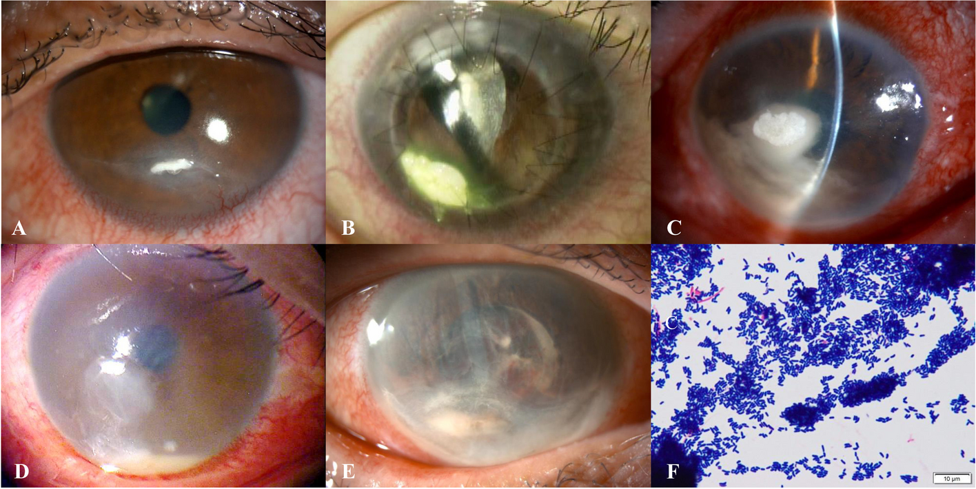
Table 1 Patient characteristics and medicationsFindingsCase 1A 72-year-old female with a history of contact lens-related corneal ulcer in the right eye was referred to our department in February 2022 for acute corneal perforation. The patient reported right eye pain that began two months prior to presentation. On examination, best corrected visual acuity was 20/600 in the right eye. Slit lamp examination revealed a 5 mm central perforated corneal ulcer with clear peripheral cornea. The patient underwent a therapeutic penetrating keratoplasty (PKP) on the same day. The host cornea was sent for histopathologic evaluation and microbiology studies including bacterial and fungal cultures.
In the immediate post-operative period following tectonic PKP (before microbiologic and histopathologic results were available), the patient was treated with fortified vancomycin, fortified tobramycin, moxifloxacin, and prednisolone. After two weeks, the epithelial defect resolved, and the cornea appeared clear. However, the following month, the patient presented with a small endothelial plaque superiorly on the donor cornea with hypopyon that spread to the host cornea site, prompting treatment with moxifloxacin, trimethroprim and polymyxin B, natamycin, voriconazole, and doxycycline. At this time, best corrected visual acuity was hand motion.
Corneal scrapings were taken for gram staining and culture examination using the standard microbiological culture techniques. Fungal isolates were identified through their morphology, which were speciated as Paecilomyces. Additionally, in vivo confocal microscopy (IVCM) of the donor cornea exhibited signs of spores (Fig. 1). The patient underwent a second therapeutic PKP with anterior chamber washout and intracameral antibiotics and antifungals.
Fig. 1
In vivo confocal microscopy showing small hyperreflective round structures, indicative of spores (arrow)
The histopathologic assessment of the penetrating graft confirmed acute stromal necrotizing inflammation associated with fungal elements which were percolating Descemet membrane.
The epithelium and Bowman layer were focally absent and associated with extensive stromal thinning, necrosis, and a dense infiltrate of neutrophils, forming microabscesses. The Descemet membrane was focally attached and revealed folds and loss of endothelial cells. Histochemical studies, periodic acid-Schiff (PAS) and Grocott’s methenamine silver (GMS) stains highlighted groups of hyphae- and yeast-like fungal elements infiltrating the posterior lamellae (Fig. 2). The concurrent microbiology cultures from the excised cornea showed growth of mold, specifically Paecilomyces species.
Fig. 2
A Penetrating corneal graft depicting dense stromal abscess and Descemet membrane folds (black arrow). H&E, original magnification × 20. Inset: corneal stroma with collections of neutrophils mixed with cellular debris. H&E, original magnification × 400. B. Grocott methenamine silver stain highlighting branching septate hyphae and chlamydospores (white arrow) in the abscess. GMS original magnification × 400. Inset: fungal elements percolating Descemet membrane. GMS × 400
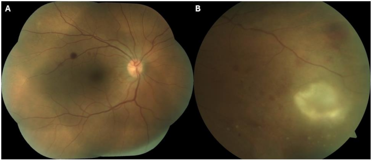
The patient continued to take antifungals and antibiotics including voriconazole, natamycin, moxifloxacin, and trimethoprim/polymixin, with no evidence of recurrence for eight weeks. However, an additional eight weeks after, the patient reported new-onset right eye pain despite the use of these antifungals and antibiotics. On examination, there was a deep stromal infiltrate in the graft host junction at 11 o’clock, accompanied by a 1.1 mm hypopyon and white cataract (Fig. 3). Visual acuity remained hand motion. Aggressive treatment for fungal keratitis was employed, which involved continuing oral voriconazole and increasing the frequency of topical voriconazole and natamycin. In addition, intrastromal amphotericin B and voriconazole injections were given on three occasions without resolution of the infiltrate.
Fig. 3
Endothelial plaque and hypopyon prior to second therapeutic PKP
Systemic therapy was switched from oral voriconazole to oral posaconazole 300 mg twice daily. Two weeks after starting posaconazole, the exam showed no endoplaque, noting resolution of fungal infiltrate. However, the patient experienced hypertensive urgency, attributed to the posaconazole, which was discontinued immediately after a 4-week course. One month after discontinuation of oral posaconazole, clinical resolution of endoplaque and stromal infiltrate was noted (Fig. 4). Furthermore, lack of fungal elements on IVCM of the cornea was confirmed monthly for three months. Due to graft failure six months after, an optical keratoplasty was performed and the failed graft was confirmed by histopathology to be free of fungal elements.
Fig. 4
Resolution of infectious keratitis without recurrence following treatment with systemic Posaconazole for patient 1
Case 2An 87-year-old female with a history of recurrent herpetic anterior uveitis of the left eye and bilateral primary open-angle glaucoma was referred to our department in March 2022 for concerns of microbial keratitis. The patient reported worsening pain and tearing in the left eye that was refractory to an antibiotic regimen of besifloxacin and erythromycin. On this initial visit, examination of the left eye revealed an inferotemporal corneal infiltrate with neovascularization and a best corrected visual acuity of 20/500. Like the previous patient, corneal scrapings obtained from the patient were taken for gram staining and culture examination using the standard microbiological culture techniques. The first corneal culture grew budding yeast with pseudohyphae. The patient was subsequently prescribed topical natamycin for fungal keratitis.
Ten days later, the culture was speciated as Paecilomyces. Broad-range fungal PCR identified Paecilomyces ilacinum species, the former name of Purpureocillium lilacinum, which belongs to the genus Purpureocillium [9]. Despite aggressive use of topical voriconazole and natamycin, there was progressive corneal thinning, worsening epithelial defect, and non-resolution of the stromal infiltrate. Therapeutic PKP of the left eye was performed with an inferiorly decentered trephination to include the entire infiltrate. Histopathology from the excised cornea was characterized by confluent microabscesses associated with hyphae-like fungal elements passing through Descemet membrane (Fig. 5).
Fig. 5
A Graft failure depicting acute necrotizing inflammation at the host-donor interface and disruption of Descemet membrane (arrow). H&E original magnification × 20. B. Deep stromal microabscesses containing numerous branching septate hyphae, highlighted with PAS-fungus, original magnification × 400
Topical antifungals were continued postoperatively in the absence of corticosteroids, but at the second postoperative week, there was a concern for intrastromal infiltrate surrounding the 6 o’clock suture. Intracameral and intrastromal voriconazole injections were administered. The suture was removed and sent for culture, which grew Paecilomyces, confirming recurrent infection. Progression of the infiltrate was noted at the sixth postoperative week (Fig. 6) and IVCM confirmed the presence of hyphae in the donor tissue (Fig. 7). Therefore, a decision was made to proceed to a second therapeutic PKP.
Fig. 6
Progression of infiltrate at 6 o’clock graft-host junction
Fig. 7
Long branching filaments demonstrate hyphae (arrow)
A repeat PKP of the left eye, as well as anterior chamber washout and administration of intracameral voriconazole was performed. At the first postoperative week, there was notable improvement in left eye visual acuity from counting fingers to 20/300. After the PKP, intracameral, intrastromal, and subconjunctival voriconazole injections were administered. Vision continued to improve in the following weeks, with examination of the left eye revealing a clear and fully epithelialized corneal graft. At the fourth postoperative week, a new infiltrate was observed in the deep stroma of the recipient rim associated with the 2–3 o’clock suture.
Despite injections of amphotericin B and voriconazole given by intracameral, intrastromal, and subconjunctival routes, the infiltrate persisted. Systemic therapy was switched from voriconazole to posaconazole 300 mg twice daily. Due to concerns for hypertension and bleeding secondary to drug interactions between posaconazole and the patient’s previously prescribed apixaban, the posaconazole dose was halved. Nevertheless, due to uncontrolled hypertension, the patient was admitted to the medical intensive care unit, where posaconazole was discontinued as it was assumed to be the cause of her refractory hypertension. After three weeks of using posaconazole, the patient’s fungal keratitis appeared to have completely resolved (Fig. 8).
Fig. 8
Resolution of infectious keratitis without recurrence following treatment with systemic Posaconazole for patient 2
Secondary graft failure was confirmed fifteen weeks after the second therapeutic PKP. An optical PKP was performed successfully, and the histopathology confirmed no evidence of fungal elements (Fig. 9).
Fig. 9
A Failed corneal graft with inflammatory pannus and stromal neovascularization. H&E × 10. B. Fragment of Bowman layer embedded in peripheral scar tissue with giant cell granulomatous reaction (arrow). Dense lymphoplasmacytic infiltrate surrounding new vessels in the stroma (asterisk). H&E × 100. C. Grocott methenamine silver stain failed to detect fungal elements. GMS × 20
Case 3A 77-year-old diabetic male with a history of right PKP for prior corneal ulcer complicated by descemetocele formation was referred in April 2022 for evaluation of persistent epithelial defect on the donor cornea. He had glaucoma of both eyes, and his right eye visual acuity was hand motion. Despite previous tarsorrhaphy, self-retained amniotic membrane, and a trial of cenegermin, the epithelial defect had failed to close for almost one year. The patient was using topical moxifloxacin 0.5% and loteprednol 0.5% chronically during this time.
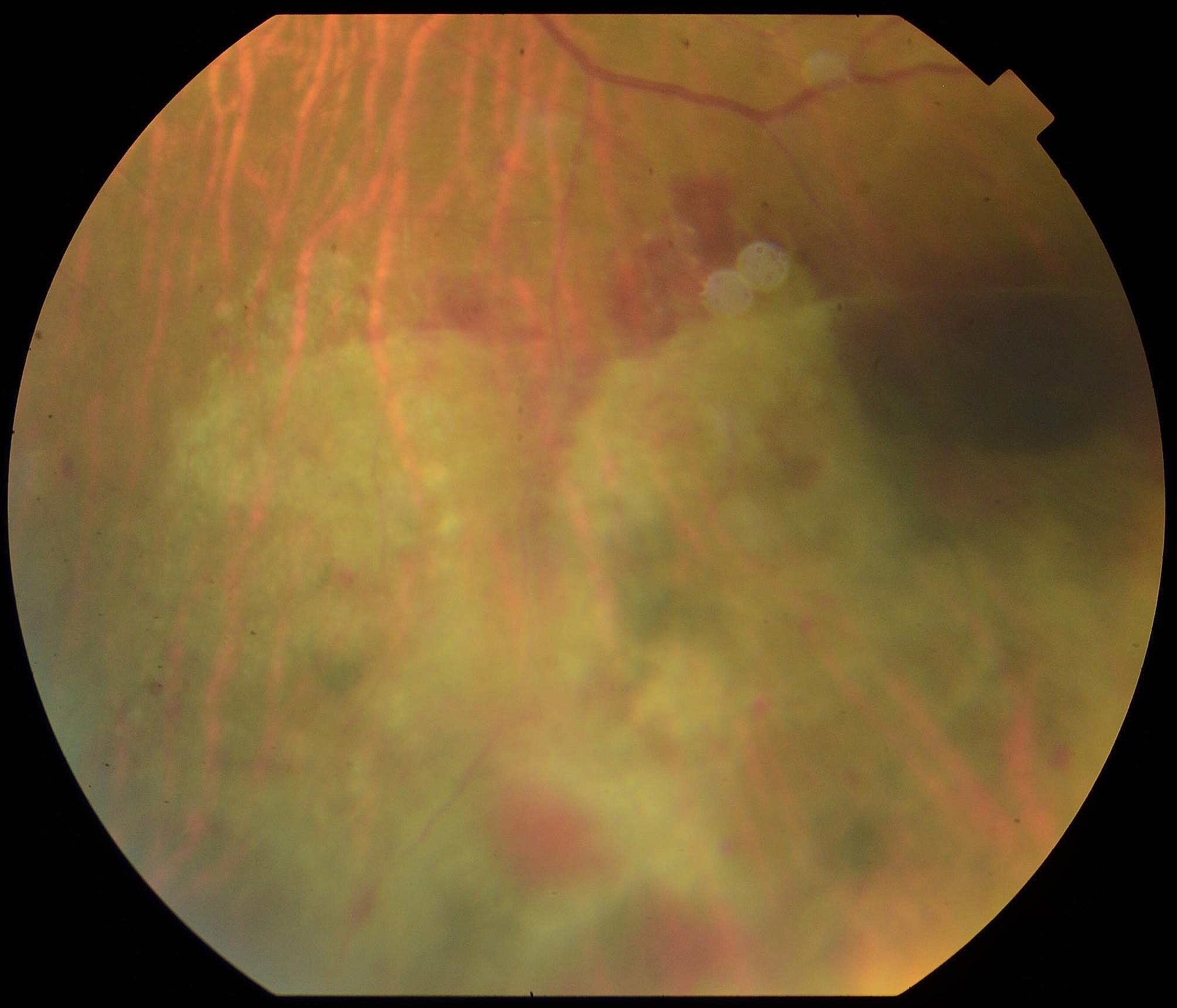
On exam, an infiltrate was noted in the bed of the epithelial defect on the donor cornea (Fig. 10). IVCM was performed over the infiltrate and showed the presence of hyphae (Fig. 11). A corneal perforation occurred within one week of starting topical natamycin and oral voriconazole, and a tectonic PKP was performed. Histopathologic evaluation of the excised cornea demonstrated numerous hyphae with focal septation, and yeast-like structures infiltrating necrotic stromal lamellae (best noted with PAS and GMS studies) (Fig. 12). Fungal culture grew Paecilomyces, identified through conventional morphologic methods. The patient began using natamycin, oral voriconazole, and moxifloxacin post-operatively without any topical steroids. Despite aggressive antifungal therapy, the cornea perforated again within one week, and a therapeutic PKP was performed. Post-operatively, the donor stroma proceeded to melt and, due to presumed poor prognosis, a Gundersen Flap and total tarsorrhaphy were performed to salvage the globe.
Fig. 10
Elevated central stromal infiltrate with epithelial defect
Fig. 11
High density of hyphae is shown
Fig. 12
A Portion of cornea with disorganization of architecture and extensive stromal necrosis. H&E original magnification × 40. B. Neutrophils infiltrating stromal lamellae associated with fungal elements (arrows). H&E original magnification × 200. C. PAS stain highlighting hyphae in abscesses (arrows). Original magnification × 200. D. Branching septate hyphae and spores in the stroma. GMS stain, original magnification × 400
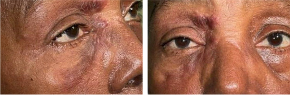
Due to concern for residual fungal organisms and the difficulty of reliable penetration of topical medication given near total tarsorrhaphy, oral posaconazole 300 mg twice daily was initiated. The patient tolerated a three-week course of posaconazole, during which the ocular surface healed appropriately. The tarsorrhaphy was opened fourteen weeks after the Gunderson flap was performed (Fig. 13) and there has been no clinical evidence of recurrent microbial keratitis.
Fig. 13
Resolution of infectious keratitis without recurrence following treatment with systemic Posaconazole for patient 3
Comments (0)