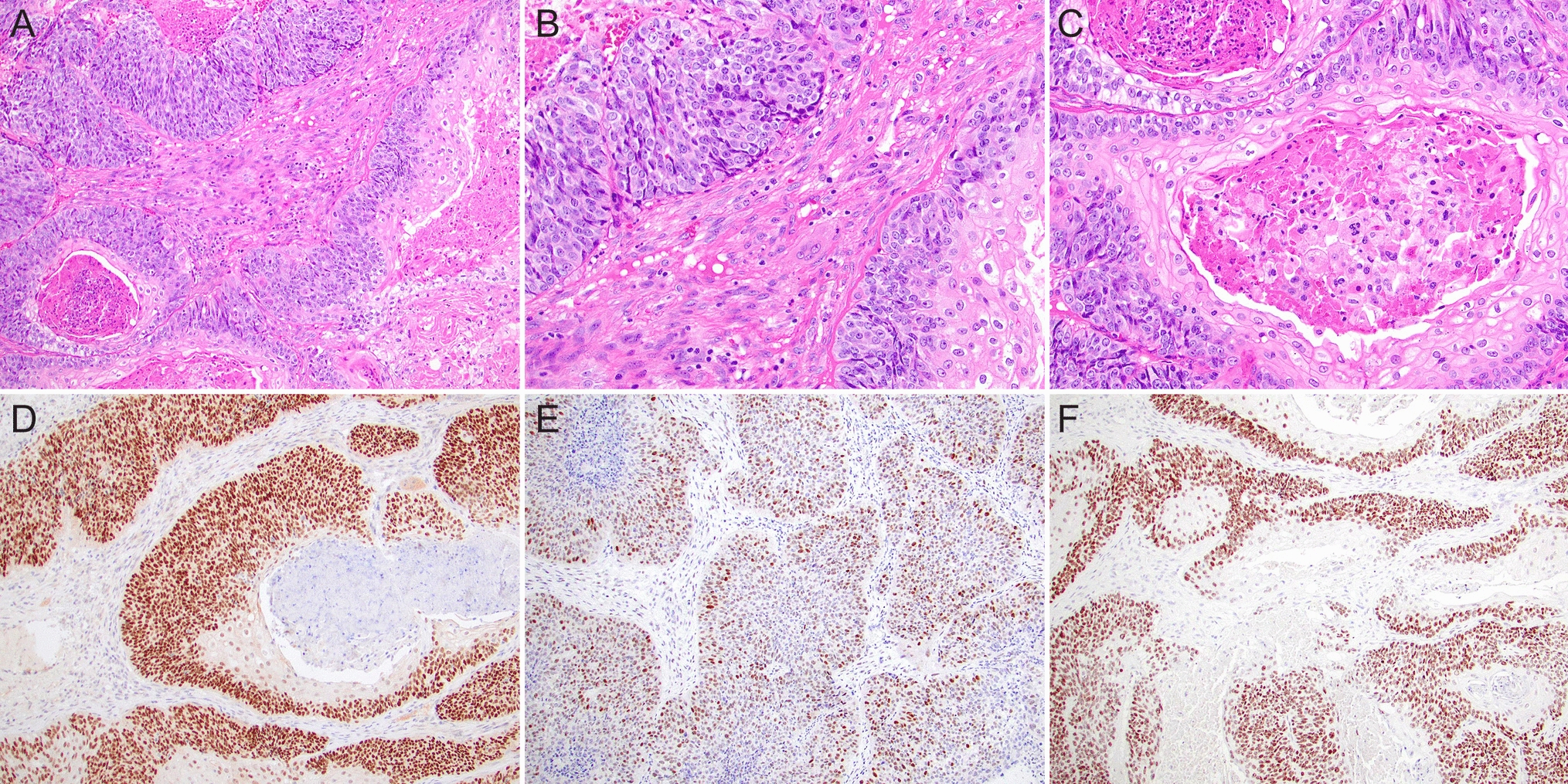
Immunohistochemical analyses were performed on formalin-fixed and paraffin-embedded (FFPE) sections. Epitope retrieval was conducted according to the manufacturer’s guidelines. The following antibodies were utilized for staining: Androgen receptor (Dako AR441; 1:200), CD117 (Dako A4502; 1:50), DOG-1 (Leica K9; 1:400), HER2 (Cell Marque SP3; 1:80), LMP-1 (Dako M0897; 1:400), ENBA2 (Leica PE2 1:100), CK5/6 (Dako D5/16B4; 1:200), p63 (Dako 4A4; 1:600), p40 (Biocare ACI 3030B; 1:50), p16 (Roche E6H4; 1:2), p53 (Leica DO7; 1:100), Rb1 (Leica 13A10; 1:50), SSTR2 (Abcam UMB1; 1:200).
To detect the non-coding EBV small RNAs, Epstein-Barr Encoding Region (EBER) in situ hybridization was performed on 4-µm thick FFPE samples using the Bond EBER probe (PB0589), anti-Fluorescein antibody (AR0222) and Bond Refine Red Detection kit (DS9390) on a Leica Bond automated staining system according to the manufacturer`s protocol (Leica biosystems).
In situ hybridization for HPV was performed using the RNAscope™ 2.5 VS Probe-HPV-HR18 (No: 312598) and Bond RNAscope Brown Detection Kit (DS9815) on a Leica Bond automated staining system according to the manufacturer`s protocol (Advanced Cell Diagnostics). Screening for E6/E7 mRNA of the HPV high-risk genotypes 16, 18, 26, 31, 33, 35, 39, 45, 51, 52, 53, 56, 58, 59, 66, 68, 73 and 82 was performed.
For HPV screening, DNA was isolated from FFPE blocks utilizing the Promega Maxwell® RSC DNA FFPE Kit. The Allplex™ HPV28 Detection kit (Seegene) including positive controls were used to test for 19 high-risk HPV genotypes (16, 18, 26, 31, 33, 35, 39, 45, 51, 52, 53, 56, 58, 59, 66, 68, 69, 73, 82) and 9 low-risk HPV genotypes (6, 11, 40, 42, 43, 44, 54, 61, 70). Amplification and detection were performed on a CFX 96 real-time PCR detection system (BioRad) and a Ct value of ≤ 43 was considered a positive result.
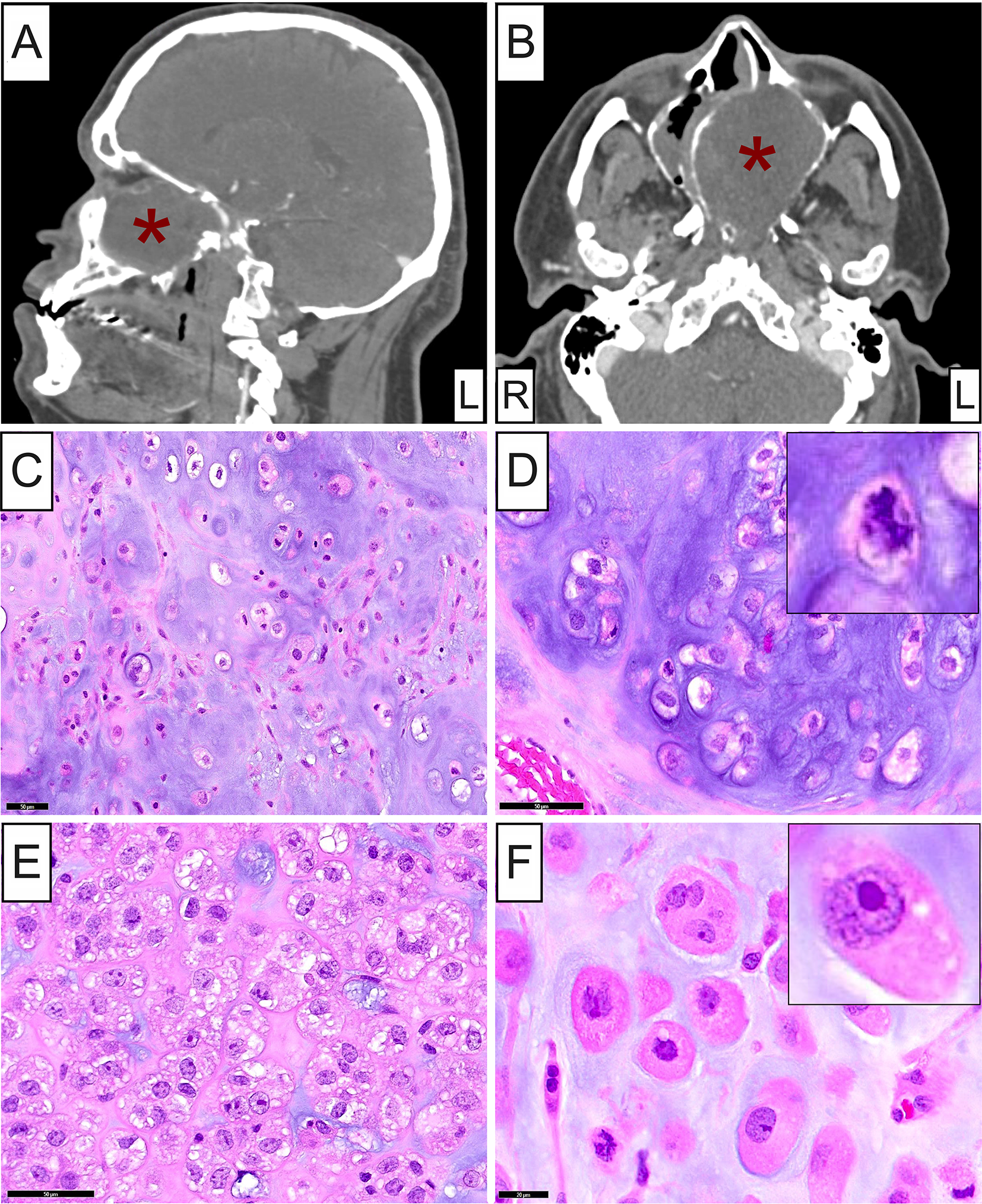
Fluorescence in situ hybridization using the FISH HER2 staining kit (TA9217; Vysis/Abbott), which includes fluorescently labeled DNA probes covering the HER2 locus (SpectrumGreen; 17q11.2-q12) and a chromosome enumeration probe (CEP) recognizing the centromeric region of chromosome 17 (SpectrumAqua;17p11.1-q11.1). Deparaffinization, pretreatment and proteinase digestion were performed on the Leica Bond slide-staining system, and samples were incubated for 18 h at 37 °C. The fluorescent signals on at least 25 cell nuclei were first independently analyzed by 2 trained lab technicians and were later independently validated by 2 trained pathologists. Samples with a HER2/CEP17 ratio ≥ 2 or HER2/cell ratio ≥ 6 were considered positive for HER2 amplification (Wolff et al., 2018).
Nucleic acid isolation, library preparation, next generation sequencing, and data analyses for the SalvGlandDx panel were performed as described in [11].

留言 (0)