
記住我


Cancer Imaging 2023, 23(1):P1
Learning Objectives
To recognise key imaging findings from Contrast-Enhanced Computed Tomography (CECT) of emergency situations due to intra-abdominal haemorrhage from spontaneous rupture of visceral tumours.
Content Organisation
Spontaneous intra-abdominal haemorrhage is the abnormal presence of blood into the abdominal cavity in absence of traumas. It causes either haemoperitoneum or haemoretroperitoneum depending on haemorrhagic site. Although non-traumatic intra-abdominal bleedings are globally rare, spontaneous tumour rupture is one of the most frequent noxae. Clinical presentation is typically non-specific, and a conclusive diagnosis should always be made with imaging findings. In the setting of emergency, multi-phase Contrast-Enhanced Computed Tomography (CECT) is the procedure of choice due to its low temporal resolution and its capability to detect even small amounts of intra-abdominal haemorrhage. In this paper we illustrate the main anatomical and clinical features of visceral tumours that are more prone to spontaneous rupture (hepatocarcinoma, hepatic adenoma and haemoangioma, adrenal tumours, kidney tumours, splenic sarcomas, etc.). Then, we describe key CT findings of spontaneous tumour rupture such as presence of a clot in the haemorrhagic site (sentinel clot sign), contrast extravasation and time-dependent density change of haematoma in different CT phases.
Conclusion
CECT scanning is a pivotal technique for intra-abdominal haemorrhage from spontaneous visceral tumour rupture. In these situations, independently from tumour type, it displays an almost unique series of findings to achieve either a conclusive diagnosis or a correct therapeutical management.
P2 CT assessment of advanced ovarian cancer: findings precluding primary cytoreductive surgeryStephan Edey, Anum Pervez, Olwen Westerland, Audrey Jacques, Sultana Hasso, Savithri Rajkumar, Rahul Nath, Gautem Mehra, Ahmad Sayasneh, Sarah NatasGuy's and St. Thomas' Hospital, London, United Kingdom Correspondence: Stephan Edey (sarahnts.6@gmail.com)Cancer Imaging 2023, 23(1):P2
Learning objectives
Advanced ovarian cancer: demonstration of specific sites of disease on CT imaging which preclude primary debulking surgery, in the light of new ultraradical surgical techniques.
Content organisation
Primary cytoreductive surgery (debulking) is the treatment of choice in advanced ovarian cancer, followed by adjuvant chemotherapy.
Improved survival is associated with optimal debulking with the aim of as little residual disease as possible. This has led to newer aggressive ultraradical surgical techniques; however, despite this, certain sites of disease may still be unresectable. These should be recognised preoperatively to ensure patients do not undergo inappropriate potentially morbid surgery, with the alternative to downstage the disease with neoadjuvant chemotherapy.
This pictorial review will show examples of key sites of disease spread which preclude primary debulking surgery. We will focus on CT which still forms the majority of preoperative imaging staging.
Sites include:
Infiltrative mesenteric root disease
Confluent porta hepatis and coeliac axis disease
Diffuse small bowel serosal disease or extensive segmental sites of bowel involvement
Multi site hepatic parenchymal disease
Invasion or encasement of large vessels (IVS/aorta/iliac)
Multi-site and confluent thoracic disease
Conclusions
Despite new ultra radical cytoreductive surgery for patients with advanced ovarian cancer, certain sites of disease remain unresectable. It is important to recognise these to avoid inappropriate surgery.
P3 Abdominal emergencies in oncologic patients: "Elementary, my dear Watson"Eleni Tsakirmpaloglou1, Georgia Mingou1, Anatoli Stoforiadi2, Reggina Goulimari2, Melanie Sahinidou1, Olga Nikolaidou1 1Radiology Department, General Hospital, "G. Papanikolaou", Thessaloniki, Greece; 2Radiology Department, General Hospital of Xanthi, Xanthi, Greece Correspondence: Olga Nikolaidou (olganikolaidou@hotmail.com)Cancer Imaging 2023, 23(1):P3
Learning Objectives
To introduce the prevalence of abdominal complications in oncologic patients.
To depict the commonest abdominal entities leading to the emergency department.
To highlight the importance of imaging and especially computed tomography in the detection of oncology emergencies.
To illustrate the imaging features of primary or secondary complications derived from abdominal malignancies.
Content organisation
Oncologic emergencies consist of any acute and potentially life-threatening events, derived from the direct effects of the underlying condition or as a post-therapy complication. Approximately, 40% of cancer patients present to the emergency department with gastrointestinal symptoms.
Oncologic emergencies can be categorised as metabolic, haematologic, and structural based. The latter remain of paramount importance to be recognised and pointed out by the radiologists, as imaging remains the standard method of choice in detecting such conditions. Abdominal emergencies are usually derived from the primary tumour. Nevertheless, in patients with advanced cancer stage, III or IV, may be an indirect consequence related to its systemic manifestations.
The gold standard and modality of choice in the evaluation of cancer patients is Contrast-enhanced computed tomography (CT), providing important findings in major abdominal complications. Thus, CT multiplanar reconstruction offers additional information in structural based complications.
We will point out the imaging characteristics of acute abdominal oncologic conditions involving the gastrointestinal and the hepatobiliary tract, the vascular system and lastly the urinary tract.
Conclusion
Oncologic emergencies are not exceptional and may be life-threatening. Therefore, they demand paramount awareness to get identified and to establish the correct diagnosis, promptly.
P4 Deep invasion volume of primary nasopharyngeal carcinoma is a strong predictor of outcomeQi Yong H Ai1,2, Ann D. King2, Ho Sang Leung2, Lun M. Wong2, Frankie K.F. Mo2 1The Hong Kong Polytechnic University, Kowloon, Hong Kong; 2The Chinese University of Hong Kong, Shatin, Hong Kong Correspondence: Qi Yong H Ai (hemis.ai@polyu.edu.hk)Cancer Imaging 2023, 23(1):P4
Aim
The primary tumour volume of nasopharyngeal carcinoma (NPC) shows only a weak association with outcome and little impact on T-staging. However, the volume of deep tumour invasion, especially as a ratio to the superficial component, may be a marker of aggressiveness and predictor of outcome.
Materials and Methods
The MRI of 743 patients with NPC were retrospectively evaluated. Three primary tumour volume measurements were obtained, total volume (Vtotal), deep invasion volume (Vdeep) and ratio of deep to total volume (Vratio). Volumes were correlated with 5-year disease-free survival (DFS) using cox regression with step-forward approach to select the optimal V-related predictor. The optimal V-related predictor, together with other confounding factors (T, N, overall stage, sex, age and treatment) were then added into the multivariate model to identify the independent predictors for predicting DFS. Hazard ratio (HR) was calculated. A p < 0.05 indicated statistically significant.
Results
Vratio outperformed the other two volume measurements for predicting DFS (HR = 3.56, p <0.01), a higher ratio had poorer outcome. The multivariate analysis showed that Vratio (HR = 3.18) and N stage (HR = 1.94) (p < 0.01) were the only two independent predictors for DFS in the multivariate analysis.
Conclusion
Primary NPCs that show greater volume of deep invasion relative to the superficial component have a poorer outcome and this ratio outperforms Vtotal and T-staging.
P5 Dual energy CT applications in oncologic imagingNils Grosse Hokamp, Thorsten PersigehlUniversity Hospital and University of Cologne, Cologne, Germany Correspondence: Nils Grosse Hokamp (nils.grosse-hokamp@uk-koeln.de)Cancer Imaging 2023, 23(1):P5
Objective
The objective of this educational exhibit is to provide an overview of dual-energy CT (DECT) and its applications in oncologic imaging. It aims to highlight the advantages of DECT in improving tissue characterisation, functional analysis, and reducing metal artifacts.
Content Organisation
This abstract is organised into three sections. The first section introduces technology and reconstructions from DECT and sets the stage for discussing applications in oncologic imaging.
The second section discusses how DECT enables improved tissue characterisation by differentiating between tumour tissue, healthy tissue, and necrotic or haemorrhagic areas. It also explores the functional information provided by DECT through the generation of iodine maps, facilitating the assessment of tumour vascularity and treatment response. Furthermore, it emphasises DECT's ability to reduce metal artifacts, which is particularly relevant in oncology patients with metallic implants or prior surgeries.
The final section concludes the abstract by summarising the key points. It underscores the value of DECT in oncologic imaging, highlighting its potential to improve diagnostic accuracy, treatment planning, and monitoring of oncology patients. It also emphasises the importance of continued advancements in DECT technology and glimpses at further innovation enabled by photon counting CT.
Conclusion
Dual-energy CT is a powerful tool in oncologic imaging that offers numerous advantages. Its ability to improve tissue characterisation, provide functional information through iodine mapping, and reduce metal artifacts makes it an invaluable resource for oncology patients. In light of increasing availability of DECT reconstructions, knowledge of fundamental concepts and common pitfalls is beneficial.
P6 Chemotherapy-Related Cognitive Impairment (CRCI) in breast cancer: an ALE meta-analysis of neuroimaging studiesSonya Utecht, Linda Larson-PriorUniversity of Arkansas for Medical Science, Little Rock, USA Correspondence: Sonya Utecht (sutecht@uams.edu)Cancer Imaging 2023, 23(1):P6
Aim
Worldwide over 1.8 million cases of breast cancer are diagnosed per year, with critical treatments leading, in many cases, to chronic cognitive dysfunction in affected individuals. Chemotherapy-related cognitive impairment (CRCI) or “chemo-brain” is a disruptive side effect of adjuvant chemotherapy plaguing many breast cancer survivors. To better understand the effects on brain network organisation, several neuroimaging studies have examined individuals suffering from CRCI. However, it can be difficult to draw conclusions from these studies as many have small sample sizes, disagreements on anatomic terminology, and varied foci. The goal of this study is to perform a meta-analysis of these studies to provide a clearer understanding of those functional brain networks underlying the cognitive dysfunction in breast cancer patients.
Methods
Using a PRISMA framework, search queries were performed using PubMed and Google Scholar with a variety of terms including “neuroimaging”, “CRCI”, “functional connectivity”, “chemotherapy", and “breast”. After filtering, 43 potential studies were identified, 7 of which had relevant image coordinate data and were entered into analysis in our pilot study. Activation Likelihood Estimation meta-analyses were performed using the GingerALE toolkit.
Results
Coordinate data from these studies were fed into the GingerALE software system. The hippocampus, amygdala, and bilateral insula showed significant differences between the 184 chemotherapy-positive breast cancer survivors and the 132 chemotherapy-naive controls.
Conclusion
The resulting meta-analyses provide evidence of decreased connectivity in regions associated with the default mode network, as well as increased connectivity in hippocampal regions associated with reduced neuropsychological test results and memory function.
P7 Influence of the “Streamline Phenomenon” on the laterality of hepatic metastases from colorectal cancerEduardo De Araujo, Eva Rolim, Rubens ChojniakAc Camargo Cancer Center, Sao Paulo, Brazil Correspondence: Eduardo De Araujo (eduardo.mparaujo@gmail.com)Cancer Imaging 2023, 23(1):P7
Aim
To discuss the concepts behind the theory of streamline flow in the distribution of liver metastases depending on the origin of the primary lesion in the colon and the impact of this phenomenon on liver metastatic recurrence-free survival in patients undergoing liver metastasectomy.
Materials and Methods
Retrospective study, in patients with colorectal neoplasia (adenocarcinoma) from 2016 to 2023. A total of 144 patients in the study who were divided into two groups according to the origin of the colon lesion side, right (45) and left (99) by counting the number of metastases in each hepatic lobe by sectional imaging methods. Follow-up of 58 patients to assess liver recurrence after colectomy and liver surgery according to surgical laterality (ipsilateral, contralateral and both lobes).
Results
The mean number of metastases in the right hepatic lobe was 5.22 (right colon) and 4.50 (left colon); and mean of 2.22 in the left lobe (right colon) and 3.42 (left colon), without significant differences, but with numerical differences (OR= 19) when in the right colon. There was no difference in the liver recurrence curve regardless of surgical laterality [(14) ipsilateral; (11) contralateral; (33) both lobes].
Conclusions
There was no significant difference in the laterality of liver metastasis. However, it was observed that isolated metastases tend to follow the laterality of the primary lesion, especially when the primary lesion is on the right.
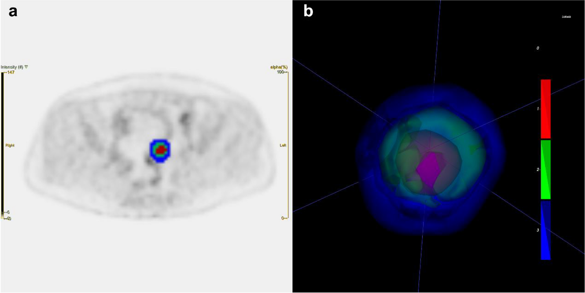
Fig. 1 (abstract P7). P8 F-18 FDG, F-18 PSMA, and Cu-64 DOTATATE PET/CT: One patient, three cancersRyan Rahman, Fathima Palot Manzil, Joshua EichhornUniversity of Arkansas for Medical Sciences, Little Rock, USA
Correspondence: Ryan Rahman (jmeichhorn13@gmail.com)
P8 F-18 FDG, F-18 PSMA, and Cu-64 DOTATATE PET/CT: One patient, three cancersRyan Rahman, Fathima Palot Manzil, Joshua EichhornUniversity of Arkansas for Medical Sciences, Little Rock, USA
Correspondence: Ryan Rahman (jmeichhorn13@gmail.com)Cancer Imaging 2023, 23(1):P8
Learning Objectives
To review the impact of appropriate PET radiotracer selection in identification and differentiation of malignancy and metastatic disease based on the patient’s primary tumour type.
Content Organisation
Selection of radiotracers is fundamental to the proper evaluation and diagnosis of malignancy, whether primary or metastatic. The choice of radiotracer is often chosen based on a combination of cancer history, suspicion for certain cancer types, and the indication for PET/CT – whether for surveillance or staging purposes.
We will discuss three well-known radiotracers in PET/CT imaging: F-18 FDG, F-18 PSMA, and Cu-64 DOTATATE; this will include:
A brief overview of the mechanism of each radiotracer uptake and imaging protocol
Indications for specific radiotracer selection
Advantages and disadvantages of each radiotracer
We will discuss a case utilising all three of these radiotracers in a single patient with multiple primary malignancies to highlight:
Real-life selection and application of these aforementioned PET radiotracers based on clinical suspicion in conjunction with patient history
The importance of follow-up imaging after false-negatives in PET surveillance
Conclusion
F-18 FDG, F-18 PSMA, and Cu-64 DOTATATE radiotracers are common tools in the radiologist’s arsenal for visualisation of primary malignancy and metastatic disease on PET/CT scans. Knowing the underlying mechanism, in accordance with benefits and limitations of each tracer, allows for patient-specific selection. This can be especially crucial when there is suspicion for multiple underlying primary malignancies.
P9 AI for prostate MRI: results from a large multi-centre, multi-vendor external validation studyAarti Shah1, Nadia Moreira da Silva2, Michael Yeung2, Francesco Giganti3, Lucy Davies2, Paul Burn4, Richard Hindley5, Nikhil Vasdev6,7, John Hayes6, Sophie Squire1, Alison Bradley8, Giles Maskell8, Adrian Andreou9, Sidath Liyanage10, Mark De Bono10, Raj Persad11, Jon Aning11, Nimalan Sanmugalingam12, Tristan Barrett12,13, Mark Hinton2, Antony Rix2, Evis Sala14 1Hampshire Hospitals NHS Foundation Trust, Winchester, United Kingdom; 2Lucida Medical Ltd, Cambridge, United Kingdom; 3University College London, London, United Kingdom; 4Somerset NHS Foundation Trust, Taunton, United Kingdom; 5University of Winchester, Winchester, United Kingdom; 6East and North Herts NHS Trust, Stevenage, United Kingdom; 7University of Hertfordshire, Stevenage, United Kingdom; 8Royal Cornwall Hospitals NHS Trust, Truro, United Kingdom. 9Royal United Hospitals Bath NHS Foundation Trust, Bath, United Kingdom; 10Mid and South Essex NHS Foundation Trust, Southend, United Kingdom. 11North Bristol NHS Trust, Bristol, United Kingdom; 12Cambridge University Hospitals NHS Foundation Trust, Cambridge, United Kingdom; 13University of Cambridge, Cambridge, United Kingdom; 14Policlinico Universitario A. Gemelli, IRCCS, Dept. of Radiology, Rome, Italy Correspondence: Aarti Shah (Aarti.Shah@hhft.nhs.uk)Cancer Imaging 2023, 23(1):P9
Aim
Evaluate how an AI decision support system for MRI in prostate cancer generalises to multi-centre datasets including multiple scanner models, vendors, field strengths and imaging protocols.
Methods AI-based software for analysis of prostate MRI was developed using data from PROSTATEx and five sites in a diagnostic study (794 patients, 34% csPCa). It was subsequently evaluated on blinded external validation data (mpMRI, 252 patients, 31% csPCa) from six sites, including one unseen site/scanner. Exclusions included prior treatment and image quality issues. The software automatically outputs scores intended to identify Gleason score (GS)≥3+4 csPCa. csPCa was confirmed by biopsy (GS≥3+4/PI-RADS ≥3), with PI-RADS 1/2 patients with no biopsy assumed negative. Performance was evaluated using ROC analysis, with 95% confidence intervals estimated by bootstrapping.
Results
At the pre-determined threshold, AI identified patients with csPCa with sensitivity 94% (95% CI 88-99%), specificity 57% (49-64%), and NPV 95% (90-99%) on the blinded external validation dataset. AUC was 0.85 (0.80-0.90). Per-site AUC ranged from 0.70-0.98, with pooled AUC 0.86±0.11.
Reporting radiologists had per-patient sensitivity 99% (95% CI 96-100%) due to the assumed ground truth, specificity 73% (67-80%), NPV 99% (98-100%), and AUC 0.95 (0.92-0.97) on this dataset. In a 2019 Cochrane meta-analysis of 12 major studies (37% csPCa), radiologists identified patients with GS≥3+4 csPCa with sensitivity 86% and specificity 42%.
Conclusion
In a large external validation, this AI system shows comparable performance to radiologists in major expert studies, indicating promising potential for AI to support PCa detection in clinical practice.
P10 Integrating clinical data with AI to optimise decision-making in prostate MRIAntony Rix1, Nadia Moreira da Silva1, Jobie Budd1, Michael Yeung1, Francesco Giganti2, Lucy Davies1, Paul Burn3, Richard Hindley4,5, Nikhil Vasdev6,7, Alison Bradley8, Giles Maskell8, Adrian Andreou9, Sidath Liyanage10, Raj Persad11, Jon Aning11, Tristan Barrett12,13, Mark Hinton1, Anwar Padhani14, Evis Sala15 1Lucida Medical Ltd, Cambridge, United Kingdom; 2University College London, London, United Kingdom; 3Somerset NHS Foundation Trust, Taunton, United Kingdom; 4Hampshire Hospitals NHS Foundation Trust, Winchester, United Kingdom; 5University of Winchester, Winchester, United Kingdom; 6East and North Herts NHS Trust, Stevenage, United Kingdom; 7University of Hertfordshire, Stevenage, United Kingdom; 8Royal Cornwall Hospitals NHS Trust, Truro, United Kingdom; 9Royal United Hospitals Bath NHS Foundation Trust, Bath, United Kingdom; 10Mid and South Essex NHS Foundation Trust, Southend, United Kingdom; 11North Bristol NHS Trust, Bristol, United Kingdom; 12Cambridge University Hospitals NHS Foundation Trust, Cambridge, United Kingdom; 13University of Cambridge, Cambridge, United Kingdom; 14Paul Strickland Scanner Centre, Northwood, United Kingdom; 15Policlinico Universitario A. Gemelli, IRCCS, Dept. of Radiology, Rome, Italy Correspondence: Antony Rix (Aarti.Shah@hhft.nhs.uk)Cancer Imaging 2023, 23(1):P10
Aim
Evaluate accuracy of multi-modal decision support models for prostate cancer combining imaging AI, clinical data and PI-RADS scores.
Methods
MRI, clinical history, histopathology and PI-RADS data were obtained retrospectively from a five-site, multi-vendor, multi-scanner study. 352 patients were used for training, and 235 patients (Gleason grade group (GGG)≥2, prevalence 34%) for held-out test. GGG≥2 was used as ground truth with MRI-negative patients assumed negative. Multi-modal models combining scores from separately-developed multi-stage MRI analysis AI software, clinical data, and original PI-RADS scores were trained. Sensitivity, specificity and AUC were evaluated at patient level on held-out data, and compared to AI-score and PI-RADS assessment alone.
Results
PI-RADS scores identified patients with GGG≥2 with sensitivity 1.00 (95% CI 1.00-1.00), specificity 0.67 (0.61-0.75) and AUC 0.94 (0.91-0.97).
AI detected patients with GGG≥2 with sensitivity 0.97 (0.93-1.00), specificity 0.55 (0.47-0.62) and AUC 0.88 (0.84-0.92), using bpMRI data.
Combining AI score and TZ-PSA density (PSAD) gave sensitivity 0.95 (0.90-0.99, p < 0.001), specificity 0.70 (0.63-0.77, p < 0.001) and AUC 0.90 (0.85-0.93, p = 0.25).
Combining AI, PSAD and PI-RADS gave sensitivity 0.99 (0.96-1.00, p < 0.001), specificity 0.83 (0.77-0.89, p < 0.001) and AUC to 0.96 (0.93-0.98, p = 0.003).
TZ volume based PSAD had modest additional benefit compared to whole-prostate PSAD. Other variables offered <5% specificity improvements or non-significant benefits. Findings with bpMRI and mpMRI AI models were similar.
Conclusion
Combining PI-RADS, PSAD and AI scores offers considerable specificity improvement, at similar sensitivity, compared to AI or PI-RADS assessments alone. This could substantially benefit selection of patients for biopsy using MRI.
P11 Lung carcinoma with atrial invasionJH Wong, Z HaronInstitut Kanser Negara, Putrajaya, Malaysia Correspondence: JH Wong (jhueyw@gmail.com)Cancer Imaging 2023, 23(1):P11
Learning objectives
To review the radiological signs of atrial invasion and pathways of spread by lung carcinoma on computed tomography (CT).
Content organisation
Cardiac invasion by lung carcinoma is a rare occurrence, however, potentially life-threatening. The predisposition for cardiac involvement depends on the histological subtype, tumour location and stage of disease.
We will present the imaging features of atrial invasion by lung carcinomas, including
We will review the pathways of tumour spread such as lymphatic spread, haematogenous spread, direct invasion and transvenous extension.
We will also discuss the potential limitations of CT in diagnosing atrial invasion.
Conclusions
Recognition of radiological findings on CT of atrial invasion by lung cancers is important as awareness of this rare entity is essential for timely diagnosis and appropriate treatment planning in patients with lung carcinoma.
P12 Radiomics for the differential diagnosis between ameloblastomas and odontogenic keratocysts on panoramic radiographyKuo Feng Hung1, Qi Yong H Ai2 1Applied Oral Sciences & Community Dental Care, Faculty of Dentistry, The University of Hong Kong, Hong Kong, Hong Kong; 2The Hong Kong Polytechnic University, Hong Kong, Hong Kong Correspondence: Kuo Feng Hung (hungkfg@hku.hk)Cancer Imaging 2023, 23(1):P12
Aim
To compare the performance of radiomics models built with and without an ensemble technique in discriminating between ameloblastomas and odontogenic keratocysts (OKCs) on panoramic radiography (PR).
Methods
86 pre-operative PRs with ameloblastomas (n = 31) or OKCs (n = 55) were retrospectively included. Radiomics features were extracted from labelled lesion regions, and the dataset was split into training and testing sets (7:3 ratio). Z-score normalisation was applied to all features. For the ensemble technique, a five-fold cross-validation was conducted in the training set to select the best-performing classifier among seven machine learning classifiers. During each fold, feature selection was performed using t-tests (p < 0.05) and LASSO, and each classifier was trained using the selected features. An ensemble approach was utilised to aggregate the best-performing classifier trained corresponding to the five-fold cross-validation through their weighted sum. The performance of the ensemble model was tested on the testing set using AUC and their 95% CIs. For the non-ensemble technique, the classifier identified as the best-performing classifier was trained using features selected by t-tests and LASSO. Hyperparameter tuning was performed using grid search and five-fold cross-validation. The model’s performance was assessed on the testing set.
Results
Among seven classifiers, the ensemble support vector machine (SVM) outperformed others in discriminating between ameloblastomas and OKCs on PR. The ensemble model achieved an AUC of 0.89 (95%CI 0.74-0.99) while the non-ensemble SVM achieved an AUC of 0.67 (95%CI 0.44-0.89).
Conclusion
The ensemble model's performance in discriminating between ameloblastomas and OKCs on PR was more reliable than the non-ensemble SVM.
P13 The role of alternate diagnosing imaging in the era of LI-RADS for diagnosing hepatocellular carcinomaWHK Chiu1, BCK Chow1, JC Ng2, KDD Leung1, WTV Chan3 1Queen Elizabeth Hospital, Hong Kong, Hong Kong; Kwong Wah Hospital, Hong Kong, Hong Kong; 3Tuen Mun Hospital, Hong Kong, Hong Kong Correspondence: WHK Chiu (keith.chiu@ha.org.hk)Cancer Imaging 2023, 23(1):P13
Aim
The Liver Imaging Reporting and Data System (LI-RADS) has improved and standardised reporting of liver lesions. Unlike other guidelines, the use of more than one imaging modality to achieve non-invasive diagnosis of Hepatocellular Carcinoma (HCC) appears limited. This study aims to examine whether combining Computed Tomography (CT) and Magnetic Resonance Imaging (MRI) improves the performance of LI-RADS in HCC detection.
Materials and Methods
Between 2020 and 2022, consecutive patients at risk for HCC who underwent CT and MRI within 4 months of each other for liver lesion characterisation were retrospectively analyzed. Two radiologists rated all treatment-naive lesions in consensus using LI-RADS v2018. Inter-modality agreement and diagnostic performance were evaluated using composite outcomes as ground truths.
Results
In total, 66 patients (median age 62 [IQR 58-70years], 26 [39.4%] female) with 115 lesions (median SLD 1.2, IQR 0.8-1.8cm, 17 [14.8%] HCC) were included. LI-RADS major features showed good inter-modality agreement (rho=0.74, p < 0.0001) and ancillary features changed LI-RADS classifications in 18 (15.6%) lesions. The highest combined LI-RADS classification has an accuracy of 91.3% when LR4/5 were considered to be HCC. Using two imaging modalities significantly improve sensitivity compared to CT alone (82.4% vs 41.2%, p = 0.03) without compromising specificity (92.9% vs 97.9%, p = ns). No significant improvement in sensitivity nor specificity was seen compared to MRI alone (76.5% and 94.9%, p = ns).
Conclusion
For indeterminate liver lesions on CT, performing MRI could improve HCC detection and avoid diagnostic delays while no such benefit is seen if the lesion was indeterminate on MRI.
P14 Value of metabolic tumour volume as a risk stratificator for supplementing staging in NSCLCAlexander Brose1,2, Jochem König3, Gabriele Anja Krombach2, Mathias Schreckenberger4, Jutta Kappes5, Matthias Miederer1,4 1Department of Translational Imaging in Oncology, National Center for Tumour Diseases (NCT/UCC) Dresden, Germany: German Cancer Research Center (DKFZ), Heidelberg, Faculty of Medicine and University Hospital Carl Gustav Carus, University of Technology Dresden (TUD), Helmholtz-Zentrum Dresden-Rossendorf (HZDR), Dresden, Germany; Department of Diagnostic and Interventional Radiology, University Hospital Giessen, Giessen, Germany; 3Institute of Medical Biostatistics, Epidemiology and Informatics (IMBEI), University Medical Center Mainz, Johannes Gutenberg-University Mainz, Mainz, Germany; Department of Nuclear Medicine, University Medical Center Mainz, Johannes Gutenberg-University Mainz, Mainz, Germany; Department of Internal Medicine/ Pulmonary Medicine, Catholic Hospital Koblenz-Montabaur, Koblenz, Germany Correspondence: Alexander Brose (abrose312@gmail.com)Cancer Imaging 2023, 23(1):P14
Aim
Whole-body metabolic tumour volume (MTVwb) has proven to be an independent prognostic value for non-small cell lung cancer (NSCLC) in randomised controlled trials. We hypothesised that its implementation could also be beneficial in routine clinical use to supplement staging of NSCLC.
Methods
We performed a single-centre retrospective analysis on 251 patients with histologically proven NSCLC who underwent FDG-PET/CT for initial staging in the years 2017 to 2019. Two readers determined baseline-MTVwb as suggested by the PERCIST criteria in a semiautomatic fashion. Agreement between readers was measured by intraclass correlation coefficient (ICC). Cox regression and Kaplan-Meier survival analyses were performed to determine the prognostic value of UICC stage and MTVwb.
Results
Median overall survival (OS) was 40 ± 4.7 months (95% CI 30.8 – 49.4). Agreement between the two readers for MTVwb was almost perfect (ICC 0.983). Both, UICC stage and MTVwb were independent prognostic parameters in the cox regression analyses (p < 0.001). When stratified into risk groups by dividing the MTVwb into quartiles (5.4, 24.4 and 109.0 mL), survival analysis showed that higher MTVwb was associated with worse OS, equally to the corresponding UICC stage. When stratified by UICC stage, higher MTVwb still showed worse overall survival in locally advanced disease and metastatic disease (p < 0.05).
Conclusion
MTVwb as suggested by PERCIST is easy to reproduce and its prognostic value is comparable to the UICC stages. Combined with the TNM staging system, it could be used for further improvement of pretreatment assessment in patients with locally advanced and metastatic NSCLC.
P15 MRI surveillance of liver metastasis in high-risk uveal melanomaChloe McMurray, Sarah MacPhersonNHS Greater Glasgow and Clyde, Glasgow, United Kingdom Correspondence: Chloe McMurray (chloe.mcmurray4@nhs.scot)Cancer Imaging 2023, 23(1):P15
Aim
Up to 50% of patients with uveal melanoma develop metastatic disease, most frequently in the liver (77-94%). Surveillance methods vary across centres. A consensus statement was released for metastatic surveillance in Scotland in 2019 – high risk patients are recommended to have baseline MRI liver with contrast, followed by 6-monthly MRI liver without contrast.
This study aimed to identify compliance with consensus statement guidelines on liver metastasis surveillance for high-risk uveal melanoma patients.
Materials and Methods
A Radiology Information System (RIS) search using key words identified patients with high-risk uveal melanoma over a 10-year period (2012-2022) who had liver MRI. Retrospective analyses were performed of the use of MRI with and without contrast before and after the introduction of the consensus statement. Caldicott and local RIS approval was granted.
Results
Prior to the consensus statement, 29.5% had MRI for metastatic surveillance, all of which had contrast. 30% of these were performed due to suboptimal views with ultrasound.
16 high risk cases were identified following the consensus statement. 19% had surveillance as per the recommendation. 69% had MRI follow-up, however, these were performed with contrast. Of this cohort, 12% developed liver metastases.
P16 Morphological and functional parameters of MRI in PET/CT-proven local pancreatic cancer recurrencesDusan Saponjski1,2, Aleksandra Djuric Stefanovic1,2 1University Clinical center of Serbia, Belgrade, Serbia; 2Faculty of Medicine, University of Belgrade, Belgrade, Serbia Correspondence: Email: saponjski.d@gmail.comCancer Imaging 2023, 23(1):P16
Introduction
Prognosis of pancreatic adenocarcinoma is very unfavourable. Only 15-20% of carcinoma is operable at the time of diagnosis. During postoperative follow-up, recurrent tumours are found in 36-63% of patients, approximately 11 months after surgery.
Material & Methods
Abdominal MRI examination was performed according to the standard protocol, the existence of local recurrence was noted, as well as morphological (localisation and size), and functional parameters in the form of ADC values (diffusion coefficient). PET/CT measured maximal standardised values of radiopharmaceutical uptake (SUVmax), as well as size and localisation of the lesions. MRI and PET/CT findings were compared and correlated using Wilcoxon's equivalent pair test (Z) and Sperman's (rS) correlation coefficient.
Objective
To analyse whether MRI findings agree and correlate with PET/CT as a reference standard.
Results
23 patients were included in study and elevated serum tumour marker values (CA19-9) were found in 16 (70%). Ten patients (43.5%) had local recurrence between SMA and SMV, and another 10 patients had local recurrence around PV in the hepatoduodenal ligament. Only 2 patients had local recurrence in the projection of anastomosis and 1 had a "muff" around SMA. Average ADC value was 1.49x10⁻³ mm2/s. Average SUVmax was 7.8g/ml. Maximal recurrence diameter measured on MRI and PET/CT did not differ significantly (p = 0.375). A negative correlation was found between lesion size and ADC values (p = 0.005) and a positive correlation between size and SUVmax (p < 0.001).
Conclusion
MRI is a reliable method in detecting local pancreatic cancer recurrence, using morphological and functional parameters.
P17 Colocolic intussusception caused by multiple grouped Vanek’s tumours: a case reportDusan SaponjskiUniversity Clinical Center of Serbia, Belgrade, Serbia. Faculty of Medicine, University of Belgrade, Serbia Correspondence: Dusan Saponjski (saponjski.d@gmail.com)Cancer Imaging 2023, 23(1):P17
Introduction Inflammatory fibroid polyp (IFP) is a benign polypoid tumour arising from the submucosa of the gastrointestinal tract. It is most commonly located in the gastric antrum, small bowel and rarely in the colon and oesophagus. Clinically it presents with abdominal pain and gastrointestinal bleeding, but in certain cases bowel intussusception or obstruction can be seen. The condition is often discovered incidentally on imaging or endoscopy. Treatments include surgery or endoscopic resection. Herein we report a very rare case of colocolic intussusception, in a middle-aged male, caused by multiple IFP.
Case outline
A 63-year-old male patient was admitted to our clinic as an emergency with abdominal pain, loss of appetite and absence of stool and gas for the last two days. On imaging studies (abdominal ultrasound and computed tomography) colocolic intussusception in the right iliac fossa was observed. Patient underwent emergency surgery with right hemicolectomy. After making full recovery patient was discharged on the sixth postoperative day.
Conclusion
Inflammatory fibroid polyps (IFPs) are a rare cause of colic intussusception and as such cannot be definitively differentiated from intussusceptions caused by malignant lesions; therefore radical surgery is often advised.
Consent to publish had been obtained from the patient.
P18 Developing a day-case Radiologically-inserted Gastrostomy (RIG) serviceRishi Chavda, Neel Raja, Steven Jepson, Bhavini BillimoriaUniversity Hospitals of Leicester NHS Trust, Leicester, United Kingdom Correspondence: Neel Raja (neelraja@doctors.org.uk)Cancer Imaging 2023, 23(1):P18
Aim
RIG catheters are an invaluable adjunct in providing nutritional support for patients unable to tolerate oral intake and in whom percutaneous endoscopic gastrostomy is not a viable option. Within our centre, all patients undergoing RIG insertion were admitted, with inpatient stays of up to seven days. Our study aimed to determine if procedures could be carried out safely as a day-case. Barriers to transitioning to day-case were explored, of which post procedure analgesia requirements and complications of insertion were acknowledged.
Materials and Methods
A pilot study including ASA grade I or II patients requiring RIG insertion was performed. Data was collected retrospectively utilising electronic patient records. Post procedure analgesia requirements for day-case procedures were analysed, ensuring adequate analgesia on discharge. Follow up data was collected on complication rates including mortality, major morbidity and readmission for pain.
Results
18 day-case RIG procedures were performed in total with 100% technical success. There were no immediate or delayed complications, nor readmissions for pain. One patient was re-admitted with a minor complication of cellulitis around the RIG site 18 days post-procedure. The 30-day all-cause mortality rate was 0.
Conclusions
Our pilot study regarding the feasibility of performing day-case RIG procedures in a carefully selected patient cohort has demonstrated clear safety and economic advantages with successful pre-procedural planning of discharge analgesia. Given this, the service will be continued and shared with neighbouring trusts.
P19 Sorting the imaging: improved metadata indices of the cancer imaging archiveTracy Nolan, Michael Rutherford, Kirk Smith, Lawrence TarboxUniversity of Arkansas for Medical Sciences, Little Rock, USA Correspondence: Tracy Nolan (tnolan@uams.edu)Cancer Imaging 2023, 23(1):P19
Aim
The Cancer Imaging Archive (TCIA) is an NCI-supported open-science repository of projects called Collections, serving access to 40TB of medical imaging data to 10,000 users across 100 nations each month. Improving access to TCIA metadata is vital as the sophistication of medical imaging interpretation via informatics techniques evolves.
The purpose of this project was to survey and index Collection-level imaging-supporting details as part of an infrastructure migration project.
Method
Each TCIA Collection landing page holds imaging and metadata; however understanding a Collection through its metadata requires traversing multiple interfaces and interpreting unstructured content.
A survey of 200 Collection pages (by June 2023) was conducted using manual (browser-based interface, GUI) and programmatic interface (API) review of summaries, imaging files, detailed descriptions, linked research articles and external content. This survey was tabulated to index the presence of details including:
Inclusion/Exclusion/Treatment description (20%)
Image processing provenance (40%)
Code (18% with links)
Funding acknowledgment (48%)
Results
Labels at the Collection level for these indices are then applied to Collection pages serving data through GUI and API. This approach will continue to enhance TCIA, with the potential to be applied retrospectively.
Conclusion
Enhancing the data abstraction used for searches, addresses the cancer imaging community's growing demand for more refined comparisons within TCIA. These advanced search filters both:
assist TCIA's users in constructing new cohorts to download from existing TCIA Collections
provide opportunities to structure contextual metadata during TCIA’s users upload and curation of new TCIA Collections.
P20 Boosting cancer therapy: a supervised clustering based on interpretable machine learning optimised mesenchymal cell separationPolat Goktas1,2, Ricardo Simon Carbajo1,2 1UCD School of Computer Science, Dublin, Ireland; 2CeADAR: Ireland’s Centre for Applied Artificial Intelligence, Dublin, Ireland Correspondence: Polat Goktas (polat.goktas@ucd.ie)Cancer Imaging 2023, 23(1):P20
Aim
We aimed to improve the characterisation and separation of Mesenchymal Stem Cells (MSCs) used in cancer cell therapies through an efficient, interpretable machine learning approach. This method differentiates MSCs under varying serum conditions, a crucial factor influencing their therapeutic efficacy.
Materials and Methods
Human MSCs derived from bone marrow were cultivated under healthy (serum-containing) and stressed (low-serum containing) conditions, reflecting varied environments in cancer treatment. These cells were then analyzed through Bright-field (BF) images using a tree-based machine learning model framework. We employed a 2-D discrete Fourier (DFT) module and used Shapley Adaptive eXplanations (SHAP) values in a supervised clustering approach, eliminating the need for potentially damaging chemical staining.
Results
The DFT module application increased the accuracy of the Random Forest model from 80.15% to 93.26%, enhancing our ability to differentiate MSCs in diverse serum conditions, especially reducing false-negative results. We effectively identified MSC characteristics on label-free images by transforming raw data into SHAP values. Accordingly, we employed a supervised clustering method to convert the raw data into SHAP values using Random Forests, enhancing the differentiation and separation of MSCs on these label-free images.
Conclusions
Our machine learning methodology offers a significant step forward for real-time, automated characterisation of MSCs, crucial in enhancing the precision and effectiveness of cell-based cancer therapies. This label-free assessment approach promises to greatly enhance the viability and effectiveness of cell-based therapies.
P21 Reliability analysis of a previously proposed MRI radiomics model for the detection of nasopharyngeal carcinomaLun M Wong1, Qi-Yong Ai2,1, Ann D King1 1The Chinese University of Hong Kong, Shatin, NT, Hong Kong; 2The Hong Kong Polytechnic University, Hung Hom, KL, Hong Kong Correspondence: Lun M Wong (lun.m.wong@cuhk.edu.hk)Cancer Imaging 2023, 23(1):P21
Background
Despite well-known challenges in reliability of radiomics analysis, current studies often prioritise exploring new radiomics applications rather than investigating the reliability of existing radiomics signatures in the literature. In this study, we aimed to validate the reliability of an MRI radiomics signature we previously proposed and trained to discriminate between nasopharyngeal carcinoma (NPC) and benign hyperplasia in an expanded dataset.
Methods
An ensemble radiomics model previously trained on a dataset consisting of 221 early-stage T1 NPC patients and 221 benign hyperplasia patients was applied in this study to an detect NPC inan additional validation dataset, comprising 284 and 163 patients with NPC of all stages and benign hyperplasia, respectively. An automatic segmentation algorithm for nasopharynx lesion we previously developed was used to generate the lesion segmentation for the ensemble model. The performance was evaluated using receiver operator characteristics (ROC) analysis.
Results
The previously proposed model attained better areas under curves (AUCs) of 0.91 (95%CI:0.87-0.93) in the additional validation data when compared to that in the original study of 0.85 (95% confidence interval [CI]:0.82–0.89).
Conclusion
In conclusion, the previously proposed and trained MRI radiomics showed promising reproducibility and reliability with better performance in a new set of data in terms of AUC, which could be attributed to the addition of all NPC stages rather than just T1 NPCs that are most difficult to discriminate from benign hyperplasia.
P22 Facing a dilemma: leiomyosarcoma or leiomyoma?Antonella Kovic, Miguel Nazar, Emilia Martinez, Enzo Casali, Silvina De LucaHospital Alemán, CABA, Argentina Correspondence: Antonella Kovic (sdeluca@hospitalaleman.com)Cancer Imaging 2023, 23(1):P22
Learning objectives
Harnessing the potential of MRI in addition to morphological features on nonenhanced postcontrast sequences, DW imaging and ADC measurement may have a potential ability to differentiate uterine sarcomas from benign leiomyomas.
Content organisation
Leiomyomas are the most common benign smooth muscle tumours of the uterus, they occur in perimenopausal as well as reproductive-age woman. On the opposite side, leiomyosarcomas are the malignant spectrum, leiomyosarcomas are the most common uterine sarcomas.
Leiomyosarcomas and leiomyomas share a similar clinical presentation as rapidly growing uterine tumours.
MRI is widely accepted as the standard for preoperative evaluation, leiomyosarcomas commonly presenting as large uterine masses with haemorrhage and necrosis, with high signal on T1-weighted and T2-weighted images and the presence of unenhanced pocket-like areas on contrast-enhanced sequences.
Sarcomas and cellular leiomyomas show high signal intensity on DWI images, whereas ordinary leiomyomas and most degenerated leiomyomas showed low signal intensity. The mean ADC value of sarcomas was 1.17 +/- 0.15, which was lower than those of the normal myometrium (1.62 +/- 0.11) and degenerated leiomyomas (1.70 +/- 0.11).
Conclusion
It is crucial to be able to differentiate between leiomyomas and leiomyosarcomas on imaging, as therapeutic management is different.
P23 MRI assessment for bladder cancer: unveiling the VI-RADS classificationAntonella Kovic, Miguel Nazar, Emilia Martinez, Silvina De LucaHospital Alemán, CABA, Argentina Correspondence: Antonella Kovic (sdeluca@hospitalaleman.com)Cancer Imaging 2023, 23(1):P23
Learning objectives
留言 (0)