
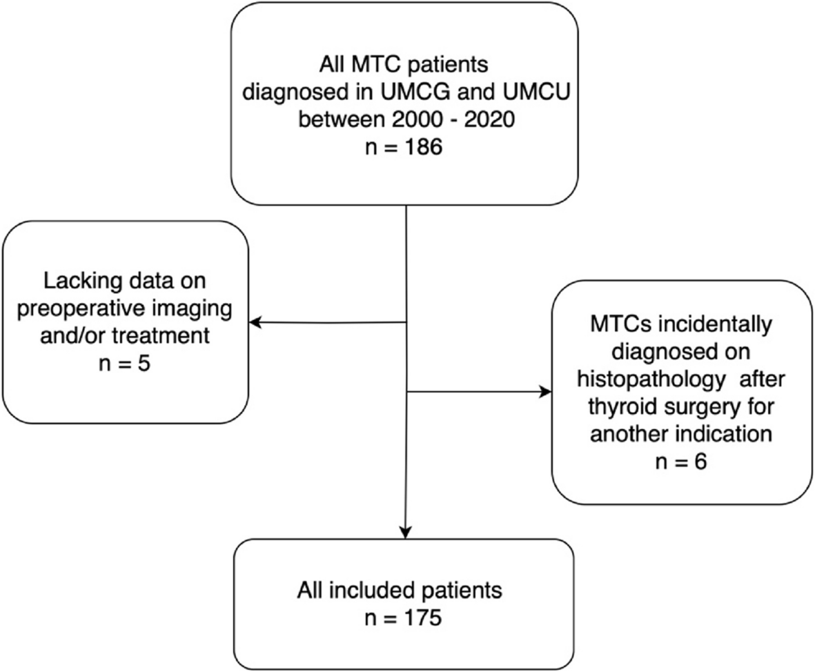
Remember me
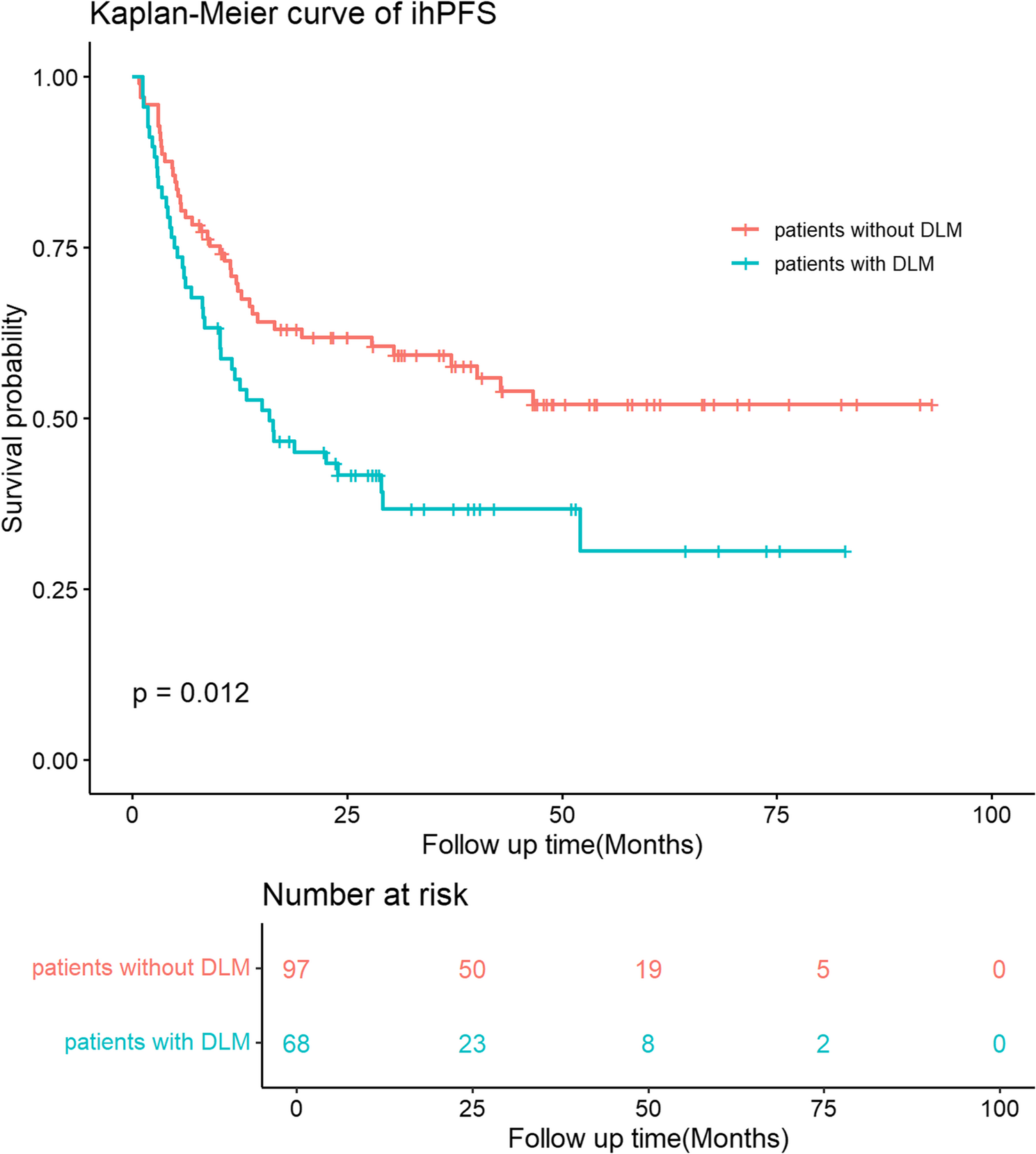


Participant characteristics are shown in Table 2. The median age of the participants at the time of WB-MRI was 55 years (45–63 years), and 1120 participants (54.3%) were men. The median body mass index of the participants was 23.9 kg/m2 (21.6–26.5 kg/m2). In total, 466 participants (22.6%) and 196 participants (9.5%) had a history of hypertension and hepatitis B carrier, respectively. Fifty-one (2.5%) participants had cancer cured for ≥ 5 years. Family history of cancer was reported in 863 participants (41.8%). In this study, 28 participants (1.4%) underwent additional imaging studies that included CT, and 7 participants (0.3%) underwent additional imaging studies that included dedicated MRI. Histopathology examinations were performed on 42 participants (2%) at a median interval of 36 days (19–55 days) after WB-MRI. Cancers were confirmed in 24 participants (1.2%).
Table 2 Characteristics of the participants, WB-MRI results, and additional investigationsCancer detection based on ONCO-RADS categoriesThe kappa value for interobserver agreement for the ONCO-RADS category was 0.927 (95% confidence interval [CI]: 0.89–0.964). On a per-participant basis, 0 (0%), 1984 (96.1%), 37 (1.8%), 35 (1.7%), and 8 (0.4%) of 2064 participants had WB-MRI findings for the ONCO-RADS categories of 1, 2, 3, 4, and 5, respectively. The cancer prevalence rates were 0.1%, 5.4%, 42.9%, and 75% for ONCO-RADS categories 2, 3, 4, and 5, respectively.
The results of further investigation according to the body region and ONCO-RADS category are shown in Table 3. ONCO-RADS category ≥ 4 was observed in the head (n = 2, 0.1%), neck (n = 7, 0.3%), chest (n = 12, 0.6%), abdominal (n = 14, 0.7%), pelvic (n = 8, 0.4%), and bone (n = 1, < 0.1%) regions, without significant differences in prevalence rates among the regions (p = 0.355), except for the head and bone regions (p = 0.002). Cancers were confirmed in the head (n = 2, 0.1%), neck (n = 3, 0.1%), chest (n = 9, 0.4%), abdominal (n = 7, 0.3%), and pelvic (n = 4, 0.2%) regions, without significant differences in prevalence rates among these regions (p = 0.139). One participant had findings for ONCO-RADS category 4 in the chest and abdomen, which were confirmed as lung adenocarcinoma and renal urothelial carcinoma.
Table 3 Results of further investigation according to regions and ONCO-RADS categoriesThe number of incidental lesions confirmed by further investigations is summarized in Table 4. In the head region, all 16 lesions with ONCO-RADS category 3 were confirmed to be benign, and 2 lesions with ONCO-RADS category 4 were malignant (Fig. 1). All participants received neck ultrasound as a part of the cancer screening program, and one histopathology-confirmed 7-mm papillary thyroid carcinoma was not visible on MRI. All 7 neck lesions with ONCO-RADS category 3 were benign. Of the 7 ONCO-RADS-category-4 neck lesions, 2 did not undergo further investigation, 2 (29%) were papillary thyroid carcinoma (Fig. 2A-C), and 3 were benign, of which 2 were salivary gland tumors (Fig. 2D-F) and 1 was a thyroid nodule.
Table 4 Number of incidental lesions confirmed by further investigationsFig. 1
Imaging findings with Oncologically Relevant Findings Reporting and Data System category 4 in the head region. Axial T2 fluid-attenuated inversion recovery (A) and coronal T2-weighted (B) images show a hyperintense area in right temporal lobe of 41-year-old woman. Lesion was confirmed to be anaplastic astrocytoma after surgical resection. In 61-year-old man, axial T2-weighted (C) and contrast-enhanced T1-weighted (D) images reveal left tonsil lesion with heterogeneously high signal intensity, which was diagnosed as squamous cell carcinoma after biopsy
Fig. 2
Imaging findings with Oncologically Relevant Findings Reporting and Data System category 4 in neck region. Axial T2-weighted (A) and contrast-enhanced T1-weighted (B) images show solid thyroid nodule (arrows) in right lobe of 63-year-old woman. Corresponding color Doppler ultrasound image (C) reveals 1.3-cm hypoechoic and hypervascular lesion with histopathology diagnosis of papillary carcinoma. In 67-year-old man, solid lesion (arrowheads) with adjacent daughter nodule was detected in right parotid gland, demonstrating hyperintensity on T2-weighted image (D) and contrast enhancement on T1-weighted image (E). Lesion was hypoechoic on ultrasound image (F) and was determined to be Warthin’s tumor by fine-needle aspiration cytology
Of 2064 participants, 785 (38%) underwent chest CT within a 30-day interval from WB-MRI. One 9-mm pure ground-glass lung nodule undetected by MRI was proven to be adenocarcinoma. Of 10 ONCO-RADS-category-3 lesions in the chest region, 9 (90%) were benign. One ONCO-RADS-category-3 lung lesion, considered to be inflammatory consolidation on MRI, appeared as a 23-mm part-solid ground-glass nodule on CT and was confirmed to be malignant. Of the 12 chest lesions with ONCO-RADS category ≥ 4, 5 were lung adenocarcinomas and 2 were thymomas (Fig. 3).
Fig. 3
Imaging findings with Oncologically Relevant Findings Reporting and Data System category 4 in chest region. Sagittal T2 half-Fourier single-shot turbo spin echo (HASTE) (A) and coronal contrast-enhanced T1 gradient echo (GRE) (B) images reveal 17-mm subpleural nodule (arrows) in upper lobe of left lung in 62-year-old man. Lesion is seen on corresponding coronal CT scan lung window (C) and was confirmed as lung adenocarcinoma after surgical resection. In 69-year-old woman, 5.8-cm anterior mediastinal mass with hyperintensity was noted on T2 HASTE image (D) and contrast enhancement on T1 GRE image (E). Corresponding axial CT scan (F) demonstrates calcification in mass and thymoma was diagnosed after surgical resection
In the abdominal region, 4 ONCO-RADS-category-4 lesions that did not undergo further examination included two 3-cm suprarenal lesions, one 4-cm pancreatic cystic lesion, and one 8-mm gastric submucosal lesion. Of the 14 abdominal lesions with ONCO-RADS category ≥ 4, 7 (50%) were confirmed as cancers (Fig. 4A-C). Of the 8 pelvic lesions with ONCO-RADS category ≥ 4, 4 (50%) were prostatic adenocarcinoma (Fig. 4D-F), 1 was endometrial polyp, and 3 were prostatic lesions that did not undergo further examination. In the bone region, one ONCO-RADS-category-3 lesion was spinal meningioma, and one ONCO-RADS-category-4 lesion was a paraspinal neurogenic tumor.
Fig. 4
Imaging findings with Oncologically Relevant Findings Reporting and Data System (ONCO-RADS) category 4 in abdominal region and ONCO-RADS category 5 in pelvic region. In 75-year-old man, 3.6-cm exophytic mass (arrows) from lesser curvature of stomach is depicted on axial T2-weighted image (A) and T1-weighted images before (B) and after (C) contrast enhancement. Diagnosis of stromal cell tumor was given after laparoscopic resection. Axial T2-weighted image (D) revealed focal lesion (arrowheads) in left transition zone with low signal intensity, measuring more than 1.5 cm. Lesion shows marked diffusion restriction on diffusion-weighted imaging (E) and corresponding apparent diffusion coefficient map (F) and was classified as ONCO-RADS category 5. Prostate cancer was confirmed in 58-year-old man after biopsy directed by cognitive MRI-transrectal-ultrasound fusion technique
Factors associated with WB-MRI findings for ONCO-RADS category ≥ 4The results of univariable and multivariable Firth logistic regression analyses of the associations between participant characteristics and WB-MRI findings for ONCO-RADS category ≥ 4 are presented in Table 5. In the multivariable model, older age (OR: 1.035, 95% CI: 1.004–1.068, p = 0.029), history of hypertension (OR: 2.051, 95% CI: 1.086–3.868, p = 026), history of hepatitis B carrier (OR: 2.584, 95% CI: 1.222–5.467, p = 0.013), and prior surgery (OR: 3.787, 95% CI: 1.992–7.197, p < 0.001) were independently associated with the WB-MRI findings of ONCO-RADS category ≥ 4.
Table 5 Univariable and multivariable Firth logistic regression analyses for factors associated with ONCO-RADS ≥ 4
Comments (0)