
記住我


This study was an observational exploratory monocentric study conducted at the Université de Lorraine, in the laboratory “Stress, Immunité et Pathogènes” (SIMPA, UR 7300), and at the University Hospital of Nancy from January 2021 to December 2022. The study was registered with clinicaltrials.gov under the number NCT05264571 (retrospectively registered 22 February 2022). This study was an ancillary study of a non-interventional prospective cohort study (the pBDG2 study, prospective evaluation of 1.3 β-d-glucan in the PF for the diagnosis of intra-abdominal candidiasis in the critically ill patients NCT03997929). The pBDG2 study included critically ill patients with intra-abdominal infections requiring abdominal surgery and allowed the constitution of a biological collection of peritoneal fluid samples.
Abdominal samplesThe samples used in this study consisted of PF obtained from critically ill patients with intra-abdominal infections, and ascitic fluid (AF) obtained from critically ill patients with decompensated cirrhosis of non-infectious origin. AF has the advantage to be the accessible sterile, biological fluid with the closest composition to PF. These samples were obtained after routine analysis, and the remaining samples were selected for use in this study. All selected samples underwent microbiological analysis to detect the presence of bacteria and fungi.
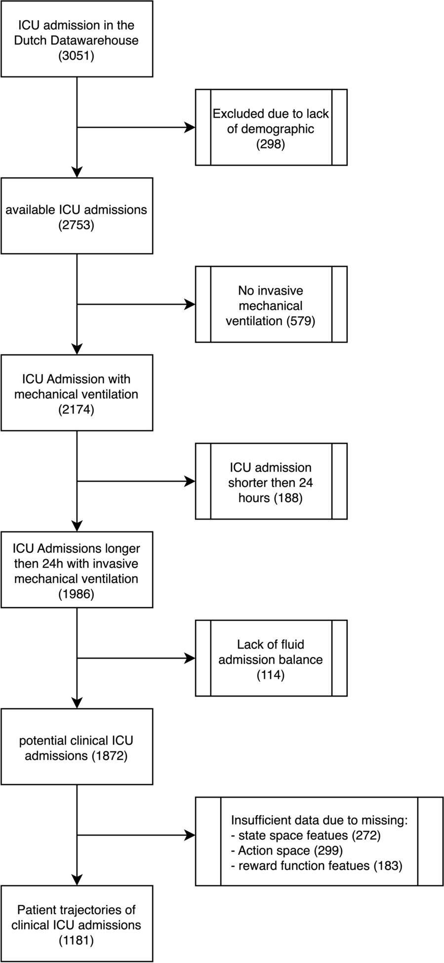
To be eligible for selection, PF samples had to meet the following criteria: absence of Candida infection, derived from the initial abdominal surgery (excluding relaparotomy), obtained from the University Hospital of Nancy, and having a minimum volume of 5 mL.
Strains and mediaThe SC5314 reference C. albicans strain was used for all experiments, as its growth and genome are well-described in the literature [22]. For the evaluation of metabolic production, clinical strains of various bacteria were utilized.
For all experiments (phenotype, molecular, metabolic) three different media were employed: two as controls (Sabouraud and AF) and the PF. Sabouraud medium (SBD) served as a control medium for assessing growth, morphology, and metabolic profile. AF was obtained as a non-infected clinical sample from humans, and its lack of infection was confirmed through microbiological analysis (direct examination, conventional culture) and cytology (neutrophil count < 250/mm3). PF, on the other hand, could exhibit different characteristics and might be infected with various bacteria, both positive and negative gram. Since AF and PF were collected consecutively, they were stored at − 20 °C until each experiment period. All analyses were conducted in duplicate or triplicate.
Phenotypic approachInoculation of C. albicans in the different mediaC. albicans SC5314 strain was inoculated in triplicate into one mL of the three different types of media (SBD, AF, PF) at an optical density (OD) of 0.3 nm, corresponding to approximately 3.106 colonies of C. albicans per millilitre (C. albicans/mL) of media. Control wells containing only the media without C. albicans were also included on the same 24-well plate to confirm the absence of contamination. All OD measurements were performed at 600 nm with a UV/Visible spectrophotometer P4 from VWR® (Radnor, Pennsylvania, United States).
Observation of C. albicans in each mediaThe 24-well plate was inoculated and incubated at 37 °C with shaking at 300 RPM for 24 h. First, the morphology of C. albicans was observed under a light microscope at hourly intervals for the initial 8 h, followed by observations at H16 and H24. The analysis of morphology was conducted by ME, in collaboration with the members of the SIMPA laboratory. The results were subsequently validated by MM, an expert in mycology, who was blinded to the sample types. Any discrepancies were resolved through consensus or consultation with a third independent researcher. Second, C. albicans growth was assessed by inoculating 10 μL from each well onto SBD dextrose agar, and the colonies were counted after 24 h of incubation at 37 °C. To facilitate counting, 1000-fold dilutions of each inoculated well were prepared beforehand.
Composition of ascitic and peritoneal fluidEach liquid received biochemical measurements (protein, glucose, pH) and cell counts using flow cytometry.
Molecular approachC. albicans inoculation in the different media followed the same protocol as for phenotypic evaluation, with an overnight culture of 24 h before gene expression analysis.
Genes of interestThe primers utilized for reverse transcription and quantitative polymerase chain reaction (RTq-PCR) analysis can be found in supplementary material (Additional file 1: Table S1). The five virulence genes of interest are UME6, ALS3, SFL2, HWP1 and ECE1. Basically, UME6 and SFL2 have a role in Candida filamentation. ALS3, HWP1, and ECE1, have a role in adhesion and epithelial cells damage. Please refer to supplementary material for the precise role of each gene.
RNA extractionAfter the 24-h overnight culture, the samples were centrifuged to retain only the cell pellet. The cell pellet was then washed twice with 10 mL of phosphate-buffered saline (PBS). Subsequently, the PBS was removed, leaving behind only the cell pellet.
RNA extraction was performed using the FASTPREP® lysis technology (MP Biomedicals, Santa Ana, California, United States), following the manufacturer’s program specifically designed for Candida cells. To assess the quality and measure the RNA concentration, each sample was analyzed using a Nanodrop 2000 system (Thermo Scientific, Waltham, MA, United States). Total RNA extraction was carried out using the Monarch Genomic RNA Purification Kit (New England Biolabs® Inc., Ipswich, MA, United States) in accordance with the manufacturer’s instructions.
Reverse transcriptionReverse transcription was performed using the QuantiTect Reverse Transcription kit from Qiagen® (Germantown, Maryland, United States) following the manufacturer's instructions. The samples were prepared in two steps: genomic DNA removal and reverse transcription. The reverse transcription process was carried out using a thermocycler, following the specified protocol. The steps involved were as follows: annealing at 25 °C for 3 min, reverse transcription at 45 °C for 10 min, and inactivation at 85 °C for 5 min.
Amplification by PCRThe qPCR was performed in MicroAmp Optical 96-Well Reaction Plates (Applied Biosystems) using the CFX96 Real-Time PCR System (Bio-Rad, Marnes-la Coquette, France). Please refer to supplementary material S1 for the details regarding the primers and cycling protocol. Relative transcript levels and the fold change of SFL2, UME6, ALS3, HWP1 and ECE1 were determined following the ΔΔCT method [23]. All analyses were performed in duplicate with negative control samples. The level of expression was compared among the PF against AF level expression, to ensure the comparability in a clinical sample.
Metabolic approach using the calscreener™ technologyThe calscreener™ technologyThe metabolic evaluation was conducted using the calScreener™ technology (Symcel AB, Solna, Sweden) (Fig. 1) [24]. This technology utilizes isothermal calorimetry to measure the heat flow generated by living cells in real time. It can be applied to various microorganisms and media types. The calScreener™ device provides continuous real-time data over an extended duration. Different parameters can be evaluated directly from the curve, such as time to activity, time to peak or decay time. Analysis of the heat production is carried out using the dedicated software, calView® (Symcel AB, Solna, Sweden). Each specific pathogen generates a distinct growth pattern in the kinetic data, enabling identification of the pathogen type [25]. This technology utilizes 48-well microtiter cell culture plates, accommodating 32 biological samples and 16 thermodynamic internal reference positions.
Fig. 1
Peritoneal fluid selection and type of analysis. Metabolic approach has been performed with the calscreener™, picture of the equipment was reproduced with permission from Symcel AB (Solna, Sweden). PF: peritoneal fluid; RTqPCR: reverse transcription quantitative polymerase chain reaction
Metabolic analysisFor the inoculation of C. albicans in the different media, the same protocol was followed as for the phenotypic and molecular evaluation. The overnight culture of C. albicans for 24 h was used before transferring the samples into the calScreener™. It should be noted that all AF and PF samples used were confirmed to be free of pathogens to serve as controls for the added inoculum. Each culture was then appropriately diluted, either 1/100 or 1/1000, in each medium as per the recommendations provided by Symcel. Subsequently, 200 µL of each medium, in duplicate, were transferred to the calScreener™ sample handling system. Real-time measurement of the heat produced from each calWells was carried out at 37 ± 0.001 °C in the calScreener™ up to 48 h. The presence of an active pathogen is associated with a metabolic activity > 5 microW, according to the manufacturer. The remaining samples in the microtitre plate were incubated at 37 °C for 24 h to ensure the presence of expected pathogens.
Statistical analysisDue to the exploratory and observational nature of this study, a predetermined calculation of the required sample size was not performed. The sample size was based on the availability of PF samples during the study period. Descriptive statistics were computed using GraphPad Prism version 9 (Boston, Massachusetts, United States). The statistics included counts, means, standard deviation, medians, quartiles, and interquartile ranges (IQR), as deemed appropriate for the analysis.
To compare the heat production between samples during the metabolic evaluation, ordinary one-way ANOVA and Mann–Whitney test were employed. This statistical analysis was used to assess any significant differences in heat production among the samples. A p value < 0.05 was considered significant.
留言 (0)