
記住我


A 27-year-old male contact lens wearer was referred to our outpatient ophthalmology clinic for the first time by his primary care ophthalmologist with an urgent case of keratitis of presumed bacterial origin of the right eye. Symptoms started four days before the referral. Treatment with a combination eyedrop containing gentamicin and dexamethasone was prescribed by the patient’s primary care ophthalmologist. The patient started taking the eye drops 4–6 times a day before referral. The last documented corrected visual acuity prior to the onset of the keratitis was 1.0. His initial visual acuity at presentation with his own glasses was 0.1 (1.00 logMAR) in the affected right eye and 1.0 (0.00 logMAR) in the unaffected left eye. Ophthalmic examination revealed a mucopurulent infiltrate in the cornea approximately the size of 25% of the cornea involving the limbal region (Fig. 1A). Stromal melting had already occurred and the corneal thickness at the thinnest point was 320 µm (Fig. 2A). In summary, based on the history and appearance, the first clinical suspected diagnosis was a corneal ulcer caused by P. aeruginosa; other bacteria or fungi were also considered for differential diagnosis. Due to the severity and size of the findings, the patient was admitted to inpatient care. Furthermore, a corneal ulcer scraping, a conjunctival swab, and the contact lens case were sent to microbiology for direct staining, culture, and antimicrobial susceptibility testing. A combination preparation eyedrop of polymyxin-B, neomycin, and gramicidin as well as moxifloxacin eye drops were started and administered by the nurse on an hourly alternating basis at day and night. In literature, the combination of polymyxin-B, neomycin and gramicidin [4] as well as moxifloxacin [5] have been described as highly effective for the treatment of bacterial corneal ulcers. With this therapy, we used commercially available antibiotic eye drops with good clinical effectiveness, a broad-spectrum antibacterial activity, and few side effects. In addition, atropine eye drops 0.5% were used throughout the inpatient stay (all eye drops including concentrations are shown in Table 1).
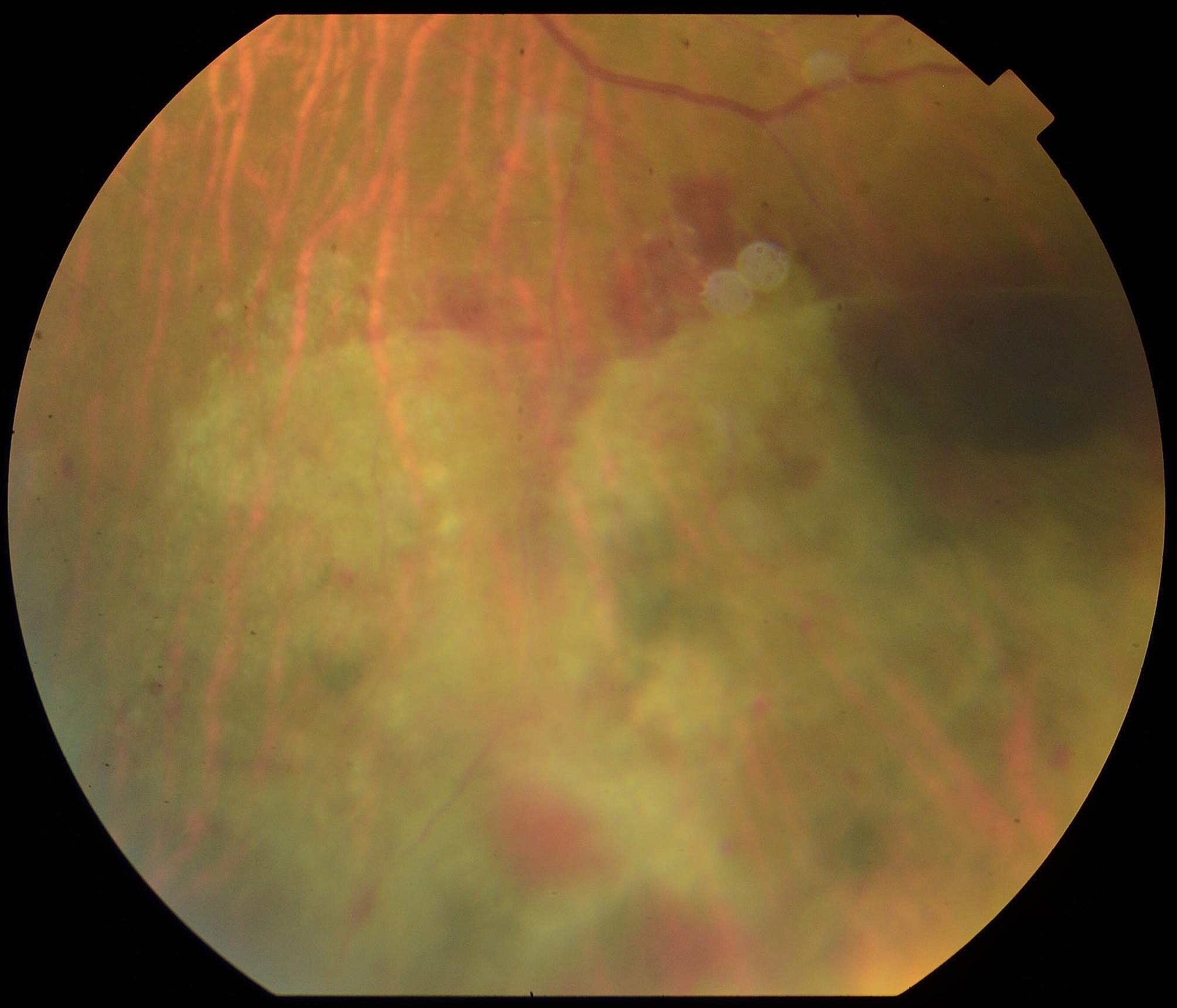
Fig. 1
Photographic images of anterior eye segment of the right eye: A) Day 1; B) Day 3; C) Day 7; D) Day 15; E) Day 20; F) after 4 months
Fig. 2
Anterior segment optical coherence tomography of the right eye: A) Day 8, thinnest corneal point 317 µm; B) after 8 months, corneal thickness 356 µm
Table 1 Concentration of administered topical eyedropsThe next day, a rapid enlargement occurred, so gentamicin eye drops were added. These eye drops have a good spectrum of efficacy in the gram-negative area.
On the third day, the size of the infiltrate increased to approximately twice its initial size (Fig. 1B). The differential diagnosis of a possible fungal infection was covered with hourly voriconazole 1% eye drops.
On the sixth day, there was a further marked worsening of the findings with new hypopyon formation. Topical levofloxacin was started and administered every hour alternating with gentamicin instead of moxifloxacin and the combination preparation of polymyxin-B, gramicidin, and neomycin.
A microbial detection with antibiogram (Table 2) of the corneal and conjunctival swab was available during the course of the sixth day and revealed a MDR P. aeruginosa. Susceptibility testing revealed multiple resistance to ureidopenicillins, cephalosporins, fluoroquinolones, and macrolides. The Pseudomonas was only sensitive to meropenem (minimal inhibitory concentration: < 0,25 mg/l) and colistin. Staphylococcus lugdunensis with a resistance to colistin was found in the contact lens case of the same eye. No fungi were detected.
Table 2 Antibiotic sensitivity testing of Pseudomonas aeruginosa in the corneal swab and Staphylococcus lugdunensis in the corneal lens case of the right eye; R resistant, I intermediate, S = sensibleBased on the antibiogram, the use of meropenem and colistin eye drops was considered. Although polymyxin B has structural similarities to colistin, a worsening of the corneal findings was observed during the therapy with Polymyxin B. In addition, the Staphylococcus lugdunensis from the contact lens case was resistant to colistin. Therefore, meropenem eye drops were chosen for further treatment. Meropenem eye drops 50 mg/ml were prepared by the hospital pharmacy. In addition to the local antibiotics, adjunctive systemic meropenem administration was started after the patient was fully informed about the individual curative trial and the off-label use. A subsequent ocular hypertension was successfully treated with a carbonic anhydrase inhibitor (dorzolamide eye drops) and a beta blocker (timolol eye drops). Meropenem eye drops were applied hourly and systemic meropenem was used 1 g three times a day. In addition, systemic voriconazole was given as intravenous infusion (720 mg for the first two infusions and then 480 mg twice daily). This resulted in a rapid improvement over the following days (Fig. 1C: day 7; Fig. 1D: day 15). Additionally topical steroids (dexamethasone) were administered two days after the onset of clinical improvement and systemic therapy was discontinued at the same time after a total administration of 7 days. The patient was discharged on day 20 on topical therapy (dexamethasone 4x/day, gentamicin 4x/day, levofloxacin 4x/day, meropenem 4x/day) with the ability to count fingers (Fig. 1E: day 20). In addition to meropenem eye drops, gentamicin and levofloxacin were continued due to the intermediate susceptibility of P. aeruginosa to these agents and the improvement in corneal status with the combination therapy. The meropenem eye drops continued to be prepared by our hospital pharmacy and provided to the patient during his outpatient treatment. The attempt to discontinue all antibiotics during the course was successful. In total, meropenem eye drops were given for 18 days (hourly for 10 days; every 2 h for one day; 4x/day for 7 days) and systemic meropenem for 7 days (1 g 3x/day). The visual acuity increased continuously. Emergency keratoplasty with a presumably poor prognosis due to extensive neovascularization was avoided.
After four and eight months, high irregular astigmatism (Fig. 3, follow-up after 8 months) and stromal scarring were evident (Fig. 1F, follow-up after 4 months; Fig. 2B, follow-up after 8 months). Visual acuity with the patient's old glasses was 0.2 (0.70 logMAR) and the intraocular pressure was 18 mmHg in the affected eye. At this point, further therapeutic options would have been a correction with glasses or contact lenses, a deep anterior lamellar keratoplasty (DALK), or a penetrating keratoplasty. In our opinion, the scar was too large for phototherapeutic keratectomy without sufficient residual thickness after laser ablation. Despite these findings and limitations, the patient was satisfied with his current state of his vision. At this time, he preferred not to undergo corneal transplant. He also decided not to get new glasses and stopped wearing contact lenses. After eight months, the patient presented with stable findings and visual acuity. The corneal thickness at the previous thinnest point was 356 µm (Fig. 2B). A follow-up visit is planned to reevaluate visual improvement for example by wearing contact lenses or by surgical options. The option of a corneal transplantation for visual improvement is still available in the future.
Fig. 3
Corneal tomography (Pentacam®) of the right eye after 8 months
留言 (0)