
Remember me
The SARS-CoV-2 WA1 was serially passaged in Vero E6 cells in DMEM with 1% antibiotics/antimycotics (Gibco, Carlsbad, CA, USA) from 36 °C to 21 °C, with 1–2 passages at each temperature. Then, 5 additional passages were performed at 21 °C to further adapt the virus. Every passage of infected Vero E6 cells was frozen and thawed once, vortexed, and clarified by centrifugation.
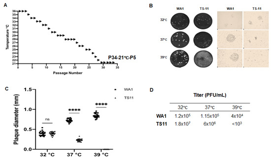
2.3. Plaque Assay and Selection of the Temperature-Sensitive SARS-CoV-2 ClonesFor plaque assay, monolayers of Vero E6 cells were inoculated with 10-fold serial dilutions of SARS-CoV-2 for 1 h. Then, the virus inoculum was removed, and the cell monolayers were washed three times with PBS. The cell monolayers were overlaid with 1.5% agarose (or 1% methylcellulose) in minimal essential medium (MEM) supplemented with 1% antibiotics/antimycotics, 1% HEPES, 1% non-essential amino acids and 2% heat-inactivated FBS. To clone the P34-21 °C-P5, the plaque assay was performed with 1.5% agarose overlay and individual plaques were picked up and cultured in Vero E6 cells at 32 °C for 4 days to produce a working virus stock. The above-selected virus clones were titrated using plaque assay at 32 °C for 4 days, and at 37 °C and 39 °C for 3 days. The diameter of 20 plaques was measured for each temperature/virus.
2.4. Multiple-Step Growth Kinetics of SARS-CoV-2 TS11To evaluate the multi-step growth kinetics, Vero E6 cells were infected with a virus at a MOI of 0.01 for 1 h at 32 °C, and shifted and incubated at one of the respective temperatures 32 °C, 37 °C or 39 °C for 1 h, 24 h, 48 h, 72 h and 96 h. Then, the infected cells and supernatants were frozen and thawed once before harvesting. Then, the samples were vortexed, centrifuged, and the supernatants were tested for infectious titers on Vero E6 cells by plaque assay (overlayed with 1% methylcellulose) at 32 °C for 4 days.
2.5. RNA Extraction and Reverse Transcription-PCRTotal cellular RNA was extracted using Trizol reagent (Invitrogen, Carlsbad, CA, USA) following the manufacturer’s instructions. We used the E and RNA-dependent-RNA polymerase (RdRp) gene-specific reverse transcription (RT)-qPCR assays to investigate the virus replication efficiency and to determine genomic equivalent titers, respectively. The positive-sense (+) RNA of SARS-CoV-2 was reverse transcribed using the E gene-specific reverse primer and SuperScript® IV Reverse Transcriptase (Thermo Fisher, Waltham, MA, USA). The cDNA was subjected to quantitative PCR using SYBR green PCR mix (Life Technologies, Carlsbad, CA, USA) according to the manufacturer’s instructions. The subgenomic (sg) (+) E RNA was amplified with forward primer 5′-CGATCTCTTGTAGATCTGTTCTC-3′ and reverse primer 5′-ATATTGCAGCAGTACGCACACA-3′ [23]. The β-actin gene of Vero E6 cells was an internal control and amplified with forward primer 5′-AGGCTCTCTTCCAACCTTCCTT-3′ and reverse primer 5′-CGTACAGGTCTTTACGGATGTCCA-3′ [24]. Relative quantification is presented as fold changes relative to the control using the 2ΔΔCT threshold method. TaqMan real-time RT-PCR (RT-qPCR) targeting the SARS-CoV-2 RdRp gene was performed using OneStep RT-PCR Kit (QIAGEN, Valencia, CA, USA) with primers and probe described previously [25]. To differentiate WA1 and TS11, conventional one-step RT-PCR was performed with primers (forward primer 5′- CTGACGGCGTAAAACACGTCTATCAGTTAC-3′ and reverse primer 5′- CTCCATTCTGGTTACTGCCAGTTGAATCTGAG-3′) with an annealing temperature of 55 °C. This pair of primers were designed in this study and covered the 371-nt-del in the ORF7b-ORF8 genes of SARS-CoV-2 and the product sizes for WA1 and TS11 were 769 bp and 398 bp, respectively. We used the OneStep RT-PCR Kit (QIAGEN) for this assay. 2.6. Next Generation Sequencing (NGS) and Sanger SequencingSARS-CoV-2 was sequenced by NGS as published [26]. Previously extracted RNA underwent 1st- and 2nd-strand cDNA synthesis (NEBNext Ultra II Non-Directional RNA Second Strand Synthesis Module; NEB, Ipswich, MA, USA), followed by sequencing using two different clinically validated amplicon-based methods. Some samples were analyzed using the SARS-CoV-2 Research Panel primers (ThermoFisher, Waltham, MA, USA) on the Ion Chef-S5 sequencer (ThermoFisher, Waltham, MA, USA) per the manufacturer’s conditions. Cultured virus and other samples were analyzed using the COVIDSeq kit (Illumina, San Diego, CA, USA) per the manufacturer’s conditions and sequenced on the NextSeq 550 sequencer (Illumina, San Diego, CA, USA). Strain typing by the two different assays was cross-validated using a set of 20 SARS-CoV-2-positive samples and 5 SARS-CoV-2-negative samples. Analysis tools include custom pipelines utilizing GATK and Mutect2 (Broad Institute) to determine variant percentages, and Dragen SARS-COVID variant detection (Illumina, San Diego, CA, USA). Viral sequences were strain-typed using NextStrain criteria (https://clades.nextstrain.org (accessed on 17 October 2022)). Pango lineages were determined according to https://pangolin.cog-uk.io (accessed on 23 April 2021) [27]. Sanger sequencing with SARS-CoV-2-specific primers was performed to confirm vague sequence regions generated by NGS. 2.7. Experimental Infection of Syrian Hamsters with SARS-CoV-2All SARS-CoV-2 hamster studies were approved by the Ohio State University Institutional Animal Care and Use Committee. Syrian hamsters were purchased from Charles River (Wilmington, MA, USA). We conducted 2 experimental trials.
In the 1st trial, we tested whether the TS11 could cause clinical disease in hamsters. Eighteen 5–6-week-old male hamsters were randomly assigned to the SARS-CoV-2 WA1 group (n = 9, 3 hamster/cage) and the SARS-CoV-2 TS11 group (n = 9, 3 hamsters/cage). WA1 and TS11 groups were housed in the same animal room in the BSL3 facility, mimicking the natural conditions of vaccinated individuals who are subject to SARS-CoV-2 exposure at variable time points post-vaccination. The hamsters were anesthetized in the same box and sampled in the same biosafety cabinet (BSC) in the sequence of the TS11 group first followed by the WA1 group after cleaning the BSC using CaviCide spray (Metrex, Orange, CA, USA), waiting for 5 min, and changing gloves between handling the two groups of hamsters. The hamsters were inoculated intranasally (IN) with 4 × 10⁴ plaque-forming units (PFU) (in 60 µL) of WA1 or TS11. The hamsters were monitored for clinical signs daily. Their body weights and ear temperatures (in °F; transferred to °C) were measured, and nasal washes were collected before viral inoculation, daily for 1–6 days post-inoculation (dpi) and every other days during 7–13 dpi. Three hamsters in the same cage of each group were euthanized on 3 dpi, 6 dpi and 13 dpi. During necropsy, nasal turbinates, trachea, lungs, bronchoalveolar lavage fluid (BALF), and small intestinal tissues were harvested and frozen at −80 °C for the detection of infectious viral titers in each tissue.
As TS11-inoculated hamsters were subsequently infected with WA1, a second experiment was carried out following decontamination. To remove environmental viruses, BSL3-Ag and necropsy spaces, which were used to house and handle the hamsters, were cleaned, sanitized and decontaminated using chlorine dioxide gas. No projects were run following the decontamination and prior to our trial. During this hamster study, no participating personnel worked with the WA1 strain. The animal cages were emptied, autoclaved and washed prior to reuse.
In this second trial, we tested the pathogenicity and protective efficacy of TS11 against the challenge with a virulent SARS-CoV-2 D614G strain 14B in Syrian hamsters. Thirty 7–8 week-old male hamsters were randomly assigned to mock-challenge (Mock-C) and TS11 immunization-challenge (TS11-C) groups and immunized intranasally with culture medium and TS11 (8 × 10⁴ PFU per hamster), respectively. At 21 dpi, both groups were challenged intranasally with 14B (3 × 10⁵ TCID50 per hamster). Hamsters were observed for clinical signs daily. The body weights were measured and nasal washes were collected daily during the first four days after inoculation and every two or three days thereafter [from 5–20 dpi/5–12 days post-challenge (dpc)]. At 2 dpi, 6 dpi, 20 dpi, 23 dpi/2 dpc and 33 dpi/12 dpc, 3 hamsters/cage/group were euthanized to measure virus loads and conduct the histopathological examination. During necropsy, URT wash samples were collected by flushing the UTR from the upper trachea with 0.5 mL of culture medium and collected the effluents from the nostrils. Nasal turbinates, trachea, lungs and BALF (from the right lung), and testicles were harvested and frozen at −80 °C for the detection of infectious viral titers in each tissue. The left lungs were fixed in formalin solution for histopathological examination. Blood samples to measure viral neutralizing antibody titers were collected before vaccination and at euthanasia.
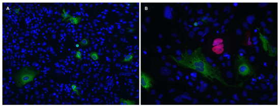
2.8. Histopathological Evaluation and Immunohistochemistry (IHC) for the Detection of SARS-CoV-2 AntigenThe left lung was fixed in 10% neutral formalin. Tissues were embedded, sectioned (3.5 µm), and stained with Gill’s hematoxylin and eosin (H&E) for light microscopic examination as described previously [28]. The Mock-C hamsters at 2 dpi or 12 dpc, histopathologic lesions mainly consisted of (1) accumulation of necrotic cells and inflammatory cells in alveolar, or bronchial or bronchiolar lumens; (2) thickening of alveolar septa by type 2 pneumocyte hyperplasia and inflammatory cell infiltration; and (3) vascular abnormalities (Supplemental Figure S1) Vascular lesions included (1) the presence of hypertrophied endothelial cells, (2) the aggregates of inflammatory cells beneath or within the endothelial cell layer, or within the vascular wall, (3) the hypereosinophilia and degeneration of the tunica media, or (4) perivascular edema and/or lymphocytic cuffing (Supplemental Figure S1), consistent with a previous report [29]. The major histopathologic lesions were used as parameters to evaluate the severity of pneumonia, as described previously with slight modifications [30]. Briefly, the severity of alveolitis and bronchitis/bronchiolitis or thickening of alveolar septa was evaluated based on the percentage of the affected area in each microscopic area (×25) of a pulmonary section, as follows: 0, no lesions; 1, affected area ≤10%; 2, affected area 10–50%; and 3, affected area ≥50%. However, the severity of vascular lesions was evaluated, as follows: 0, no lesions; 1, focal or multifocal, mild lesions; 2, multifocal, moderate lesions; and 3, multifocal, severe lesions. For each parameter, mean values of all scores from three different areas in each cranial, middle, and caudal region of the left lung were calculated and then cumulated (ranging from 0 to 9).The formalin-fixed lung tissue sections were tested by IHC for the detection of SARS-CoV-2 antigen, as previously described with slight modifications [31]. A recombinant monoclonal antibody against SARS-CoV-2 N protein (Kerafast, Boston, MA, USA) was used as the primary antibody and a non-biotin polymerized horseradish peroxidase system (BioGenex Laboratories, San Ramon, CA, USA) was used for visualization as brown staining. Stained tissues were counterstained with hematoxylin. SARS-CoV-2 antigen-positive IHC scores were computed by estimating the intensity and frequency of IHC-positive cells in each given microscopic area (×100) of a pulmonary section based on the following criteria: 0, no positive cells; 1, low numbers of positive cells showed focal or multifocal, mild staining; 2, moderate numbers of positive cells showed multifocal or multifocal to coalescing, moderate staining; and 3, high numbers of positive cells showed diffuse strong staining. Mean values of all scores from three different areas in each cranial, middle, and caudal region of the left lung were calculated (ranging from 0 to 3). 2.9. Measurement of Infectious Viral Titers and Serum Viral Neutralizing (VN) Antibody TitersHamster tissues were weighed and homogenized for 30 s at 4.0 m/s, using the TissueLyser II (Qiagen) in 500 µL DMEM containing 2% FBS and 1% Antibiotic-Antimycotic. The nasal washes, tissue homogenates, URT washes and BALF samples were clarified by centrifugation at ~ 2000× g for 5 min and the supernatants were collected for measurement of infectious virus titers in median tissue culture infectious dose 50 (TCID50) using 96-well plates. Briefly, Vero E6 cells were seeded into 96-well plates one day before the testing. The cell monolayers were washed twice before inoculation (100 µL of 10-fold serial dilutions of samples per well and four replicates per dilution). The plates were incubated at 32 °C for the TS11 mutant or at 37 °C for the 14B strain. Viral cytopathic effects (CPEs) were observed at 3 dpi and virus titers were calculated by the Reed-Muench method [32].Serum VN antibody titers were measured by TCID50-reduction neutralization assay. To prepare the serum-virus mixture, 4-fold serially diluted serum samples were mixed with an equal volume of SARS-CoV-2 14B strain (100 TCID50 of virus/well for the final inoculation). The serum-virus mixtures were incubated at 37 °C for 1 hr before inoculation of the Vero E6 cell monolayers, with four replicates per dilution. Virus control, medium control, and positive and negative serum controls were included. The plates were incubated at 37 °C for 3 days. Viral CPEs were observed and the absence of CPEs indicates that the virus was neutralized. All serum samples were tested at the same time. VN antibody titers were calculated by the Reed-Muench method [32]. 2.10. Statistical AnalysisThe statistical analyses were performed using GraphPad Prism, version 8. The comparison of values of TS11 and WA1 were analyzed by Student’s t-test. RNA and infectious viral titers were analyzed by one-way ANOVA followed by Dennett’s test. A p value of less than 0.05 was considered significantly different.
4. DiscussionHere, we present our results on the development and characterization of a cold-adapted temperature-sensitive variant of SARS-CoV-2 (TS11) that shows attenuated phenotype both in vitro and in Syrian hamsters. Compared with its parent virus, TS11 has sequence alterations in the furin cleavage site of S and other genes including nsp3 (Table 1). The nsp3 of SARS-CoV-2 is the largest viral protein and is an essential component of the replication/transcription complex [34]. The nsp3 SARS-Unique Domain (SUD) includes three distinct subdomains: macrodomain II (Mac2), macrodomain III (Mac3), and domain preceding ubiquitin-like domain 2 (Ubl2) and papain-like protease 2 (PL2pro) (DPUP) [35]. Previously, Deng et al. [36] reported a temperature-sensitive mouse hepatitis virus (MHV), a betacoronavirus (tsNC11) whose phenotype was induced by the mutations in its nsp3 suggesting a similar mechanism for TS11 which showed changes in Mac2, Mac3 and DPUP. Mac2 is dispensable for the SARS-CoV replication/transcription complex, while Mac3 is necessary. The Mac3 domain of SARS-CoV nsp3 can interact with DNA or RNA G-quadruplexes (G4) and is essential for SARS-CoV replication [37]. It has been shown that Mac2 and Mac3 of SARS-CoV-2 also interact with G4 structures [38]. Recently, the nsp3 Mac2 of both SARS-CoV and SARS-CoV-2 have been shown to interact with human poly(A) binding protein interacting protein 1 (Paip-1), a component of the cellular translation machinery [39]. For SARS-CoV, this interaction between Mac2 and Paip1 stimulates viral RNA translation but does not affect the translation of cellular mRNAs. The M494K mutation of SARS-CoV-2 TS11 located in Mac2 changes the amino acid methionine with an S-methyl thioether side chain to lysine with a positively charged polar side chain. This M494K mutation may influence the interactions between Mac2 and Paip1, thereby influencing the viral RNA translation. The A579V mutation is in the Mac3 domain. The Ubl2 and PL2pro domains are conserved in all CoVs. The exact functional role of the Ubl2 domain is not clear, while the PL2pro possesses proteolytic, deubiquitinating, and deISGylating activities [35]. The T763M is a non-conservative mutation within the PL2pro domain and might alter its functions. It appears that T1456I mutation in the nsp3 ectodomain (3Ecto) is not critical for the domain functionality since the conserved cysteines and the two glycosylation sites (Asn1431 and Asn1434) essential for 3Ecto functions were retained [35]. Whether some or all of these mutations in the nsp3 of SARS-CoV-2 TS11 mutant are essential to induce temperature-sensitivity remains to be determined using reverse genetics.The furin cleavage site of the SARS-CoV-2 WA1 S protein plays a critical role in the efficient infection of the LRT [40]. A SARS-CoV-2 WA1 variant lacking the furin cleavage site had decreased replication in a human respiratory cell line, was attenuated in both hamsters and K18-hACE2 transgenic mice [41], and had reduced transmission in ferrets [42]. Therefore, the 12-amino acid deletion including the furin cleavage site may contribute to the attenuated phenotype of SARS-CoV-2 TS11 in hamsters. However, we observed that despite the attenuated phenotype, TS11 was still capable of efficient transmission to the WA1-inoculated hamsters starting at 2 dpi, which was similar to the transmission of WA1 to the TS11-inoculated hamsters. The orf8 of SARS-CoV-2 is a glycoprotein that is secreted as homodimers [43]. It can interact with the IL17 receptor A to modulate IL-17 signaling [44], decreasing IFN-β production [45] and downregulating the surface expression of MHC class I to evade immune responses [46].A SARS-CoV-2 variant carrying a 382-nucleotide-deletion in ORF8 was first detected in 23.6% (45/191) of samples in Singapore in January 2020 [47]. It exhibited dramatically higher replicative fitness in vitro than the wildtype SARS-CoV-2. This 382-nucleotide-deletion was also found to be associated with lower concentrations of inflammatory cytokines and a milder disease [48]. Subsequently, variable length of the ORF7b and/or ORF8 deletions in SARS-CoV-2 variants were found in patients in Taiwan [49], Bangladesh [50], Australia, and Spain [51]. While orf8 is a rapidly evolving accessory protein and is involved in immune responses, its expression is not essential for SARS-CoV-2 infection and transmission [52]. Therefore, the partial deletion of orf7b and the complete deletion of orf8 may contribute to the attenuated phenotype of SARS-CoV-2 TS11 in hamsters. The nsp16 of CoVs is a conserved SAM-dependent 2′-O-methyltransferas (2′-O-Mtase) to form a cap-1 structure, which is critical for the evasion of host innate immunity [53,54]. Although TS11 does not have mutations in the catalytic tetrad (K-D-K-E) of the enzyme, the 69H of nsp16 is near the S-Adenosyl methionine [55]-binding pocket [56] (Figure 6A). In TS11 mutant, the 69H with a positively charged side chain is replaced by tyrosine with a non-charged polar side group, which could reduce the SAM affinity of nsp16 (Figure 6B). Therefore, the H69Y substitution in TS11 nsp16 could reduce the enzyme activity, resulting in enhanced innate immune responses and attenuation. However, whether these mutations found in the TS11 lead to attenuation remains to be determined using a reverse genetics system.Our hamster studies showed that SARS-CoV-2 TS11 is attenuated in these animals. The TS11-inoculated hamsters shed lower infectious viruses in the lungs and BALFs than the WA1-inoculated hamsters. Moreover, these TS11-inoculated hamsters did not show any weight loss during the entire infection phase, whereas WA1-inoculated hamsters had up to 6% weight loss. In addition, priming the hamsters’ immunity with a high dose of TS11 prevented the virulent WA1 disease during a mixed infection. In contrast, TS11 failed to alleviate the WA1-induced disease when the hamsters were first inoculated with a high dose of WA1. This protective effect of prior TS11 inoculation may be due to several possibilities that lead to the inhibition of WA1 replication: (1) As different groups of hamsters were housed in different cages, the TS11-inoculated hamsters probably were infected with a low dose of WA1 through the contaminated box used for anesthesia; (2) Prior TS11 infection generated a large amount of TS11 viruses that can interfere with the replication of subsequent low amount of WA1 in hamsters; and (3) since TS11 has a mutation in nsp16 and deletions spanning the orf7b and orf8 accessory proteins that can blunt innate immune responses, prior TS11 infection may induce strong and lasting host innate immune responses, allowing the host to control replication of subsequent WA1 exposure. On the other hand, when large amounts of WA1 infected the hamster first, virus production may effectively overcome the host innate immune responses rendering TS11 cross-protection less effective. These alternate hypotheses need to be addressed more directly in future studies.
Nevertheless, considering that vaccinated humans can be exposed to and infected with wildtype SARS-CoV-2 in the real world, this property of TS11 is a favorable feature for a LAV. The attenuated phenotype of TS11 was further confirmed in the second hamster trial. Additionally, TS11 infection induced protective immunity, including the generation of moderate VN antibody titers, and efficiently protected the hamsters from disease following challenge with a high dose of virulent heterologous D614G strain 14B. Therefore, TS11 is a promising LAV candidate and deserves further evaluation and development.
In the second hamster study, the TS11-B variant was not generated by a recombination event during the co-infection with two parental strains (WA1 and TS11) because there was no WA1 virus after facility decontamination. Instead, the genome of TS11-B was similar to two clones TS15 and TS28, indicating that TS11-B is an intermediate between WA1 and TS11 during the cold-adaptation process. Recent studies indicate that viruses exist in the form of extracellular vesicle-cloaked viral clusters and free virus aggregates in addition to free single virions [57]. Although our TS11 virus stock was purified by two cycles of plaque assays and TS11-B was undetectable by the differentiation RT-PCR and NGS, it is possible that the TS11 inoculum contains TS11-B at an extremely low level. On the other hand, H#20 may be an outlier individual in which the extremely low amount of TS11-B outcompeted the dominant TS11 during replication resulting in the observed weight loss during the later stage (4–6 dpi) of acute infection. The host factors involved in this process necessitate future investigation. Our results also highlight the importance of using infectious clones to prepare large quantities of homogeneous virus inoculum. The limitation of this study is that SARS-CoV-2-infected Syrian hamsters do not develop fever, so we could not assess whether TS11 replicates in the lungs when the animal has a fever, which can be tested in other animal models such as non-human primates that develop fever following SARS-CoV-2 infection [58].
Comments (0)