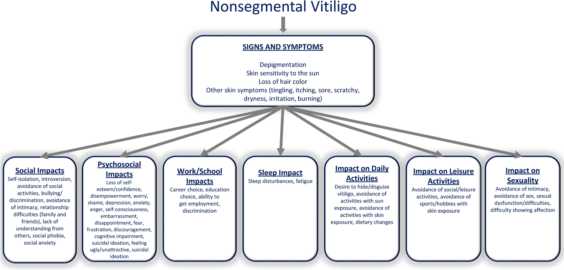
BoNT/A is a foreign antigen that can trigger antibody production, causing secondary botulinum toxin treatment failure [19, 20]. Many factors can influence the production of BoNT/A antibodies, such as the total units of BoNT/A administered and the interval between each treatment [22]. The aim of this study was to address whether the route of injection impacts antibody production.
Our study revealed that the levels of total antibodies against BoNT/A were higher in the sera of the intradermal group than those in the intramuscular group for most types of BoNT/A (Fig. 1a). This might be because the dermis contains professional antigen-presenting cells, such as dermal dendritic cells, which effectively process and present antigens to activate the immune response [30]. This finding is consistent with other reports regarding different immune responses provoked by vaccines via different routes of administration [31, 32]. Surprisingly, we observed the unexpected outcome that LetiA did not show a greater IgG response intradermally and intramuscularly. We repeated the entire experiment; however, the results were the same (Fig. 1). These results were generated from average antibody levels against two LetiA sources. When we explored the raw data, we found that LetiA from one source (A1) can provoke higher levels of hIgG specific to whole molecules of LetiA when injected intradermally compared with intramuscular administration. However, LetiA from another source (A2) provoked the same levels of hIgG specific to whole molecules of LetiA derived from both intradermal and intramuscular administration. These results might reflect the heterogeneity of LetiA production (Figure S2). When we explored antibodies against three functional BoNT/A sites, there was no significant difference in the percentages of specific hIgG between the intradermal and intramuscular groups (Fig. 2a). The finding that total hIgG specific to the whole BoNT/A molecule comprises hIgG against both functional and non-functional sites indicates that intradermal injection might induce higher levels of hIgG against non-functional sites than intramuscular injection. The high levels of these specific hIgGs may bind to non-functional sites of BoNT/A, leading to immunological clearance by immune complexes, which may affect the ability of a given dose to yield an acceptable therapeutic effect of BoNT/A [25]. However, injecting BoNT/A directly into this immunologically active environment provides a more efficient pathway for antigen uptake and presentation to the adaptive immune system than the relatively less immune-cell-rich intramuscular space. This likely explains the generation of a detectable binding antibody response. However, the critical insight for the clinician is that this immune response appears to be qualitatively insufficient to cause treatment failure. Even our findings support the continued favorable safety and efficacy profile of intradermal BoNT/A; they also advocate clinical vigilance. Clinicians should remember to use the minimum effective dose, adhering to recommended treatment intervals, and watch for any subtle changes in treatment response over the long term.
We also observed the effect of different BoNT/A preparations on antibody production. Interestingly, the levels of total hIgG against BoNT/A derived from the IncoA intramuscular group declined after day 14 and reached half of the baseline level (Fig. 1b). This reduction might be due to the contraction phase of the immune response, which entails a reduction in antibody production after antigen clearance. However, the total hIgG levels of the intramuscular groups treated with OnaA, AboA, PraboA, and LetiA were sustained at the same level from day 0 (Fig. 1c–f). This could be because antigens introduced via the intramuscular route can trigger the formation of basal antibodies [33]. This finding raises the possibility that a pure botulinum toxin, such as IncoA, may induce a weaker antibody response than a BoNT/A, which contains complexing proteins. The presence of complexing protein in certain formulations of botulinum toxin A (Table S5) may elicit an immune response, as illustrated in Fig. 1. It induced elevated antibody responses for extended durations (a minimum of 3 months) relative to the baseline level (day 0). These proteins can be recognized as foreign by the immune system. This recognition may enhance the development of polyclonal antibodies binding to both the neurotoxic and the associated proteins. Patients who develop these antibodies at significant levels may encounter a reduced response or total resistance to future therapies, a condition known as secondary treatment failure.
Our study demonstrated the trends in the production of hIgG specific to three functional sites of several BoNT/A types before and after treatment. The results revealed that BoNT/A caused a peak in the production of specific hIgG about 30 days after BoNT/A administration and that specific hIgG had declined slightly by day 180 (Fig. 2a). AboA, PraboA, and LetiA induced significant production of specific hIgG on day 30 compared with the specific hIgG levels on day 0 (Fig. 2d–f). In contrast, the other BoNT/As (IncoA and OnaA) did not induce specific hIgG production on day 30 (Fig. 2b and c, respectively). Around day 90, the specific hIgG trend started to decline. Our results were in line with the findings of another study that suggested a second BoNT/A injection should be given at a > 90-day interval to lower the probability of antibody-induced treatment failure [25, 26]. Therefore, administering BoNT/A again within 90 days of a previous injection may diminish its effectiveness and increase the production of antibodies.
Our study revealed that the route of administration of OnaA, AboA, PraboA, and LetiA had little impact on the induction of hIgG specific to complexing proteins (Fig. 3). The levels of anti-complexing proteins in our cohort were not as high as expected, and even complexing proteins might act as adjuvant-like effects to provoke immune responses. This may be because of any of these possibilities: First, modern BoNT/A formulations are highly purified with fewer immune-stimulating proteins, leading to a weaker immune response. Second, the small therapeutic doses used are likely too low to trigger the robust reaction typically seen in vaccination. Finally, repeated low-dose administration might induce immunological tolerance, causing the body to actively suppress an antibody response. Although there is a minimal likelihood that anti-complexing protein antibodies may result in secondary BoNT/A treatment failure, if this does occur, patients might need to receive BoNT/A injections in larger doses to compensate for immune-complex clearance and sustained treatment results [34].
In this study, the percentages of most antibodies against functional sites and complexing proteins did not exceed their cutoffs (Figs. 2 and 3, respectively). These results correlated with all subjects reporting that they were still satisfied with their BoNT/A treatments. However, in previous studies of subjects with failed BoNT/A treatment, the percentage of antibodies exceeded the cutoff values [24, 25]. Our results were also consistent with earlier findings indicating that the percentage of specific hIgG against the three functional sites and complexing proteins of BoNT/A strongly influences the outcomes of BoNT/A treatment [24, 25]. Although the injection route did not affect the percentage of specific hIgG against functional sites and anti-complexing protein antibodies, intradermal injection of BoNT/A showed a trend toward increasing the production of total hIgG against BoNT/A (Fig. 1).
The novelty of the study lies in its direct, prospective, and comparative investigation of intradermal versus intramuscular injection routes across multiple, distinct BoNT/A formulations within a human clinical setting. To our knowledge, this specific multivariable comparative approach has not been previously reported and addresses a clear gap in clinical study. The core finding of the study is an important piece of information for clinicians who employ this technique, particularly in Asian countries. Nevertheless, our study has several limitations. The whole history of previous BoNT/A treatments for each participant was not accessible, and this inevitable limitation may have influenced the results. This unknown history of BoNT/A exposure, including cumulative dose and specific formulations used, likely explains the wide variation in immune responses seen across individuals. A higher cumulative dose could cause the body to recognize more parts of the toxin molecule and broaden the immune reactions. Conversely, a long and regular history of low-dose injections might have paradoxically induced immunological tolerance in other individuals, making them less responsive. The hyporesponsiveness could be mediated by induction of regulatory T cells (Treg). Furthermore, exposure to different historical formulations, some potentially more immunogenic than others, could affect their responses to the treatment used in the current study. To mitigate this potential confounding variable, we assessed each individual’s antibody level prior to the study injection and established this as their personal baseline for all subsequent comparisons. Second, the participant number in each subgroup was limited. The primary objective of this study was to compare the effect of intradermal versus intramuscular administration on antibody responses against BoNT/A, for which the overall sample size (n = 60 per administration route) was adequately powered for analysis. However, in the subgroup analysis, the small sample size could have been limiting for detecting small or moderate differences specifically between the various BoNT/A formulations. Consequently, head-to-head comparisons of formulations with larger sample sizes should be explored in a future study dedicated to comparing these specific formulations to confirm the trends observed here. Third, all participants in this study were Thai with Fitzpatrick skin phototypes III–IV, which are associated with moderate to high epidermal melanin content. Melanin has been suggested to modulate cutaneous immune responses through its antioxidant properties and potential influence on local cytokine environments. In darker skin, variations in dermal architecture, baseline inflammatory activity, and density of immune cells, such as Langerhans and dermal dendritic cells, may alter antigen processing following intradermal injection [30]. Although these factors were not directly measured in our study, they could partly explain differences in immune kinetics compared with populations of lighter or darker skin phototypes. Consequently, our findings may be most directly applicable to individuals with similar phototypes, and further studies including a broader range of skin types are warranted to clarify the role of melanin and other dermatological variables in modulating botulinum toxin immunogenicity. In addition, as BMI and sex have been shown to exert minimal influence on antibody response, these variables were neither collected nor included in the present study’s analysis [35]. Moreover, the results from the inhibition and absorption ELISA employed in this work had been previously assessed with the frontalis test [26, 27]. Interestingly, our results from inhibition ELISA (to determine hIgG specific to functional epitopes of BoNT/A) and absorption ELISA (to determine hIgG specific to complexing proteins) with their cutoff points were well correlated with Toxogen mouse protection assays (data not shown, manuscript in preparation). Lastly, the immunological kinetic response of specific hIgG production was constrained by the absence of specific hIgG level assessments on days 60, 120, or 150. Although we cannot comprehensively draw conclusions regarding the tendency and timeline of specific hIgG levels, our findings still emphasize the importance of injection route and timing in BoNT/A administration.


Comments (0)