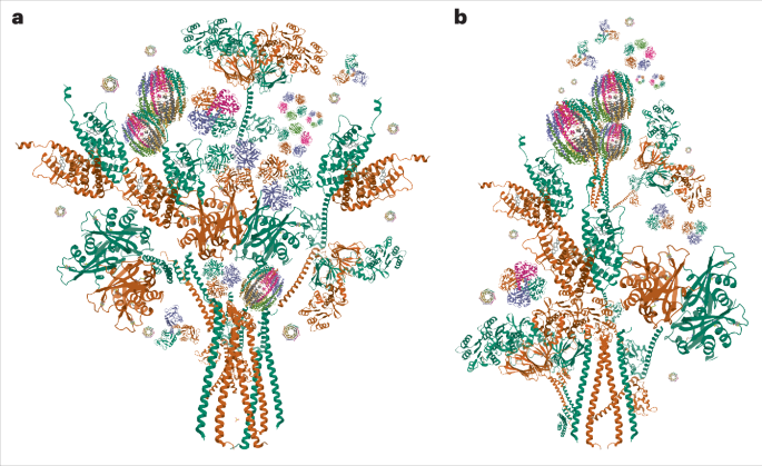
Ferlin structures
Ferlins, such as dysferlin, myoferlin and otoferlin, are membrane proteins involved in calcium-dependent vesicle fusion, and how they interact with membranes remains a mystery.
To determine the high-resolution structure of a human ferlin, Cretu et al. expressed and purified myoferlin and dysferlin; the proteins remained stable and capable of binding calcium and negatively charged lipids. Unlike earlier models, authors found no evidence of C2 domain-mediated dimerization. Using cryo-electron microscopy (cryo-EM), they resolved the structures of human myoferlin and dysferlin in calcium and lipid-bound states. Initial cryo-EM of lipid-free ferlins revealed flexible N- and C-terminal domains, limiting resolution. Authors found that nanodiscs and anionic lipids stabilized myoferlin–lipid complexes, enabling high-resolution (2.4–2.9 Å) structures. Contrary to previous predictions of an extended ‘beads-on-a-string’ arrangement, their cryo-EM maps showed that lipid-bound myoferlin adopts a compact, elliptical ring (about 150 × 90 Å) surrounding a central cavity.

Comments (0)