
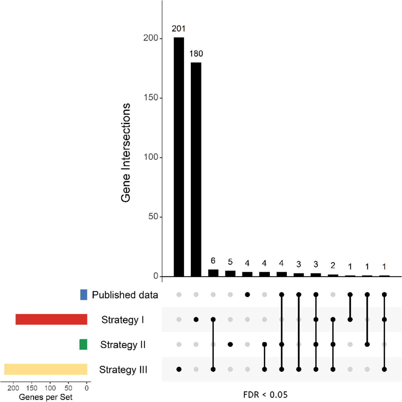
Remember me

The data presented in this study was obtained from participants enrolled in NIH protocol 10N0143, Natural History Study of Patients with Syringomyelia. Consent was obtained from all patients. Nine patients from this study underwent neurosurgical treatment for syringomyelia associated with arachnoiditis. Surgical specimens of arachnoid tissue were collected from these patients during surgery for treatment of syringomyelia and/or associated arachnoiditis and embedded in paraffin for future tissue processing.
Image AcquisitionTissue samples were later deparaffinized and underwent multiplex immunohistochemical (MP-IHC) staining for visualization of nuclear (DAPI) and immune cell (CD4, CD8, CD20, CD68, IBA1) markers. We followed a well-accepted MP-IHC method (Riggle et al., 2020). Briefly, the arachnoid tissue sections were de-paraffinized, then exposed to the following primary antibodies: Goat IgG anti-CD4 (R&D Systems) to identify helper T-cells, Mouse IgG2b anti-CD8 (Thermo Fisher Scientific) to identify cytotoxic T-cells, Mouse IgG2a anti-CD20 (Thermo Fisher Scientific) to identify B-cells, and Mouse IgG3 anti-CD68 (Thermo Fisher Scientific) in combination with Chicken IgY anti-IBA1 (Cedarlane Labs) to identify infiltrating macrophages. After washing off excess primary antibodies, sections were incubated with the appropriately cross-absorbed secondary antibodies, facilitating their binding with the species-specific epitopes of the primary antibodies and subsequent visualization of the markers. These secondary antibodies (purchased from either Thermo Fisher Scientific or Jackson ImmunoResearch) were conjugated to one of the following spectrally compatible fluorophores: Alexa Flour 488, Alexa Flour 647, Alexa Flour 594, Alexa Flour 546, Alexa Flour 790, Alexa Flour 680, or IR800CW. The image samples analyzed in this study were acquired from whole images of arachnoid tissue sections. The initial tissue samples were imaged using the Axio Imager.Z2 slide scanning florescence microscope (Zeiss) equipped with a × 20/0.8 Plan-Apochromat (Phase-2) non-immersion objective (Zeiss), a high-resolution ORCA-Flash 4.0 sCMOS digital camera (Hamamatsu), a 200W X-Cite 200DC broad band lamp source (Excelitas Technologies), and filter sets (Semrock) customized to detect different fluorophores. Image tiles (600 × 600 um viewing area) were individually captured at 0.325 micron/pixel spatial resolution, and the tiles were stitched into whole specimen images using the ZEN 2 image acquisition and analysis software program (Zeiss). Pseudocolored stitched images were overlaid in Imaris 9.2.1 (Bitplane) as individual layers to create multicolored merged composites.
To standardize the pixel dimensions of each image sample for counting, sections of each stained tissue image were selected to cover an area of 25,000,000 square pixels. For larger tissue images, multiple non-overlapping sections were selected as different samples (from one patient specimen). Image sections were chosen such that areas with zero, low, medium, and high colocalized cell populations were included in the counting analysis. 27 arachnoid tissue image samples were collected from 9 patients for analysis.
The tissue in each image sample had been stained for the nuclear marker DAPI as well as several immune cell markers: CD4, CD8, CD20, CD68, IBA1. A colocalized cell was defined as a cell that was co-stained for the nuclear DAPI marker and an immune cell marker. Colocalized cell counts were conducted for each immune cell marker type individually; cells that were colocalized for DAPI, CD68, and IBA1 were also quantified. Two independent observers performed the colocalized cell counts on the tissue samples, using a manual and semi-automated colocalized cell counting method. The automated method did not require user input (Fig 1).
Fig. 1
Representative images of nuclear (DAPI), red blood cell (RBC), and immune cell markers in a tissue sample following multiplex immunohistochemistry (MP-IHC). (a) Overview image of an arachnoid tissue section with an area of 25.4 mm2, illustrating the spatial distribution and colocalization of the marker analyzed in this study. Panels (b–i) show higher magnification views of the same tissue, highlighting specific marker expression. The scale bar in panel (a) applies only to panel (a), while the scale bar in panel (i) applies to panels (b–i). Both scale bars represent 80 µm
Image Pre-ProcessingFor the manual and semi-automated counting methods, images were pre-processed to streamline counting and, if necessary, improve visibility of the staining. For each immune cell marker, composite images of the sample tissue stained with the nuclear marker and respective immune marker were prepared from the individual channel images. Each composite image was prepared by first opening the nuclear and immune marker channel images on ImageJ. Each channel image was converted into a 16-bit image; if the brightness of the staining was too low, the Adjust Brightness and Contrast feature on ImageJ was used to increase brightness and contrast. The "Merge Channels" ImageJ function was used to merge the nuclear and immune marker composite image. In each composite image, the nuclear channel (DAPI) image was the green channel, and the respective immune marker channel image was the red channel. For the analysis of DAPI/CD68/IBA1 colocalized cells, a composite image was created from the three channels, with the green channel displaying the DAPI staining; the red channel displaying the CD68 staining; and the blue channel displaying the IBA1 staining. Composite images were prepared for all 27 samples, and each immune cell marker for those samples (CD4, CD8, CD20, CD68, IBA1). Each observer independently pre-processed the image samples. To prevent discrepancy in counts due to different image pre-processing, each observer used the same composite image for the manual and semi-automated counting methods (Fig 2).
Fig. 2
Flowchart illustrating the image preprocessing steps. The original images (a and e) are converted into 16-bit black-and-white images (b and f), followed by adjustments to brightness and contrast (c and g). These preprocessed images are then merged to create a composite image, with DAPI (nuclear stain) displayed in green and the immune cell marker in red (d). The scale bar in panel (g) is equal to 50 µm and applies to panels (a-d)
Manual Counting MethodThe multi-point tool on ImageJ was used to perform the manual colocalized cell counts. For each composite image containing a specific immune and nuclear marker, the observer zoomed in until individual cells were clearly distinguishable. The observer then identified cells co-stained with DAPI and immune markers, manually marking each identified cell with a multi-point. After reviewing the entire image sample, the observer recorded the total number of multi-points as the count of colocalized cells for that particular immune marker. This process was repeated for DAPI/CD68/IBA1 colocalized cells, except the observer was looking for cells co-stained for all three markers.
Semi-Automated Counting MethodThe Colocalization Object Counter ImageJ plugin, developed by Lunde and Glover (Lunde & Glover, 2020) was used to perform the semi-automated colocalized cell counts. Access to the plugin can be found here: https://sites.imagej.net/ObjectColocalizationPlugins/. This ImageJ plugin allows for an automated centroid-based approach to colocalized cell counting, with manual verification and correction. For each sample, the plugin and composite image were loaded into ImageJ. The nuclear channel (green) was opened first in the composite image, and the "Find 2D maxima (as multipoints)" plugin tool was clicked to automatically mark all the nuclei in the image sample with multipoints. If nuclei were overcounted or background noise was detected as counts, the "Pre blur radius" and "Noise tolerance" parameters were increased accordingly until nuclei were no longer being overcounted. With category 1 checked, the "Convert multipoint to overlays" tool was then clicked to save the marked nuclear multipoints as overlays for cells.
Next, the immune channel (red) was opened in the composite image. "Find 2D maxima (as multipoints)" was used to automatically mark all the corresponding immune cells in the image sample with multipoints. As described above, the "Pre blur radius" and "Noise tolerance" parameters were adjusted accordingly until immune cells were being properly counted. After the automatic immune cell detection, the observer then reviewed the image sample to manually add or remove any marked immune cells as needed. The checked category on the plugin tool was switched from 1 to 2. With category 2 checked, the "Convert multipoint to overlays" tool was then clicked to save the marked immune multipoints under category 2 cells.
By clicking the "Count information" button on the plugin, the observer can see the number of cells marked under category 1 (nuclear) and category 2 (immune). The plugin also outputs the number of colocalized cells, marked under category 1 and 2 (termed category 12). The observer recorded the number of category 12 cells as the count of colocalized cells for that particular immune marker.
This process was repeated for all immune markers across all samples. For DAPI/CD68/IBA1 colocalized cells, the process was the same except an additional category (category 3) was included. As described above, the DAPI multipoints were in category 1 and the CD68 (an immune marker) multipoints were in category 2. The IBA1 multipoints were marked under category 3, and the colocalized cell count was the number of category 123 cells for the image (Figs. 3 and 4).
Fig. 3
Analysis of a two-channel image containing DAPI (green) and an immune cell marker CD4 (red) using the semi-automatic counting method. These images depict cells with an ‘easy-to-count’ morphology. Panel (a) shows the composite image displaying both channels, while panels (b) and (e) display the individual green (DAPI-positive nuclei) and red (IBA1-positive cells) channels, respectively. Panels (c) and (f) illustrate the identification and plotting of 2D maxima in the green and red channels. In panels (d) and (g), the plotted maxima are converted into overlays of a defined radius, with panel (h) specifically showing the merging of overlays from both channels, where overlapping regions between channel 1 (DAPI, green) and channel 2 (CD4, red) are counted as colocalized objects. The scale bar in panel (g) is equal to 50 µm and applies to all panels
Fig. 4
Analysis of a two-channel image containing DAPI (green) and the immune cell marker IBA1 (red) using a semi-automatic counting method. The images depict cells with a "hard-to-count" morphology. Panel (a) shows the composite image displaying both channels, while panels (b) and (f) display the individual green (DAPI-positive nuclei) and red (IBA1-positive cells) channels, respectively. Panels (c) and (g) illustrate the identification and plotting of 2D maxima in the green and red channels. In panels (d) and (h), the plotted maxima are converted into overlays of a defined radius, with panel (h) specifically showing the merging of overlays from both channels. Overlapping regions between the DAPI and IBA1 channels are counted as colocalized objects. Due to the irregular and fragmented structure of IBA1-positive cells, initial counts were overestimated, as seen in panel (e). To account for this "hard-to-count" morphology, counting parameters were adjusted by setting the pre-blur radius to 1 and reducing the overlay radii from 50 to 30. These adjustments improved the accuracy of maxima detection in the green (DAPI) channel (i) and red (IBA1) channel (k), resulting in a substantial reduction in overcounting. The final refined counts are shown in panel (j). The scale bar in panel (a) is equal to 50 µm and applies to all panels
Automated Counting MethodA custom object-based colocalization analysis pipeline was run on the widely used CellProfiler software to perform the automated colocalized cell counts (Fonseca & Rosa, 2025; Jones et al., 2009; Zhang et al., Feb 2018). The CellProfiler software is available for download here: https://cellprofiler.org/releases.
The pipeline consisted of successively performing CorrectIlluminationCalculation (smoothing method = Fit Polynomial) on both channel images, CorrectIlluminationApply, IdentifyPrimaryObjects (threshold strategy = global; thresholding method = Otsu; Three classes thresholding) on both images, RelateObjects, ExpandOrShrinkObjects (shrink objects to a point) on both images, ExpandOrShrinkObjects (expand objects 5 pixels) on both images, RelateObjects, ExportToSpreadsheet. Objects within 5 pixels of each other between the 2 channels were considered colocalized by the algorithm.
Images were not pre-processed for the automated counting method. To perform the counting analysis, un-processed nuclear and immune marker channel images were inputted into the CellProfiler software. After the analysis was complete, the pipeline exported a spreadsheet of results, including the number of colocalized objects. The number of colocalized cells was the number of objects counted by the pipeline for that particular sample and marker.
The same pipeline was run for the DAPI/CD68/IBA1 colocalized cells, with the addition of a segmentation and object identification module. The pipeline consisted of successively performing the same pipeline as described above, with the additional steps ClassifyObjects, FilterObjects, IdentifyPrimaryObjects on the third channel image, RelateObjects, ExpandOrShrinkObjects (shrink objects to a point) on both images, ExpandOrShrinkObjects (expand objects 5 pixels) on both images, RelateObjects, ExportToSpreadsheet. These additional steps accounted for the extra channel to be analyzed (Fig 5).
Fig. 5
Schematic representation of the automated object-based colocalization pipeline applied to a sample image containing two distinct marker channels. The pipeline processes two separate channels (a and e) to identify and quantify colocalized objects (i), enabling the analysis of overlapping signals from the two markers. The scale bar in panel (h) is equal to 50 um and applies to panels (a-i)
Time CalculationsDuring the counting process for the manual and semi-automated methods, each observer recorded the time it took them to collect the colocalized cell counts from opening the image to saving the colocalized cell count number. Both the manual and semi-automated counting methods required image pre-processing. The time to pre-process each image sample was measured and added to the time calculation accordingly.
Initial setup of the automated pipeline took 15 min. As the images did not require pre-processing, the time it took to count each image sample was the processing time of the software. For each immune marker, the pipeline was run for all samples at the same time; the time to analyze each image was recorded by the software and outputted in the spreadsheet of results.
Statistical AnalysisA correlation analysis was conducted between semi-automated vs. manual and automated vs. manual counts to determine how accurate the semi-automated and automated methods were to the "standard" manual counting method. Paired t-tests were used to compare the absolute colocalized cell counts between semi-automated and automated counts to manual counts. The average count values between the two observers were used for all the listed statistical analyses. To evaluate inter-operator variability, paired t-tests were conducted for the measured counts between both observers. All statistical analysis was performed in Prism 10.
Comments (0)