
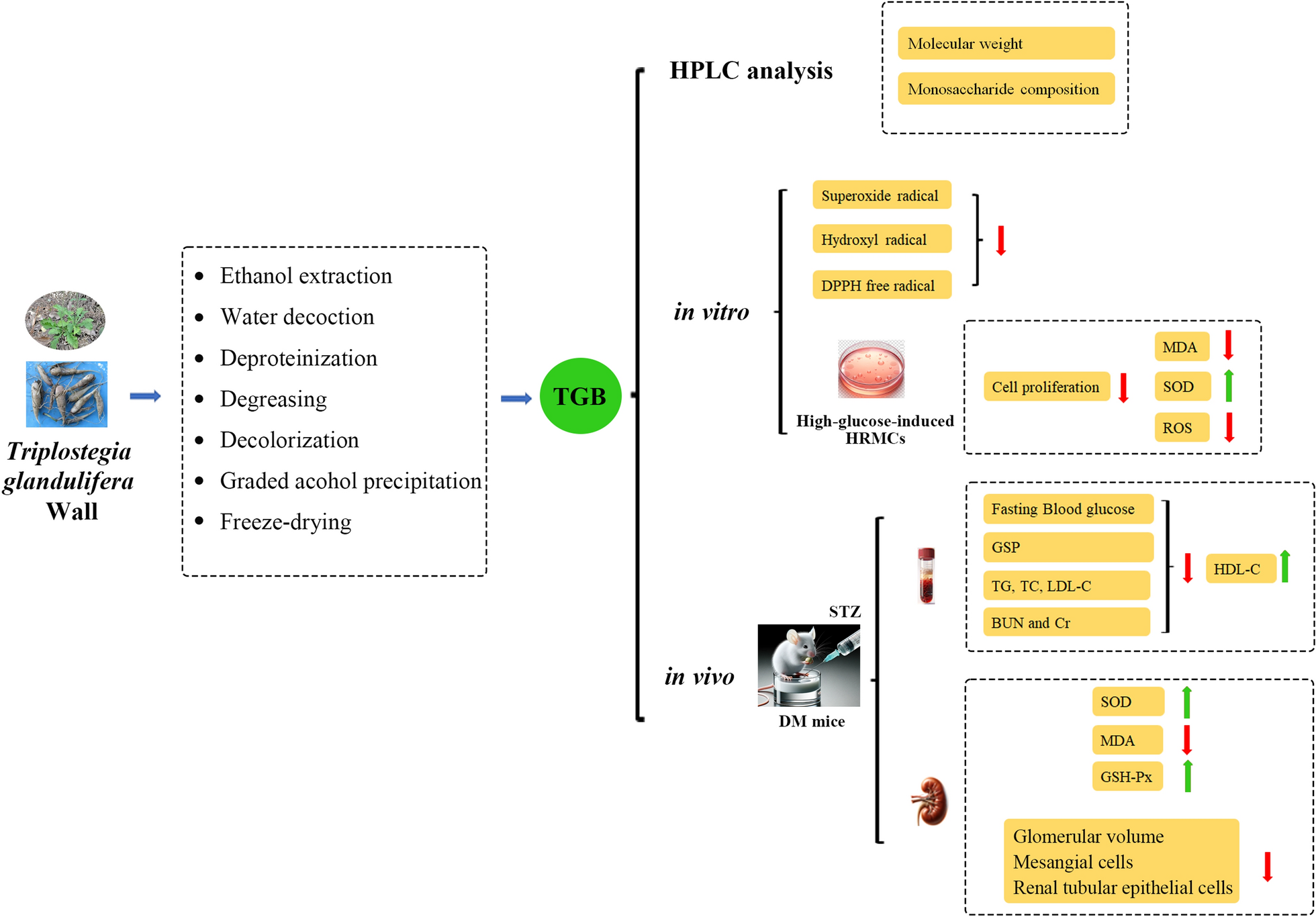
Remember me
In this paper, to clarify the taxonomic position of the strain KMM 4401, we sequenced the molecular markers, such as 28S rDNA, ITS regions and the partial TEF1 gene sequence. Approximately 950 bp fragment of the partial 28S rDNA region, about 1600 bp fragment of the ITS region, and about 1000 bp fragment of the partial TEF1 gene sequence were successfully amplified. BLAST search showed that these sequences were 99 to 100% identical with those of the non-ex-type strains Px. luzulae CBS 494.67, CBS 495.67, and CBS 935.69. Phylogenetic ML tree of the concatenated 28S-ITS-TEF1 gene sequences clearly showed that the strain KMM 4401 clusters with the strains Px. luzulae (Fig. 1).
Fig. 1
ML tree based on concatenated 28S-ITS-TEF1 nucleotide sequences showing the phylogenetic position of the strain KMM 4401 among members of the genus Paragliomastix from the family Bionectriaceae. Bootstrap values (%) correspond 1000 replications. The scale bar represents 0.01 substitutions per site
2.2 Metabolite profile of Paragliomastix luzulae КMM 4401The UPLC MS chromatogram of extract of Px. luzulae (Pl) КMM 4401 culture is presented in Fig. 2.
Fig. 2
UPLC MS chromatogram of Paragliomastix luzulae КMM 4401 (Pl) culture
In total, 27 compounds were annotated using the in-house database or MetFrag service with PubChem database (Fig. 3, Table S1). The detailed characteristics of the identified compounds are presented in the Supplementary Materials (Table S1).
Fig. 3
The secondary metabolites detected in Paragliomastix luzulae KMM 4401 monoculture
The peak #21 detected at 6.2 min at m/z 659.3271 corresponded to the molecular formula C32H50O14, the same as virescenoside R2, an isopimarane glycoside with a disaccharide moiety isolated early from Px. luzulae KMM 4401 [4]. The compound can be suggested based on an exact mass value. The peak #25 found at 9.2 min at m/z 479.2653 corresponded to either virescenosides T or U, as also confirmed by fragmentation under CID conditions. These two compounds have nearly identical structures, differing only in the position of the double bond in the aglycone. Moreover, four other peaks (#22, 24, 23 and 26) with similar exact mass (m/z 479.2618, 479.2653, 479.2618, and 479.2653) were detected at 6.4, 8.4, 9.1, and 9.6 min, respectively. All of them, have characteristic ion at m/z 317.2103 and 299.2005, arising from losses of altrose by in-source dissociation. Virescenosides T and U were earlier isolated from the KMM 4401 strain [19], and three additional peaks very likely corresponds to previously undescribed virescenosides, which are isomeric to virescenosides T and U.
The peak #9 found at 7.4 min at m/z 497.2737 corresponded to the molecular formula C26H40O9, which can be associated with isomeric virescenosides M and V that were isolated from the KMM 4401 strain [20, 21]. It was also suggested based on MS/MS fragmentation. In addition, three other peaks (#7, 8 and 10) with similar exact mass were detected at 6.8, 7.2 and 7.7 min, respectively. All these peaks, besides the molecular ion peak, have characteristic aglycone peaks at m/z 317.2103 and 335.2217, formed by in-source dissociation. Two of these peaks obviously correspond to unknown isomers of virescenosides M and V.
The peaks #11, 12, and 13 found at 6.9, 7.3, and 7.5 min at m/z 499.2909 corresponded to the molecular formula C26H42O9, which can be associated with virescenosides N [20], W and X [21] that have isomeric aglycones and were earlier described as metabolites of the KMM 4401 strain. The peak #14 found at 8.7 min at m/z 481.2803 corresponded to the molecular formula C26H40O8, which can be associated with isomeric virescenosides P [22] and S [19] that were earlier obtained from this fungal strain. Moreover, four other peaks (#15, 16, 17 and 18) were detected at 8.9, 9.4, 10.0, and 11.0 min, respectively, and have characteristic aglycone peaks at m/z 301.2159 and 319.2260, formed by in-source dissociation. Two of these peaks obviously correspond to unknown isomers of virescenosides N, W, and X [20, 21].
The peaks #19 and 20 detected at 9.6 min and 10.5 min at m/z 645.3461 and 645.3501 corresponded to the molecular formula C32H52O13, which can be associated with the isomeric compounds virescenoside R and R3. Compounds were identified based on an exact mass values and fragmentation patterns. Earlier these compounds were isolated from the KMM 4401 strain [4, 19].
The peaks #1 and 2 detected at 10.6 min and 11.5 min at m/z 483.2961 corresponded to the molecular formula C26H42O8, the same as virescenosides A [23] and O [22], respectively. Compounds were identified based on comparison of exact mass values, fragmentation patterns, and RT with whose of authentic standards.
The peak #3 detected at 12.7 min with m/z 467.2998 corresponded to the molecular formula C26H42O7, the same as virescenoside B [23]. The compound was identified based on comparison an exact mass value, MS/MS fragmentation, and RT with those of virescenoside B.
The peaks #4 and 5 found at 12.4 at m/z 467.2981 and 13.3 min at m/z 467.3015 corresponded to the molecular formula C26H42O7, which can be associated with virescenoside Q, known metabolite of the KMM 4401 strain [22], and another unknown isomeric virescenoside. This was suggested based on characteristic aglycone peaks at m/z 301.2159 and 319.2260, formed by in-source dissociation.
The peak #6 detected at 15.6 min at m/z 303.2325 corresponded to the molecular formula C20H30O2, the same as aglycone of virescenoside C [24]. The compound was identified as virescenoside C based on corresponding MS and RT data from in-house database.
The peak #27 detected at 16.2 at m/z 1176.7974 corresponded to the molecular formula C59H105N11O13, the same as 6-[(3R,4S)-3-hydroxy-N-methyl-5-oxo-L-leucine]cyclosporin A and 9-(N-Methyl-L-serine)cyclosporin A. Compound was annotated based on an exact mass value and the MetFrag service.
The peak #50 detected at 20.4 min at m/z 429.3350 corresponded to the molecular formula C28H44O3, the same as ergosterol peroxide, a usual derivative of main triterpenoid [25]. Compound was identified based on an exact mass value and RT with an in-house database.
Earlier cyclosporin A derivative was synthesized by ozonolysis of cyclosporin methyl vinyl ketone followed by reductive workup and its immunosuppressive properties was patented [26, 27]. Cyclosporin A derivative was reported in the patent as a synthetic compound [28]. So, we proposed that Px. luzulae КMM 4401 may be natural source of these synthetic derivatives of cyclosporin A and this strain should be promising for the isolation of this and others cyclosporin A derivatives for future investigations.
2.3 Metabolite profile of Paragliomastix luzulae and Penicillium hispanicum co-culture with simultaneously inoculationFor this study, the culture of Px. luzulae KMM 4689 were seeded in flasks and P. hispanicum KMM 4689 was inoculated in these flasks immediately. After that the co-culture grown for 3 weeks and then was extracted for following UPLC MS analysis.
Earlier P. hispanicum KMM 4689 axenic culture (Ph) metabolite profile was reported [18] and in this work it was confirmed by UPLC MS technique. 25 compounds were detected in Ph extracts (Supplementary Materials, Table S1).
The UPLC-MS chromatogram of the extract of Px. luzulae KMM 4401 and P. hispanicum KMM 4689 simultaneously inoculated co-culture (PlPh1) in comparison with UPLC-MS chromatogram of Ph are presented in Fig. 4.
Fig. 4
UPLC MS chromatograms of Paragliomastix luzulae KMM 4401 and Penicillium hispanicum KMM 4689 co-culture (PlPh1) (red) and Penicillium hispanicum KMM 4689 monoculture (Ph) (blue)
A total of 38 compounds were successfully identified using the in-house database and GNPS (Figs. 3 and 5, Table S1). The detailed characteristics of the identified compounds are presented in the Supplementary Materials (Table S1). 17 from these peaks were corresponded to metabolites of Px. luzulae KMM 4401: virescenosides A (#1), B (#3), O (#2), Q/its isomer (#4 and #5), S/P/their isomers (#15–18), R/R3 (#19 and #20), and T/U/their isomer (#24–26), aglycone of virescenoside C (#6), as well as one from the virescenosides M/V/their isomers (#7), and cyclosporin A derivative (#27).
Fig. 5
The secondary metabolites of Penicillium hispanicum KMM 4689 [18] detected in the co-cultures
In addition, the compounds previously reported from axenic culture of P. hispanicum KMM 4689 [18] were detected: 3β-hydroxydeoxyisoaustamide (#28), 3,4-dimethoxycinnamic acid (#29), ( +)-deoxyisoaustamide (#38), 3β-hydroxydeoxyisoaustamide (#28), austamide (#32), brevianamide F (#30), 7-hydroxy-3-(2-hydroxypropyl)-5-methylisochromen-1-one (#31), 16β,17α-dihydroxy-deoxydihydroisoaustamide (#33), 16,17-dihydroxydeoxydihydroisoaustamide (#34), 16α,17α-dihydroxy-deoxydihydroisoaustamide (#35), 16α-hydroxy-17β-methoxy-deoxydihydroisoaustamide/16β-hydroxy-17α-methoxy-deoxydihydroisoaustamide/16α-hydroxy-17α-methoxy-deoxydihydroisoaustamide (#36), deoxydihydroisoaustamide (#37), endocrocin (#39), citreorosein (#40), desoxybrevianamide E (#41), 2-chlorocitreorosein (#42), deoxy-14,15-dehydroisoaustamide (#43), emodine(#44), nephrolaevigatin D (#46), nephrolaevigatin C (#47), nephrolaevigatin A (#48), and nephrolaevigatin B (#49).
2.4 Metabolite profile of time delay co-culture of Paragliomastix luzulae and Penicillium hispanicumAnother variant of co-culture was obtained when the fungus Paragliomastix luzulae KMM 4401 was inoculated in the flasks and Penicillium hispanicum KMM 4689 culture was added in these flasks after 14 days. Then this co-culture was fermented for three weeks and then extracted for following UPLC-MS analysis.
The UPLC-MS chromatogram of the extract of Paragliomastix luzulae KMM 4401 and Penicillium hispanicum KMM 4689 co-culture with time delay inoculation (PlPh2) in comparison with UPLC-MS chromatogram of Ph are presented in Fig. 6.
Fig. 6
UPLC-MS chromatograms of Penicillium hispanicum KMM 4689 monoculture (Ph) (blue), Paragliomastix luzulae KMM 4401 and Penicillium hispanicum KMM 4689 co-culture (PlPh2) (red)
In total, 45 compounds were successfully identified or annotated using the in-house database and GNPS (Figs. 3 and 5, Table S1). These are 20 metabolites of Px. luzulae KMM 4401 including virescenosides A (#1), O (#2), B (#3), Q and its isomer (#4 and #5), virescenoside C aglycone (#6), virescenosides M/V/their isomers (#7–9), virescenoside N or W or X (#11 and #13), virescenoside S/P/their isomers (#14–18), virescenoside R/R3 (#19 and #20), virescenoside T or U or their isomer (#25) and cyclosporin A derivative (#27).
Moreover, 25 compounds earlier detected in axenic culture of P. hispanicum KMM 4689 as 3β-hydroxydeoxyisoaustamide (#28), 3,4-dimethoxycinnamic acid (#29), brevianamide F (#30), 7-hydroxy-3-(2-hydroxypropyl)-5-methylisochromen-1-one (#31), austamide (#32), 16β,17α-dihydroxy-deoxydihydroisoaustamide (#33), 16,17-dihydroxy-deoxydihydroisoaustamide (#34), 16α,17α-dihydroxy-deoxydihydroisoaustamide (#35), 16α-hydroxy-17β-methoxy-deoxydihydroisoaustamide/16β-hydroxy-17α-methoxy-deoxydihydroisoaustamide/16α-hydroxy-17α-methoxy-deoxydihydroisoaustamide (#36), deoxydihydroisoaustamide (#37), ( +)-deoxyisoaustamide (#38), endocrocin (#39), citreorosein (#40), desoxybrevianamide E (#41), 2-chlorocitreorosein (#42), deoxy-14,15-dehydroisoaustamide (#43), emodine (#44), secalonic acid D (#45), and nephrolaevigatins D (#46), C (#47), A (#48), and B (#49), as well as ergosterol peroxide (#50).
2.5 The comparative analysis of metabolite profiles of fungal culturesThe relative content of the announced compounds calculated as a ratio of the peak area to the total area of these peaks in the UPLC-MS chromatogram of Pl, Ph, PlPh1, and PlPh2 extracts was visualized in the heatmap (Fig. 7).
Fig. 7
The heatmap of a related content of compounds identified in fungal extracts Paragliomastix luzulae KMM 4401 (Pl), Penicillium hispanicum KMM 4689 (Ph) and PlPh1 and PlPh2 co-cultures. Each cell presents a peak area in UPLC-MS chromatogram
It was found that cyclosporin A derivative (#27) is the main component of Pl extract, but its amount dramatically decreased in both PlPh1 and PlPh2.
( +)-Deoxyisoaustamide (#38) is predominant not only in Ph extract, but in both PlPh1 and PlPh2. Moreover, the content of this alkaloid in PlPh1 extract increased by ~ 190%, and in PlPh2 extract by ~ 60% compared to the monoculture of P. hispanicum KMM 4689. 3β-Hydroxydeoxyisoaustamide (#28) and deoxydihydroisoaustamide (#37) are the second in content in both PlPh1 and PlPh2 extracts with an increase in concentration of ~ 170% and ~ 50% (for 3β-hydroxydeoxyisoaustamide, #28), respectively, and 68% and 12% (for deoxydihydroisoaustamide, #37). In addition, peak #46, corresponding to neprolaevigatin D, was observed only in both co-cultures, and peak #45, corresponding to secalonic acid D, was detected only in PlPh2 co-culture. At the same time, both compounds were previously described as metabolites of the axenic culture of P. hispanicum KMM 4689 [18].
The analysis of UPLC-MS data was also carried out by the principal component analysis (PCA) and both PCA plot and dendrogram are presented in Fig. 8. The PCA model determined that two principal components (PCs) were optimal for describing approximately 80% of the variation in the samples. The first PC accounted for roughly 55% of the variance, while the second PC accounted for about 24% of the variation (Fig. 8a).
Fig. 8
PCA plot (a) and dendrogram (b) of UPLC-MS data
The PlPh1 extract has minimal differences from the Ph extract in both components. At the same time, the PlPh2 is even more similar to the Ph in terms of the PC1 component but differs as much as possible from Ph in PC2. Both PlPh1 and PlPh2 differ significantly from the Pl extract in the PC1 component.
The dendrogram confirms the main conclusions of the PCA plot, showing the maximum similarity of PlPh1 and Ph extracts, as well as placing PlPh2 in the same cluster while Pl extract is in another cluster.
2.6 Bioactivity of fungal extractsThe effect of Pl, Ph, PlPh1 and PlPh2 extracts on the urease activity and the growth of Gram-positive bacteria Staphylococcus aureus, Gram-negative bacteria Escherichia coli and yeast-like fungus Candida albicans test strains is presented in Table 1.
Table 1 Antimicrobial activity of extractsThe extract of Pl culture inhibited the growth of S. aureus, E. coli and C. albicans by 53.7%, 56.4%, and 20.4%, respectively. At same time, the extract of Ph culture inhibited the growth of S. aureus and E. coli by 29.1% and 34.2%, respectively, and was inactive against C. albicans. The extract of PlPh1 did not show any activity in this test while the extract of PlPh2 inhibited the growth of S. aureus and E. coli by 18.0% and 34.2%, respectively.
The influence of these extracts on human hepatocarcinoma HepG2 and normal rat cardiomyocytes H9c2 cells are presented in Fig. 9.
Fig. 9
The influence of the extracts on the viability of HepG2 (a) and H9c2 (b) cells. The data are presented as a mean ± standard error of mean. All tests were carried out in triplicates
The viability of HepG2 was decrease by 17–30% when the extracts were used at a 100 µg/mL (Fig. 9a). The extracts Pl, Ph, and PlPh1 at a concentration of 10 µg/mL were nontoxic for HepG2 while PlPh2 caused the decrease of HepG2 viability by near 30%.
The toxic effect of the extracts on H9c2 cell viability was more significant (Fig. 9b). The extracts Pl, Ph, PlPh1, and PlPh2 used at a 100 µg/mL decreased the viability of H9c2 by 66.7%, 45.9%, 52.3%, and 42.8%, respectively. After dilution of the extracts by 10 times, all these decreased the viability of H9c2 by 8.9–18.2%.
DPPH radical scavenging activity of all extracts at a concentration of 100 µg/mL was measured. The extracts Pl, Ph, PlPh1, and PlPh2 scavenged 48.5%, 16.0%, 34.2%, and 49.7% of DPPH radicals.
Comments (0)