
記住我
Dear Editor,
Darier disease (OMIM 124200) is an inherited skin condition characterised by malodorous keratotic papules, primarily affecting the seborrheic areas on the upper trunk and scalp.1 Mutations in the ATP2A2 gene are identified as the causative factors for Darier disease.1 Darier Disease is thought to be associated with extracutaneous conditions such as diabetes, heart failure and neuropsychiatric disorders.1,2 Here, we report a case of a severe Darier disease with pterygium and scoliosis, potentially linked to a start codon mutation in ATP2A2.
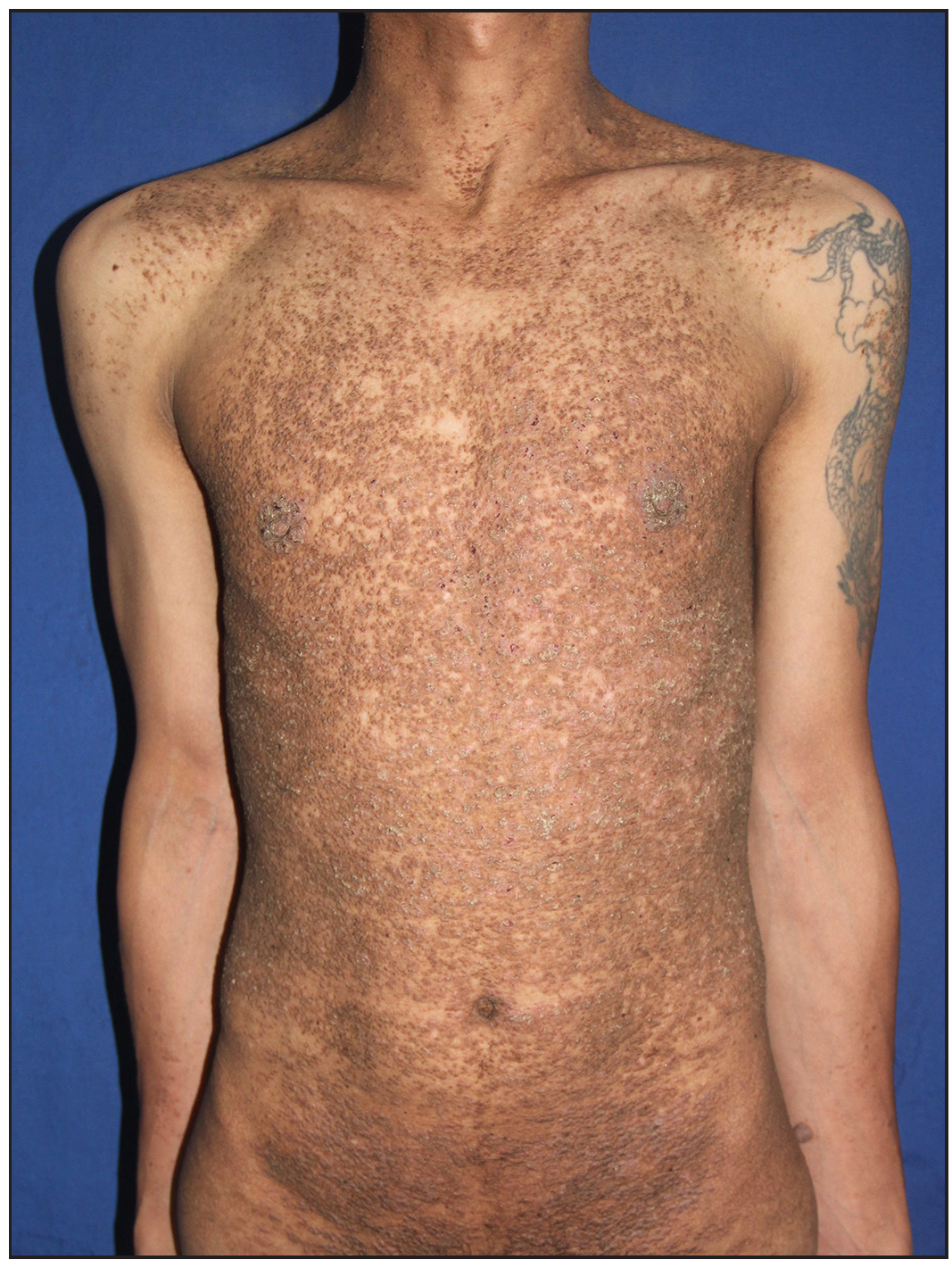
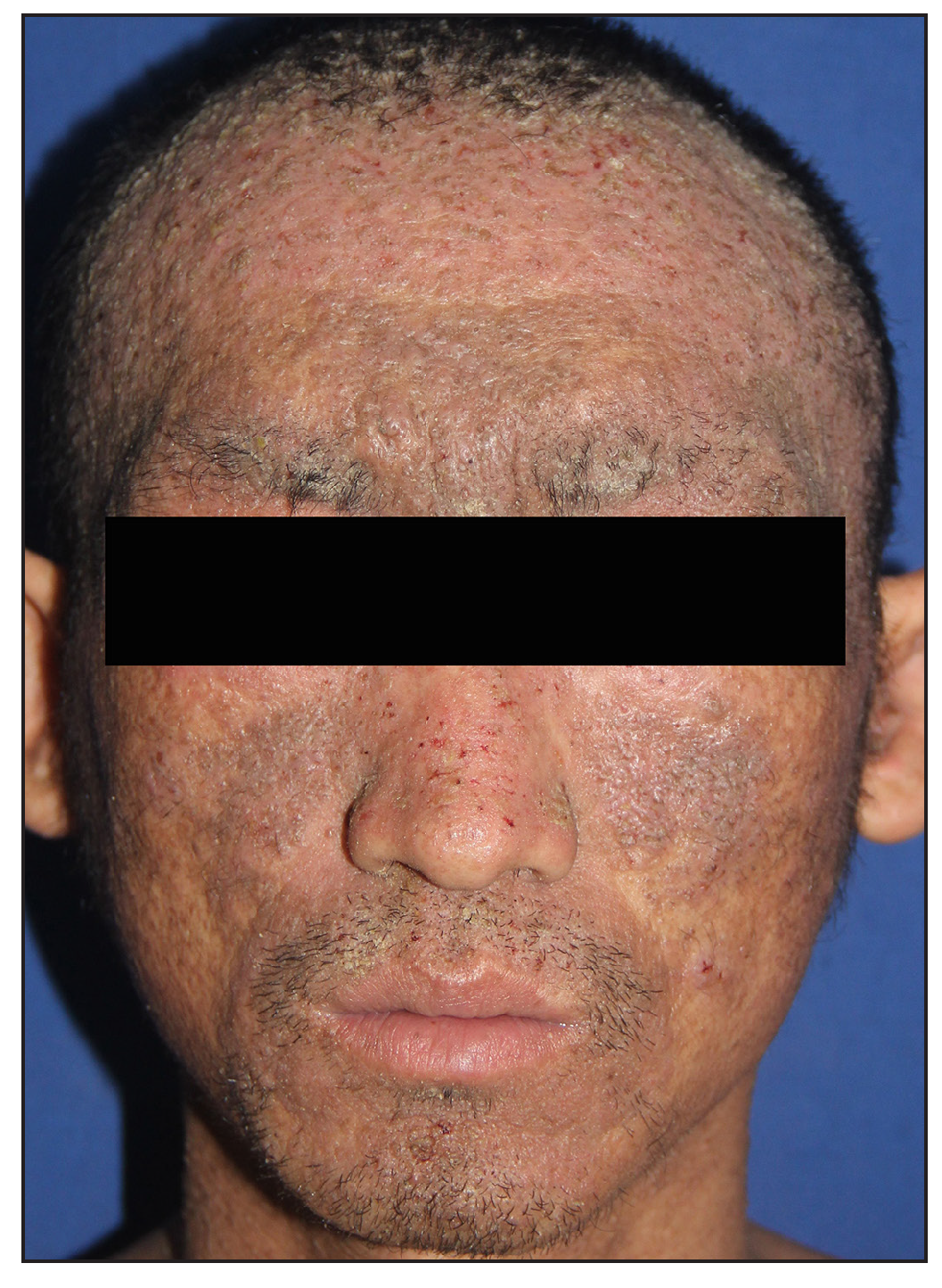
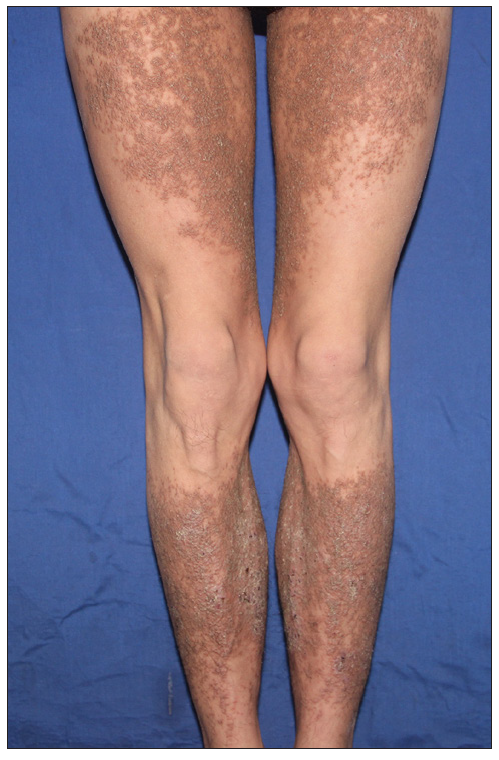
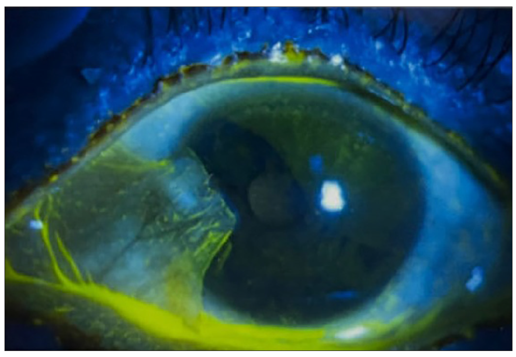
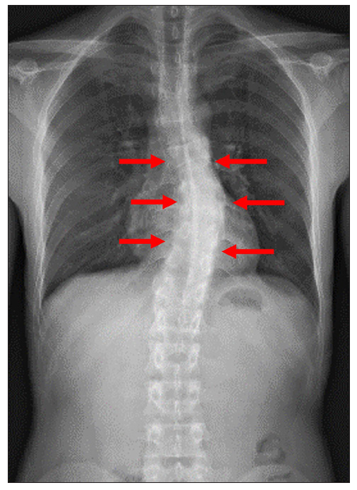
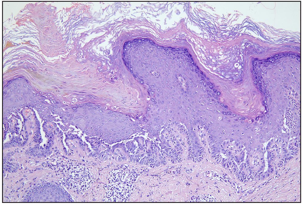
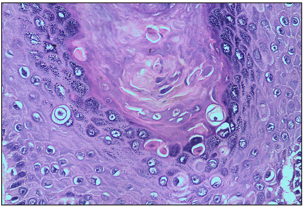
A 30-year-old Chinese man presented with generalised pruritic eruptions persisting for 16 years. These lesions initially developed on his scalp at the age of 14, progressively involving the face, trunk and extremities. He had a history of recurrent bacterial infections and suicidal behaviour. On examination, multiple erythematous and hyperpigmented warty papules and plaques were widely distributed on the body, predominantly on the scalp, face, trunk and extensors of all the limbs [Figures 1a, 1b and 1c]. Nail and oral mucosa were unremarkable. Anterior segment photography unveiled the presence of blepharitis and pterygium in both eyes, and corneal epithelial defects were observed under corneal fluorescein staining [Figure 1d]. X-ray of the spine revealed significant scoliosis [Figure 2]. Importantly, he reported no history of long-term weight-bearing or trauma, common risk factors for scoliosis, suggesting an atypical aetiology in this case. Histopathological study indicated papillated epidermal hyperplasia, acantholytic cleft formation and dyskeratotic cells in the epidermis [Figures 3a and 3b], leading to a diagnosis of Darier disease.
Export to PPT
Export to PPT
Export to PPT
Export to PPT
Export to PPT
Export to PPT
Export to PPT
Genomic DNA isolated from the proband and parents revealed a recurrent heterozygous mutation (c.1A>G, p.Met1Val) in ATP2A2, confirmed by Sanger sequencing. This mutation was absent in his unaffected parents [Figure S1]. No pathogenic mutations associated with scoliosis were identified.
ATP2A2 encodes sarcoendoplasmic reticulum Ca2+-ATPase isoform 2 (SERCA2), which transports Ca2+ back into the lumen of the endoplasmic reticulum from the cytosol.1 Therefore, inherited defects in ATP2A2 occurring in Darier disease may lead to impaired regulation of the intracellular calcium signalling. Haploinsufficiency, resulting from the presence of a dysfunctional allele, is acknowledged as the molecular mechanism underpinning Darier disease.1,2 Although more than 200 ATP2A2 mutations have been documented to be associated with Darier disease, no precise genotype–phenotype correlation has been recognised to date.1,2
In this case, we describe a severe form of Darier disease with an ATP2A2 c.1A>G mutation. Interestingly, two previous reported Darier disease cases with start codon mutation also presented with widespread severe lesions.3,4 This indicates that start codon mutations may contribute to a complete haplo-insufficiency, potentially explaining severe phenotypes.
In addition to classical features, our case also presented with pterygium and scoliosis. To the best of our knowledge, these features have not been reported in Darier disease.1,2 Ophthalmic involvement in Darier disease may not be uncommon, with a survey reporting 39% of patients experiencing eye problems, predominantly manifesting as blepharitis and dry eye disease.5 Pterygium is an ocular surface disorder presenting as a raised, wedge-shaped lesion with potential implications for vision. In our case, potential mechanisms contributing to pterygium may include chronic inflammation affecting the ocular surface epithelium and the crusting of skin lesions, which could obstruct the Meibomian gland and disrupt its normal functioning.
Skeletal changes are rarely documented in Darier disease. Bone cysts and developmental retardation were found in a few cases.6,7 The aetiology of scoliosis in Darier disease remains elusive, potentially involving a spectrum of genetic and environmental factors. In our case, no known scoliosis-related mutations were found, and the patient’s history lacked known risk factors. This suggests a possible link with SERCA2’s role in calcium homeostasis affecting osteoclast survival in the skeleton, which may contribute to scoliosis.6,7 However, further experiments are needed to verify this hypothesis.
In conclusion, we identified a start codon variant in the ATP2A2 gene in this case with severe Darier disease. Our case highlights the importance of ophthalmologic and orthopaedic evaluation in Darier disease patients. The potential associations of ATP2A2 mutations with scoliosis and pterygium warrant further investigation.
留言 (0)