Synthesis and characterization of SWCNT–COOH–CDDP complexes
Carboxyl-functionalized single-walled carbon nanotubes (SWCNT–COOH) were obtained by covalent functionalization of SWCNT through chemical oxidation. Further, SWCNT–COOH was functionalized with CDDP to obtain carboxyl-functionalized single-walled carbon nanotubes conjugated with CDDP (SWCNT–COOH–CDDP). The detailed procedure of synthesis and characterization of the complex is presented in a previous study (Badea et al. 2022).
Cell culture and treatment conditions
Human MDA-MB-231 epithelial cell line (HTB-26™, ATCC, Manassas, VA), from an adenocarcinoma of the mammary gland, was grown as monolayer cultures in 75 cm2 flasks and Dulbecco’s Modified Eagle Medium (DMEM, 31,600-083, Gibco, UK) supplemented with 3.5 g/L glucose, 1.5 g/L NaHCO3, 1% penicillin/streptomycin/amphotericin B solution (A5955, Sigma-Aldrich, St. Louis, MO, USA), and 10% fetal bovine serum (10270-106, origin South America, Gibco, Life Technologies, Carlsbad, CA, USA). The culture medium was completely refreshed once every two–three days. For the subculture procedure, cells were detached from the flask surface using a 0.25% trypsin – 0.53 mM EDTA solution and the cells were counted using a Bürker-Türk chamber. The cultures were maintained in standard conditions (37 °C, humidity, 95% air, 5% CO2) and monitored every day.
MDA-MB-231 cells were seeded in culture plates and flasks and on the second day of culture were treated with various concentrations of CDDP (0.158, 0.316, 0.632, and 1.26 µg/mL), SWCNT–COOH (0.25, 0.5, 1 and 2 µg/mL), and the same doses of SWCNT–COOH–CDDP for 24 and 48 h. For treatment, the culture medium was removed and replaced with medium containing treatment of the tested concentrations. Untreated cells were used as control.
MDA-MB-231 spheroids generation and treatment
Multicellular tumor spheroids (MCTSs) were generated from MDA-MB-231 cells in complete DMEM medium supplemented with 2.5% Matrigel (Matrigel Basement Membrane Matrix, 356237, Corning, NY) by liquid-overlay technique using low-cell-attachment Nunclon Sphera Microplates. Single MCTSs were generated in 96-well round bottom plates (cat. no. 12-566-430, 174925) in 200 µL culture medium and multiple MCTSs were obtained using 6-well plates (cat. no. 12-566-435, 174932) in 3 mL culture medium, according to our previous study (Badea et al. 2019).
MCTSs were seeded at a density of 5000 cells/well, respectively 5 × 10⁵ cells/well in 96- and 6-well plates. Starting with the third day of culture, MCTSs were exposed to the same doses of treatment as monolayers cultures for 24 and 48 h. For single MCTS, half of the culture medium was replaced with medium containing double concentrations of the tested samples. An identical procedure was followed for multiple MCTSs cultures, with the mention that the added medium contained Matrigel in a concentration of 1.25%. Untreated MCTSs represented the control.
Lactate dehydrogenase (LDH) release assay
Membrane integrity and induction of cell death by necrosis pathway were evaluated by quantifying the level of lactate dehydrogenase (LDH) enzyme activity in the culture medium of 2D and 3D cultures. The method was performed using a commercial kit Cytotoxicity Detection Kit (LDH) purchased from Roche (cat. no. 11644793001) which used a catalyst (diaphorase/NAD+ mixture) and a dye solution (Iodotetrazolium chloride (INT) and sodium lactate) as reagents. A volume of 50 μL culture medium from treated and untreated cells and MCTSs was homogenized with 50 μL reaction mixture (catalyst:dye solution 1:45) and then incubated in the dark, at room temperature, for 15 min. The absorbance of samples was measured at 490 nm, at Flex Station 3 (Molecular Devices, San Jose, CA, USA). The results were expressed as percentages (%) of control (untreated cells and MCTSs respectively).
Morphology assessment
After 24 and 48 h of treatment with the complex and free constituents, morphological changes of breast cancer cells and single MCTSs were investigated by optical microscopy using an inverted microscope Olympus IX73 (Olympus, Tokyo, Japan). The cells and MCTSs were imaged using a Hamamatsu camera (A3472-06, Hamamatsu, Japan) and CellSens Dimension software.
Live/dead staining assay
To evaluate the cell viability after treatment exposure, treated cells and single MCTSs were fluorescently labeled with calcein AM and ethidium homodimer-1 (EthD-1), following the instructions of LIVE/DEAD™ Viability/Cytotoxicity Kit (L3224, Invitrogen). Calcein AM highlighted the live cells based on esterase activity and EthD-1 distinguished dead cells with a permeable membrane. Thus, the cells were seeded in 24-well plates (4 × 10⁴ cells/well) to form monolayer cultures and in Nunclon Sphera plates (5000 cells/well) for MCTSs generation. After 24 and 48 h from treatment, the culture medium of monolayer cultures was replaced with the reaction mix (a serum-free solution containing 2 μM calcein AM and 4 μM EthD-1). In parallel, half of the culture medium of single MCTSs was substituted with a similar solution containing double concentrations of the dyes. Afterward, monolayer cultures and MCTSs were incubated for 20 min, at 37 °C and then visualized at a fluorescence inverted microscope Olympus IX73 (Olympus, Tokyo, Japan). Images were acquired using FITC and TRITC filters and CellSens Dimension software (ver. 1.11, Olympus, Tokyo, Japan).
Measurement of mitochondrial membrane potential (MMP)
Mitochondrial membrane potential (MMP) was evaluated after the treatment of 2D cultures and single MCTSs with two doses of CDDP (0.316 μg/mL and 0.632 μg/mL), SWCNT–COOH (0.5 and 1 μg/mL), and complex (same doses). For this assessment, the “Mitochondria Membrane Potential Kit” was used. This assay is based on a fluorometric method in which a decreased fluorescence is associated with apoptosis. In brief, MDA-MB-231 cells were seeded in 96-well plates to form 2D (1.2 × 10⁴ cells/well) and 3D (5000 cells/well) cultures and then treated with the samples. After 24 and 48 h, in monolayer cultures, the culture medium was discarded and the cells were incubated at 37 °C with 100 μL Dye Loading Solution for 30 min. Then, 50 μL Assay Buffer B was added to every well. After 30 min of incubation at 37 °C, the fluorescence was measured at Flex Station 3 (ex. 540 nm/em. 590 nm) and the results were calculated related to the control.
The same protocol was followed for 3D cultures, with the mention that 50 μL culture medium was kept in wells and then mixed with 50 μL Dye Loading Solution twice concentrated. Finally, the spheroids were analyzed under Olympus IX73 fluorescence microscope. The images were acquired using CellSens Dimension software and the Z-stack function. For each experimental condition, three spheroids were analyzed. Fluorescence level was quantified using ImageJ software (ver. 1.52a, NIH, Bethesda, MD, USA), indicated as corrected total cell fluorescence (CTCF) and represented related to control.
Preparation of cell lysates and estimation of total protein content
For obtaining the cell lysates, MDA-MB-231 cells were seeded as 2D cultures in flasks (106 cells/flask) and MCTSs in 6-well plates (5 × 10⁵ cells/well). After treatment with CDDP (0.316 and 0.632 μg/mL), SWCNT–COOH (0.5 and 1 μg/mL), and complex (same doses), the cells (enzymatically detached from the culture surface) and MCTSs were collected and centrifuged for 5 min, 1500 rpm. Cell pellets were washed and then resuspended in phosphate-buffered saline (PBS). Then, cells were lysed by sonication (30 s, 3 times, on ice) using a UP50H sonicator (Hielscher Ultrasound Technology, Teltow, Germany), set at 80% amplitude, 1 cycle. Cell lysates were centrifuged (10 min, 3000 rpm, 4 °C) and the supernatants were aliquoted, stored at – 80 °C, and used in the next week for biochemical analyses. Total protein content was quantitated by the Bradford assay using bovine serum albumin (BSA) standard curve (0–1.5 mg/mL) and spectrophotometric detection at 595 nm (Flex Station 3, Molecular Devices, San Jose, CA, USA) (Bradford 1976).
Measurement of reduced glutathione (GSH) content
The antioxidant defense of 2D and 3D cultures after treatment was assessed by quantifying the concentration of reduced glutathione (GSH) using the reaction between GSH and Ellman's reagent—5,5ʹ-dithio-bis-(2-nitrobenzoic acid)—DTNB with the generation of 2-nitro-5-thiobenzoic (TNB) acid that can be spectrophotometrically detected at 405 nm. First, cell lysates were deproteinized with equal volumes of a 5% solution of 5-sulfosalicylic acid and centrifuged at 10,000 rpm, for 10 min, and 4 °C. Then, 10 μL of deproteinized lysate was homogenized with 150 μL reaction mix (1.5 mg/mL DTNB in potassium phosphate buffer 0.1 M, pH = 7 and EDTA 1 mM) and incubated for 10 min at room temperature. Finally, the absorbance was read at 405 nm using a Flex Station 3 microplate reader (Molecular Devices, San Jose, CA, USA). GSH concentration of the samples was estimated by extrapolation on a 3.125–200 μM GSH standard curve and calculated related to control (% of control).
Western blot analysis
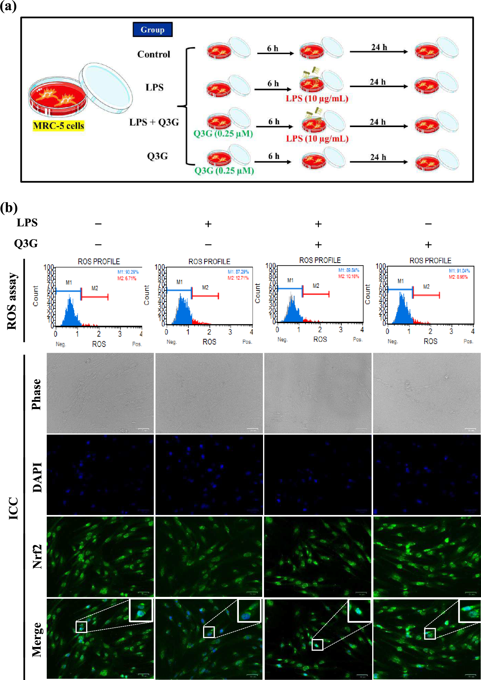
Western blot technique was used for the evaluation of various protein expressions to elucidate the mechanisms triggered in 2D cultures and spheroids of MDA-MB-231 cells after treatment with CDDP (0.316 and 0.632 μg/mL), SWCNT–COOH (0.5 and 1 μg/mL), and complex (same doses). The total protein extracts were diluted with PBS up to equal amounts of protein, and the samples were then subjected to a chemical and thermal (SDS, 5 min, 95 °C) denaturation. The proteins were separated on polyacrylamide gels (8, 10, 15%) in Tris-Gly electrophoresis buffer (0.05 M Tris, 0.05 M glycine, and 0.1% SDS), at 90 V, and then transferred onto polyvinylidene fluoride (PVDF) membranes (cat. no. IPVH00010, Merck, Darmstadt, Germany) using a Tris-Gly transfer buffer (25 mM Tris, 192 mM glycine, and 20% (v/v) methanol). The detection of protein bands was done using Western Breeze Chromogenic Anti-Mouse and Anti-Rabbit kits solutions (WB7103, WB7105, Invitrogen, Carlsbad, CA, USA). Following blocking of non-specific binding sites, the membranes were incubated overnight with primary antibody solutions anti-Nrf2 (sc-13032, Santa Cruz, CA, USA), anti-MCM2 (sc-373702, Santa Cruz), anti-caspase-3 (sc-7148, Santa Cruz), anti-caspase-8 (sc-5263, Santa Cruz), anti-caspase-9 (sc-56076, Santa Cruz), and anti-caspase-4 (sc-56056, Santa Cruz). On the next day, membranes were washed with wash solutions and incubated with anti-mouse and anti-rabbit secondary alkaline phosphatase-conjugated antibodies. Immune complexes were revealed with the BCIP/NBT chromogenic substrate and imaged using a ChemiDoc Imaging System (Bio-Rad, Hercules, CA, USA). Western blot signal intensities were quantified using the ImageLab program (version 6.1.0, Bio-Rad, Hercules, CA, USA). The β-actin protein served as a reference for normalization.
Evaluation of invasion potential
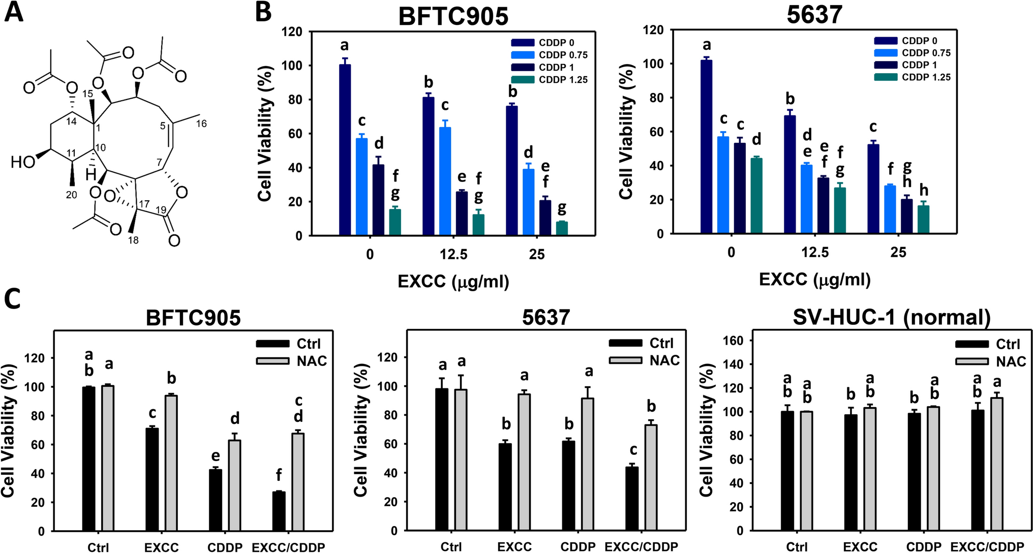
The invasion capacity of MDA-MB-231 cells was assessed after the treatment of 2D and 3D cultures with CDDP (0.316 and 0.632 μg/mL), SWCNT–COOH (0.5 and 1 μg/mL), and the corresponding doses of the complex for 24 and 48 h.
In 2D cultures, the invasion potential was evaluated following the instructions of CytoSelect™ 96-Well Cell Invasion Assay (Basement Membrane, Fluorometric Format, CBA-112, Cell Biolabs), which uses a 96-well invasion plate (composed of feeder tray, membrane chamber, and plate cover) and a polycarbonate membrane inserts (8 µm pore size) to discriminate non-invasive cells from invasive cells which were detected with CyQuant® GR dye. First, in the wells of the feeder tray were added 150 μL culture medium supplemented with 10% fetal bovine serum. In parallel, the membrane was rehydrated with 100 μL serum-free culture medium and after 1 h, the medium was replaced with 100 μL cell suspension (8 × 10⁵ cells/mL) prepared in free-serum culture medium supplemented with 5% BSA, also containing the treatment. Assays were carried out over 24 and 48 h of incubation in a humidified atmosphere of 5% CO2, and 95% air at 37 °C. The culture medium was removed from the membrane chamber, which was placed in the Cell Harvesting Tray containing 150 μL of Cell Detachment Solution/well and incubated for 30 min, at 37 °C. Then, in each well was added 50 μL of 4X Lysis Buffer/CyQuant® GR dye solution and after 20 min of incubation at room temperature, the fluorescence was measured at Thermo Scientific Appliskan (Waltham, MA, USA), ex. 485 nm/em. 535 nm. The results were calculated related to control (% of control).
The cell invasion in 3D cultures was investigated after treatment using the manufacturer’s guidelines of “96-Well 3D Spheroid BME Cell Invasion Assay” (3500-096-K, Trevigen, Inc., Gaithersburg, MD). A matrix based on basement membrane proteins, which generates a hydrogel network that embeds the spheroid and can be penetrated by invasive cells, was used. Briefly, spheroids were seeded at a density of 3000 cells in 50 μL 1× Spheroid Formation ECM in 3D Culture Qualified 96-Well Spheroid Formation Plate. The plate was centrifuged at 200×g, 3 min, room temperature, to promote spheroid formation and then incubated at 37 °C. On the third day of culture, in the pre-chilled plate was added 50 μL of Invasion Matrix/well and the plate was centrifuged at 300×g, 4 °C for 5 min and then transferred in the incubator at 37 °C for 1 h to enhance gel formation. Further, culture media with or without indicated concentrations of treatment (100 μL/well) were added and then the plates were further incubated in a 5% CO2 atmosphere at 37 °C. After 24 and 48 h, the invasion of cells into the surrounding matrix was monitored, and the spheroids were photographed using a Hamamatsu camera (A3472-06, Hamamatsu, Japan) of an Olympus IX73 microscope (Olympus, Tokyo, Japan) and CellSens Dimension software (ver. 1.11, Olympus, Tokyo, Japan). Image analysis was performed using ImageJ software (ver. 1.52a, NIH, Bethesda, MD, USA) using the instructions of the kit. Five spheroids were analyzed for each experimental condition. Cell invasion was calculated by reporting the area of treated spheroids to the area of control spheroids (% of control).
Statistical analysis
All investigations were performed in triplicate. The data were expressed as relative values in comparison with control (100%) and calculated as mean ± standard deviation. The results were statistically analyzed in GraphPad Prism (Version 8, GraphPad Software, La Jolla, CA, USA), using the two-way ANOVA method and Tukey’s multiple comparisons tests (treated cells vs. control). The values p < 0.05 (*), p < 0.01 (**), and p < 0.001 (***) were considered statistically significant.

留言 (0)