
記住我

Although virus-specific TBYS cells have been reported by accumulating evidence6,7,8,9,10,11,12,13,14,15, a detailed portrait of these specialized T cells, especially CD4+ TBYS cells, is lacking. We thus sought to analyze the kinetics, molecular characteristics and lineage relationship with other memory types of both CD8+ and CD4+ TBYS populations in the TME. To this end, congenic naive (CD45.1+CD44loCD62Lhi) P14 CD8+ T cells recognizing the lymphocytic choriomeningitis virus (LCMV) glycoprotein (GP) epitope H-2DbGP33–41 and congenic naive (CD45.1+CD44loCD62Lhi) SMARTA (SM) CD4+ T cells recognizing the LCMV GP epitope I-AbGP66–77 were adoptively transferred into C57BL/6 recipients (CD45.2+), which were then infected with LCMV Armstrong to establish acute viral infection and resultant virus-specific P14 TMEM and SM TMEM cells. On day 60 after infection, recipients were subcutaneously engrafted with syngeneic MC38 colon adenocarcinoma cells and tumor-infiltrating P14 CD8+ and SM CD4+ T cells, and TBYS populations were analyzed on days 10, 15 and 20 after tumor engraftment (Fig. 1a and Extended Data Fig. 1a). As shown, frequencies of tumor-infiltrating P14 TBYS and SM TBYS cells remained stable at the different indicated time points (Fig. 1b,c), suggesting that the infiltrating virus-specific TBYS cells were not altered at the population level during tumor progression.
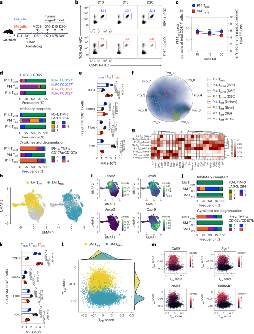
Fig. 1: Features of tumor-infiltrating virus-specific TBYS cells.
a, Schematic of the experimental design. Congenic CD45.1+ P14 CD8+ T cells and CD45.1+ SM CD4+ T cells were adoptively transferred into naive C57BL/6 recipients (CD45.2+), which were then infected with LCMV Armstrong and engrafted with MC38 cells on day (D)60 after infection. On days 10, 15 and 20 after tumor engraftment, tumor-infiltrating P14 and SM cells were analyzed. b, Flow cytometry analysis of MC38 tumor-infiltrating CD8+ (top) and CD4+ (bottom) T cells on days 10, 15 and 20 after tumor engraftment. Numbers adjacent to the outlined areas indicate percentages of CD45.1+Vα2+ P14 cells (blue) of tumor-infiltrating CD8+ T cells or CD45.1+Vα2+ SM cells (red) of tumor-infiltrating CD4+ T cells. c, Frequency of P14 TBYS cells of total MC38 tumor-infiltrating CD8+ T cells (indicated by blue dots, n = 5 mice (day 10) or n = 4 mice (day 15 and day 20)) and frequency of SM TBYS cells of total MC38 tumor-infiltrating CD4+ T cells (indicated by red cubes, n = 5 mice (day 10) or n = 4 mice (day 15 and day 20)) at the indicated time points. d, Frequencies of P14 TMEM, P14 TEX and P14 TBYS cells that express KLRG1 and/or CD127 (top; n = 4 mice (P14 TMEM), n = 3 mice (P14 TEX) or n = 5 mice (P14 TBYS)), coexpress the indicated number of inhibitory receptors (PD-1, TIM-3, LAG-3 and 2B4) (middle; n = 5 mice (P14 TMEM), n = 3 mice (P14 TEX) or n = 4 mice (P14 TBYS)) or coexpress the indicated number of cytokines (IFN-γ and tumor necrosis factor (TNF)-α) and cytotoxic degranulation markers (CD107a and CD107b) (bottom; n = 5 mice (P14 TMEM), n = 3 mice (P14 TEX) or n = 3 mice (P14 TBYS)). e, Comparison of TCF-1, Eomes, T-bet and TOX expression levels among P14 TMEM (n = 3 mice), P14 TEX (n = 3 mice) and P14 TBYS cells (n = 5 mice). MFI, mean fluorescence intensity. f, A Radviz projection of P14 TMEM, P14 TRM (kidney, liver, salivary gland (SG) and small intestine intra-epithelial lymphocyte (siIEL)) and P14 TBYS cells based on nine gene expression programs (Pro_1–Pro_9) inferred by consensus non-negative matrix factorization. A total of 38,880 P14 cells passed quality control. g, Heatmap showing selected DEGs in various P14 cells. h, UMAPs of SM TBYS (left) and SM TMEM cells (right). A total of 3,565 SM TBYS cells are colored in yellow, and a total of 5,648 SM TMEM cells are colored in blue. i, UMAPs showing expression levels of Ly6c2, Gzmb, Foxp3 and Cxcr5 in SM TBYS and SM TMEM cells. j, Frequency of SM TMEM, SM TTST and SM TBYS cells that coexpress the indicated number of inhibitory receptors (PD-1, TIM-3, LAG-3 and 2B4) (top; n = 3 mice (SM TMEM), n = 3 mice (SM TTST) or n = 3 mice (SM TBYS)) or coexpress the indicated number of cytokines (IFN-γ and TNF-α) and cytotoxic degranulation markers (CD107a and CD107b) (bottom; n = 3 mice (SM TMEM), n = 3 mice (SM TTST) or n = 5 mice (SM TBYS)). k, Comparison of TCF-1, Eomes, T-bet and TOX expression levels among SM TMEM (n = 3 mice), SM TTST (n = 3 mice) and SM TBYS (n = 5 mice) cells. TFs, transcription factors. l, Gene set scoring analysis of 3,565 SM TBYS cells and 5,648 SM TMEM cells. Density of cells in the TCM versus TRM score space annotated by cell type is depicted. m, Expression profiles of Cd69, Rgs1, Nr4a1 and Bhlhe40 of SM TBYS and SM TMEM cells in the TCM versus TRM score space. Data (b–e,j,k) are representative of two independent experiments. One-way ANOVA with Turkey’s test was used in e,k. Center values and error bars (c,e,k) indicate mean and s.e.m.
Next, we set out to compare the molecular characteristics of virus-specific CD8+ TBYS cells with their counterparts recognizing the same epitope but induced to differentiate into TMEM or TEX cells. Accordingly, congenic naive P14 CD8+ T cells were adoptively transferred into C57BL/6 recipients, which were infected with the LCMV Armstrong strain to induce the differentiation of P14 TMEM cells. Furthermore, a fraction of these infected recipients was subcutaneously engrafted with MC38 cells on day 60 after infection, and tumor-infiltrating P14 TBYS cells were analyzed on day 10 after tumor engraftment. In the scenario of tumor-specific TEX cells, congenic naive P14 CD8+ T cells were adoptively transferred into C57BL/6 recipients, which were then subcutaneously engrafted with syngeneic B16F10 melanoma cells expressing the LCMV GP (hereafter referred to as B16-GP cells)19 and killed on day 15 after tumor engraftment to analyze tumor-specific P14 TEX cells (Extended Data Fig. 1b).
Resembling P14 TMEM cells, we found that P14 TBYS cells are mainly populated by the killer cell lectin-like receptor G1 (KLRG1)loCD127hi subset1 (Fig. 1d). Furthermore, P14 TBYS cells retain an expression pattern of transcription factors similar to that of P14 TMEM cells1, as exemplified by high expression of TCF-1 and Eomes and low expression of T-bet (Fig. 1e). In contrast to TMEM cells, TEX cells are characterized by coexpression of multiple inhibitory receptors, impaired cytokine secretion and the transcription factor TOX associated with T cell exhaustion2,20,21,22,23. Although preserved in the immunosuppressive TME, P14 TBYS cells show limited expression of inhibitory receptors and TOX and exhibit polyfunctional cytokine secretion upon ex vivo re-stimulation (Fig. 1d,e). Thus, these findings confirm that CD8+ TBYS cells phenotypically and functionally resemble CD8+ TMEM cells but not CD8+ TEX cells.
To further define the molecular traits of CD8+ TBYS cells, we performed single-cell RNA sequencing (scRNA-seq) of P14 TBYS cells from the TME of MC38-engrafted mice. Conventional P14 TMEM and tissue-resident memory (TRM) cells from external scRNA-seq datasets24,25 were introduced as controls (Extended Data Fig. 1c). A Radviz projection based on nine gene expression programs inferred by consensus non-negative matrix factorization clearly distinguished P14 TMEM cells from P14 TRM cells of various non-lymphoid tissues (Fig. 1f and Extended Data Fig. 1d,e). Importantly, P14 TBYS cells were drawn toward the groups of P14 TRM cells (Fig. 1f), likely representing the acquisition of certain TRM cell features. Further unbiased hierarchical clustering confirmed that P14 TBYS cells are in close proximity with P14 TRM cells and highlighted by cytolytic activity (Gzmb, Tnf) and tissue residency (Cd69, Nr4a1)26 (Fig. 1g). The potential residency of P14 TBYS cells was also evidenced by high expression of CD69 protein (Extended Data Fig. 2a,b). Therefore, these findings suggest TRM cell features of CD8+ TBYS cells.
Next, we aimed to trace the differentiation of virus-specific CD4+ TBYS cells by performing scRNA-seq of SM TBYS cells from the MC38 TME with conventional SM TMEM cells as controls (Extended Data Fig. 1c). Consistent with previous studies27,28, SM TMEM cells adopt a bifurcation of follicular helper T cells (Cxcr5) and type 1 helper T (TH1) cells (Ly6c2, Gzmb) (Fig. 1h,i). Instead of adopting a follicular helper T/TH1 bifurcation, we found that SM TBYS cells were predominantly fated toward the TH1 lineage (Fig. 1h,i). Indeed, TH1-type SM TBYS cell differentiation was further evidenced by abundant Ly6C protein but rare expression of C–X–C motif chemokine receptor (CXCR)5 (Extended Data Fig. 2c–e). Furthermore, similar to P14 TBYS cells, SM TBYS cells also showed high expression of TCF-1 and Eomes and low expression of T-bet (Fig. 1k), suggesting the retention of memory properties. In addition, SM TBYS cells showed no features related to exhaustion, including multiple inhibitory receptors29 and TOX30, as compared to tumor antigen-specific T (TTST) cells (Fig. 1j,k); instead, SM TBYS cells exhibit a substantial capacity to secrete multiple cytokines upon ex vivo re-stimulation (Fig. 1j). These results define the memory properties of TH1-type CD4+ TBYS cells in the TME.
Previous studies stated distinct molecular signatures between central memory T (TCM) and TRM cells31,32. We therefore examined the enrichment for TCM and TRM cell gene signatures in SM TBYS cells, with conventional SM TMEM cells being introduced as controls, to explore the effector–memory state in the SM TBYS population. We found a largely overlaid TCM signature between SM TBYS and SM TMEM cells, with a noticeable dissection in the TRM signature (Fig. 1l). Indeed, genes related to TRM cells (for example, Cd69, Rgs1, Nr4a1 and Bhlhe40) were highly expressed in SM TBYS cells relative to those in SM TMEM cells (Fig. 1m); meanwhile, genes related to TCM cells (for example, Il7r, Tcf7 and Ccr7) were largely overlapping between SM TBYS and SM TMEM cells (Extended Data Fig. 2f). Distinct from SM TBYS cells, P14 TBYS cells were dominated by the gene signature of TRM cells rather than that of TCM cells (Extended Data Fig. 2g). Thus, these findings highlight a unique hybrid gene signature consisting of both TCM and TRM cell features in CD4+ TBYS cells but not in CD8+ TBYS cells.
We further explored endogenous virus-specific TBYS cells from MC38 tumors engrafted in LCMV-infected mice by quantifying interferon (IFN)-γ-expressing cells upon ex vivo stimulation with cognate viral peptides (Extended Data Fig. 3a). Consistently, we observed the presence of endogenous CD8+ TBYS cells specific to LCMV GP33–41, LCMV GP276–286 and LCMV nucleoprotein (NP)396–404 and endogenous CD4+ TBYS cells specific to LCMV GP66–77 in the MC38 tumors engrafted in LCMV-infected mice (Extended Data Fig. 3b–e). In addition to the MC38 tumor model, endogenous LCMV-specific TBYS cells were also found in the TME of B16F10-engrafted mice (Extended Data Fig. 3f–h). In sum, these findings highlight the functional memory characteristics of virus-specific CD8+ and CD4+ TBYS cells in the TME.
OV-BYTE therapy elicits anti-tumor responseGiven the abundant memory pool of virus-specific TBYS cells in the TME, we hypothesized that OV-mediated delivery of TBYS cell epitopes (hereafter referred to as OV-BYTE) to tumor cells might redirect the cytotoxicity of TBYS cells toward tumor cells and thus curtail tumor progression. For this purpose, the gene encoding LCMV GP was inserted into an NDV rSG10 strain (referred to as NDV wild type (WT)) and thus termed NDV-GP (Extended Data Fig. 4a). To assess the ability of NDV-GP to infect and deliver LCMV GP expression in tumor cells, we infected MC38 cells with NDV-GP and then detected LCMV GP expression in the infected MC38 cells. In accordance with the previously reported highly efficient transgene delivery of oncolytic NDV33,34, we found that NDV-GP substantially infected MC38 cells and expressed the LCMV GP transgene (Extended Data Fig. 4b–d). Furthermore, we also found enhanced expression of major histocompatibility complex (MHC)-I and MHC-II molecules in NDV-GP-infected tumor cells and specific killing of these tumor cells by LCMV GP-specific T cells (Extended Data Fig. 4e–g), indicating MHC presentation of LCMV GP epitopes by NDV-GP-infected tumor cells to LCMV GP-specific T cells.
Next, we aimed to assess the in vivo anti-tumor activities of NDV-GP in tumor-bearing mice that have established immune memory for LCMV. To this end, naive C57BL/6 mice were infected with LCMV Armstrong, followed by subcutaneously engraftment with MC38 cells on day 60 after infection and then intratumoral treatment with phosphate-buffered saline (PBS), NDV-WT or NDV-GP (Fig. 2a). Remarkably, NDV-GP treatment delayed tumor growth more efficiently than NDV-WT treatment (Fig. 2b) and led to ~30% complete remission on day 50 after tumor engraftment (Fig. 2c). Moreover, NDV-GP treatment did not show any therapeutic advantages over NDV-WT treatment in tumor-engrafted mice without pre-existing immune memory (Fig. 2d,e); meanwhile, the therapeutic function of NDV-GP to treat MC38 tumors engrafted in LCMV-infected mice was abolished in the presence of neutralizing antibodies specific to LCMV GP proteins (Fig. 2f,g).
Fig. 2: OV-mediated delivery of TBYS cell epitopes to solid tumor cells elicits anti-tumor response.
a, Schematic of the experimental design for b,c. C57BL/6 mice were infected with LCMV Armstrong and engrafted with MC38 cells on day 60 after infection. On days 7, 8, 9, 11 and 12 after tumor engraftment, recipients were intratumorally administered PBS, NDV-WT or NDV-GP. b,c, Tumor growth curve (b) and Kaplan–Meier survival curve (c) of MC38 tumor-bearing mice intratumorally treated with PBS (n = 6 mice), NDV-WT (n = 10 mice) or NDV-GP (n = 10 mice) as described in a. d, Schematic of the experimental design for e. Naive C57BL/6 mice were engrafted with MC38 cells and then intratumorally administered PBS, NDV-WT or NDV-GP on days 7, 8, 9, 11 and 12 after tumor engraftment. e, Kaplan–Meier survival curve of MC38 tumor-bearing mice intratumorally treated with PBS (n = 12 mice), NDV-WT (n = 8 mice) or NDV-GP (n = 9 mice) as described in d. f, Schematic of the experimental design for g. LCMV Armstrong-infected C57BL/6 mice were engrafted with MC38 cells on day 60 after infection. On days 7, 8, 9, 11 and 12 after tumor engraftment, recipients were intratumorally administered PBS, NDV-WT or NDV-GP in the presence or absence of LCMV GP-neutralizing antibodies. g, Tumor growth curve of MC38 tumor-bearing mice intratumorally treated with PBS, NDV-WT or NDV-GP with or without LCMV GP-neutralizing antibodies (NAbs) as described in f. PBS (n = 10 (control) or n = 9 (neutralizing antibodies) mice), NDV-WT (n = 9 (control) or n = 9 (neutralizing antibodies) mice) and NDV-GP (n = 9 (control) or n = 12 (neutralizing antibodies) mice). h, Schematic of the experimental design for i. C57BL/6 mice were infected with LCMV Armstrong and engrafted with MC38 cells on day 60 after infection. On days 7, 8, 9, 11 and 12 after tumor engraftment, recipients were intratumorally administered PBS, NDV-WT or NDV-NP. i, Kaplan–Meier survival curve of MC38 tumor-bearing mice intratumorally treated with PBS (n = 9 mice), NDV-WT (n = 9 mice) or NDV-NP (n = 11 mice) as described in h. j, Schematic of the experimental design for k. C57BL/6 mice were infected with LCMV Armstrong and engrafted with MC38 cells on day 60 after infection. On days 7, 8, 9, 11 and 12 after tumor engraftment, recipients were intratumorally administered PBS, Ad5-WT or Ad5-GP. k, Kaplan–Meier survival curve of MC38 tumor-bearing mice intratumorally treated with PBS (n = 10 mice), Ad5-WT (n = 9 mice) or Ad5-GP (n = 7 mice) as described in j. l, Schematic of the experimental design for m. C57BL/6 mice were infected with LCMV Armstrong and engrafted with B16F10 cells on day 60 after infection. On days 7, 8, 9, 11 and 12 after tumor engraftment, recipients were intratumorally administered PBS, NDV-WT or NDV-GP. m, Kaplan–Meier survival curve of B16F10 tumor-bearing mice intratumorally treated with PBS (n = 6 mice), NDV-WT (n = 8 mice) or NDV-GP (n = 8 mice) as described in l. n, Schematic of the experimental design for o. C57BL/6 mice were infected with LCMV Armstrong, and three plasmids (encoding Myc-tagged NICD1, Myc-tagged AKT and hyperactive sleeping beauty transposase (SB100)) were injected into the hydrodynamic tail vein on day 60 after infection. On days 7, 8, 9, 11 and 12 after tumor engraftment, recipients were intravenously administered PBS, NDV-WT or NDV-GP. o, Kaplan–Meier survival curve of mice with NICD- and AKT-induced murine intrahepatic cholangiocarcinoma as described in n. PBS (n = 6 mice), NDV-WT (n = 5 mice) and NDV-GP (n = 6 mice). p, Schematic of the experimental design for q,r. NCG mice were engrafted with PBMCs from HLA-A2-positive donors with a history of H1N1 infection. After reconstruction of human T cells, these humanized NCG mice were engrafted with A375 cells and then intratumorally administered PBS, NDV-WT or NDV-H1N1 NP on days 7, 8, 9, 11 and 12 after tumor engraftment. q,r, Tumor growth curve (q) and Kaplan–Meier survival curve (r) of A375 tumor-bearing humanized NCG mice intratumorally treated with PBS (n = 5 mice), NDV-WT (n = 6 mice) or NDV-H1N1 NP (n = 5 mice) as described in p. All data are representative of at least two independent experiments. Two-way ANOVA was used to compare tumor growth curves in b,g,q. The log-rank (Mantel–Cox) test was performed to compare survival curves among groups in c,e,i,k,m,o,r. Center values and error bars (b,g,q) indicate mean and s.e.m.
In parallel, we also constructed the NDV strain carrying the LCMV NP gene (referred to as NDV-NP) to treat MC38-bearing mice with LCMV immune memory (Fig. 2h and Extended Data Fig. 4a,h). Similarly, NDV-NP treatment led to significant tumor growth delays compared to NDV-WT treatment and generated ~30% complete regression (Fig. 2i). To test whether another non-NDV OV platform could also be used to target TBYS cells to limit tumor progression, LCMV Armstrong-infected C57BL/6 mice were engrafted with MC38 cells and then intratumorally administered the oncolytic adenovirus serotype 5 (Ad5) that carries the LCMV GP gene (referred to here as Ad5-GP) (Fig. 2j and Extended Data Fig. 4i,j). Similarly, Ad5-GP treatment effectively slowed tumor growth and resulted in marked tumor regression (~25% complete regression) compared to control treatments (Fig. 2k).
In addition to MC38 colon adenocarcinoma, the therapeutic functions of NDV-GP were also evident in a B16F10 melanoma model of weak immunogenicity (Fig. 2l,m and Extended Data Fig. 4k). Aside from transplanted tumor models, NDV-GP administration also largely improved the survival rates of mice with LCMV memory and Notch intracellular domain (NICD)- and AKT-induced autochthonous intrahepatic cholangiocarcinoma35 (Fig. 2n,o), suggesting the potential efficacy of OV-BYTE therapy in different cancer types.
To more closely examine the clinical relevance of OV-BYTE therapy, we further recruited a cohort of human leukocyte antigen (HLA)-A2-positive healthy donors with a history of influenza A (H1N1) infection (Extended Data Fig. 4l). The peripheral blood mononuclear cells (PBMCs) of these donors, which contain memory CD8+ and CD4+ T cells specific to the H1N1 NP epitope (Extended Data Fig. 4m,n), were used to develop humanized mice by engraftment of human PBMCs into NCG mice (Fig. 2p). Humanized NCG mice with an appropriate reconstruction of human T cells were engrafted with HLA-A2-matched A375 human melanoma cells to make a cell line-derived xenograft (CDX). Next, these CDX mice were intratumorally administered PBS, NDV-WT or the NDV strain expressing H1N1 NP (referred to as NDV-H1N1 NP) (Fig. 2p and Extended Data Fig. 4o). Remarkably, treatment with NDV-H1N1 NP restricted tumor growth of A375 melanoma and prolonged the overall survival of CDX mice (Fig. 2q,r). Together, these data demonstrate the potential anti-tumor functionality of OV-BYTE therapy.
OV-BYTE therapy is attributed to CD8+ and CD4+ TBYS cellsTo determine the cellular mechanism(s) underlying OV-BYTE therapy, we next analyzed the composition of important immune cells within NDV-GP-administered MC38 tumors in mice with LCMV memory (Extended Data Fig. 5a). We found that numbers of CD45+ immune cells were increased in tumors treated with NDV-GP compared with those of tumors treated with PBS or NDV-WT (Extended Data Fig. 5b). Moreover, the NDV-GP-mediated increased immune cell number was mainly attributed to T cells and dendritic cells (DCs) (Extended Data Fig. 5c). Further analysis of T cells revealed increased numbers of both CD8+ and CD4+ T cells in NDV-GP-treated tumors (Extended Data Fig. 5d). In addition, the ratio of CD8+ T cells or conventional CD4+ T cells to regulatory CD4+ T cells was largely enhanced upon NDV-GP treatment (Extended Data Fig. 5e–h). Thus, these findings suggest that NDV-GP treatment favors CD8+ and CD4+ T cell responses in the TME.
To decipher the contributions of CD8+ and CD4+ T cells to the therapeutic effects of NDV-GP administration, we carried out NDV-GP treatment with CD8+ or CD4+ T cell depletion (Fig. 3a). As shown, depletion of either CD8+ or CD4+ T cells similarly abolished the therapeutic effects of NDV-GP and led to a significant reduction in long-term survival of MC38 tumor-bearing mice with LCMV memory (Fig. 3b). To further confirm the important roles of virus-specific CD8+ or CD4+ TBYS cells in the OV-BYTE strategy, LCMV-specific memory CD8+ and/or CD4+ T cells were collected and adoptively transferred into naive C57BL/6 recipients, which were then engrafted with MC38 cells and intratumorally administered PBS, NDV-WT or NDV-GP (Fig. 3c). Compared to the NDV-GP-treated group with no T cell transfer, single adoptive transfer of memory CD8+ or CD4+ T cells resulted in limited tumor retardation in mice administered NDV-GP (Fig. 3d). Remarkably, the combined transfer of memory CD8+ and CD4+ T cells synergized to better control tumor growth in the NDV-GP-treated group than in monotransfer of memory CD8+ or CD4+ T cells (Fig. 3d). Nevertheless, the anti-tumor effects of LCMV-specific memory CD8+ and/or CD4+ T cell transfer were abolished upon PBS or NDV-WT treatment (Fig. 3d). Therefore, these observations indicate that OV-BYTE therapy can target virus-specific CD8+ and CD4+ TBYS cells and efficiently redirect their cytotoxicity toward tumor cells.
Fig. 3: Anti-tumor effects of OV-BYTE therapy are attributed to both virus-specific CD8+ and CD4+ TBYS cells.
a, Schematic of the experimental design for b. Naive C57BL/6 mice were infected with LCMV Armstrong and engrafted with MC38 cells on day 60 after infection. On days 7, 8, 9, 11 and 12 after tumor engraftment, recipients were intratumorally administered PBS, NDV-WT or NDV-GP. Meanwhile, recipients were intraperitoneally injected with depleting antibodies for CD8+ or CD4+ T cells at the indicated time points. b, Kaplan–Meier survival curve of MC38 tumor-bearing mice intratumorally treated with PBS or NDV-GP in the presence or absence of depleting antibody for CD8+ (αCD8) or CD4+ (αCD4) T cells as described in a. PBS (n = 11 mice), NDV-GP (n = 11 mice), PBS and anti-CD8 antibody (n = 9 mice), NDV-GP and anti-CD8 antibody (n = 7 mice), PBS and anti-CD4 antibody (n = 12 mice), NDV-GP and anti-CD4 antibody (n = 12 mice). c, Schematic of the experimental design for d. Splenic memory CD8+ and/or CD4+ T cells were collected from LCMV Armstrong-infected mice (day 60) and adoptively transferred into naive C57BL/6 mice. These recipients were then subcutaneously engrafted with MC38 cells 1 day after adoptive cell transfer and intratumorally treated with PBS, NDV-WT or NDV-GP on days 7, 8, 9, 11 and 12 after tumor engraftment. d, Tumor growth curves of PBS (left; n = 9 (no cells), n = 8 (CD8+ T cells), n = 8 (CD4+ T cells) or n = 8 (CD8+ T cells and CD4+ T cells) mice), NDV-WT (middle; n = 9 (no cells), n = 8 (CD8+ T cells), n = 8 (CD4+ T cells) or n = 9 (CD8+ T cells and CD4+ T cells) mice) or NDV-GP (right; n = 9 (no cells), n = 8 (CD8+ T cells), n = 8 (CD4+ T cells) or n = 5 (CD8+ T cells and CD4+ T cells) mice) -treated MC38 tumor-bearing mice receiving adoptive transfer of LCMV Armstrong-activated CD8+ and/or CD4+ T cells or receiving no cell transfer as described in c. All data are representative of at least two independent experiments. The log-rank (Mantel–Cox) test was performed to compare survival curves among groups in b. Two-way ANOVA was used to compare tumor growth curves in d. Center values and error bars (d) indicate mean and s.e.m.
OV-BYTE therapy provokes cytotoxic CD4+ TBYS cellsTo ascertain how TBYS cells respond to OV-BYTE therapy, C57BL/6 mice initially receiving transfer of congenic P14 and SM cells were infected with LCMV Armstrong and engrafted with MC38 cells on day 60 after infection. After five injections of PBS, NDV-WT or NDV-GP, P14 and SM TBYS cells were analyzed (Fig. 4a). Notably, we found that both the frequency and number of total P14 TBYS cells were comparable among PBS, NDV-WT and NDV-GP groups in the TME (Fig. 4b). Consistently, NDV-GP treatment showed no effects in boosting the proliferation of P14 TBYS cells (Fig. 4b). Similarly, the frequencies and numbers of endogenous CD8+ TBYS cells specific to LCMV GP33–41, GP91–101, GP118–125 and GP276–286 were also not influenced by NDV-GP treatment (Extended Data Fig. 5i–m). These results highlighted characteristics similar between CD8+ TBYS cells and TRM cells, both of which immediately differentiate into effector cells but with much less efficient expansion upon antigen rechallenge than TCM cells36.
Fig. 4: OV-BYTE therapy expands and provokes the cytotoxic effector functions of virus-specific CD4+ TBYS cells.
a, Schematic of the experimental design. Congenic CD45.1+ P14 CD8+ T cells and CD45.1+ SM CD4+ T cells were adoptively transferred into naive C57BL/6 recipients (CD45.2+), which were then infected with LCMV Armstrong and engrafted with MC38 cells on day 60 after infection. On days 7, 8, 9, 11 and 12 after tumor engraftment, recipients were intratumorally treated with PBS, NDV-WT or NDV-GP. On day 14 after tumor engraftment, tumor-infiltrating P14 and SM cells were analyzed. b, Frequency of P14 TBYS cells of tumor-infiltrating CD8+ T cells (left), number of P14 TBYS cells (middle) and frequency of Ki-67+ cells of P14 TBYS cells (right) from PBS (n = 5 mice), NDV-WT (n = 6 mice) and NDV-GP (n = 5 mice) -treated MC38 tumors. c, Frequency of SM TBYS cells of tumor-infiltrating CD4+ T cells (left), number of SM TBYS cells (middle) and frequency of Ki-67+ cells of SM TBYS cells (right) from PBS (n = 5 mice), NDV-WT (n = 6 mice) and NDV-GP (n = 5 mice) -treated MC38 tumors. d, UMAP analysis of P14 TBYS and SM TBYS cells upon PBS (left, 6,571 cells), NDV-WT (middle, 5,397 cells) or NDV-GP (right, 5,951 cells) treatment. e, Bubble chart showing selected genes in each cluster of P14 TBYS and SM TBYS cells as described in d. f, Volcano plot showing DEGs between SM C2 and SM C1 and SM C3 in d. FC, fold change; NS, not significant. g, Flow cytometry analysis of tumor-infiltrating SM TBYS cells from the PBS-, NDV-WT- or NDV-GP-treated group. Numbers adjacent to the outlined areas indicate percentages of TCF-1hiT-betlo cells (blue) or TCF-1loT-bethi cells (red) of SM TBYS cells. h, Frequencies of TCF-1hiT-betlo cells (left) or TCF-1loT-bethi cells (right) of SM TBYS cells upon PBS (n = 5 mice), NDV-WT (n = 6 mice) or NDV-GP (n = 5 mice) treatment. i, Flow cytometry analysis of total SM TBYS cells of the PBS- or NDV-WT-treated group and TCF-1hiT-betlo or TCF-1loT-bethi SM TBYS cells of the NDV-GP-treated group. Numbers adjacent to the outlined areas indicate percentages of GzmB+ cells of the indicated PBS- or NDV-WT-treated SM TBYS cells or NDV-GP-treated TCF-1hiT-betlo or TCF-1loT-bethi SM TBYS cells. j, Frequency of GzmB+ cells of PBS (n = 6 mice) or NDV-WT (n = 6 mice) -treated SM TBYS cells or NDV-GP-treated (n = 6 mice) TCF-1hiT-betlo or TCF-1loT-bethi SM TBYS cells. k, Ex vitro killing efficiency of SM TBYS cells from the PBS (n = 3 mice) or NDV-WT (n = 3 mice) -treated group and Ly108hiCD39lo or Ly108loCD39hi SM TBYS cells from the NDV-GP-treated group (n = 3 mice). l, Schematic of the experimental design for m. Naive CD45.2+ C57BL/6 mice receiving adoptive transfer of congenic CD45.1+ SM CD4+ T cells were infected with LCMV Armstrong and then engrafted with MC38 cells on day 60 after infection. On days 7, 8, 9, 11 and 12 after tumor engraftment, recipients were intratumorally treated with NDV-GP. On day 14 after tumor engraftment, tumor-infiltrating Ly108hiCD39lo or Ly108loCD39hi SM TBYS cells were isolated and transferred into a new cohort of C57BL/6 mice, which were not exposed to LCMV Armstrong and engrafted with MC38 cells 6 days before the SM TBYS cell transfer. These new recipients were next intratumorally administered PBS, NDV-WT or NDV-GP on day 7 to day 12 after tumor engraftment. m, Tumor growth curve of MC38 tumor-bearing mice treated with PBS, NDV-WT or NDV-GP upon Ly108hiCD39lo (n = 10 (PBS), n = 10 (NDV-WT) or n = 10 (NDV-GP) mice) or Ly108loCD39hi (n = 7 (PBS), n = 8 (NDV-WT) or n = 8 (NDV-GP) mice) SM TBYS cell transfer or no cell (n = 9 (PBS), n = 9 (NDV-WT) or n = 9 (NDV-GP) mice) transfer as described in l. i.t., intratumoral. n, Schematic of the experimental design for o. Naive WT C57BL/6 mice and naive GzmbKO mice were infected with LCMV Armstrong. On day 60 after infection, splenic CD44+CD127+ memory CD4+ T cells were isolated from WT or GzmbKO mice and then adoptively transferred into a new cohort of C57BL/6 mice. Next, these recipients were engrafted with MC38 cells and intratumorally treated with PBS, NDV-WT or NDV-GP on days 7, 8, 9, 11 and 12 after tumor inoculation. o, Tumor growth curve of MC38 tumor-bearing mice treated with PBS (n = 9 (no cells), n = 8 (WT CD4+ T cells) or n = 8 (GzmbKO CD4+ T cells) mice), NDV-WT (n = 9 (no cells), n = 10 (WT CD4+ T cells) or n = 9 (GzmbKO CD4+ T cells) mice) or NDV-GP (n = 10 (no cells), n = 8 (WT CD4+ T cells) or n = 10 (GzmbKO CD4+ T cells) mice) upon WT or GzmbKO memory CD4+ T cell transfer or no cell transfer as described in n. Data (b,c,g–k,m,o) are representative of two independent experiments. One-way ANOVA with Turkey’s test was used in b,c,h,j,k. The Wilcoxon signed-rank test was used in f. Two-way ANOVA was used to compare tumor growth curves in m,o. Center values and error bars (b,c,h,j,k,m,o) indicate mean and s.e.m.
In the scenario of CD4+ TBYS cells, we found that, in contrast to CD8+ TBYS cells, NDV-GP treatment dramatically boosted both the frequency and number of SM TBYS cells, accompanied by increased cell proliferation (Fig. 4c). Consistently, endogenous LCMV GP66–77-specific CD4+ TBYS cells were also greatly expanded by NDV-GP treatment (Extended Data Fig. 5n). Thus, a biased robust increment
留言 (0)