Animals
All mice received humane care in accordance with Canadian Council on Animal Care (CCAC) guidelines. This study was approved by the McMaster Animal Research Ethics Board. This study is reported in accordance with the ARRIVE 2.0 guidelines (Additional file 2: Appendix S1). Male and female C57BL/6 mice (Helicobacter hepaticus-free) were purchased from Charles River Laboratories (Sherbrooke, Quebec, Ontario, Canada) and placed in standard housing in the Animal Care Facility at the Thrombosis and Atherosclerosis Research Institute (TaARI) at McMaster University (Hamilton, Ontario, Canada). Mice were housed in a Helicobacter- and Norovirus-negative clean room in HEPA-filtered ventilated cages (Tecniplast Sealsafe Plus system) under 12 h dark/light cycles. Cages contained corncob bedding, nesting material, a structure (e.g. plastic igloos), and autoclaved bottled water (provided via the Avidity Life Sciences Reverse Osmosis 8600 system).
Diet restriction model of aging
Starting at approximately 8 months of age, the mice were placed on diet restriction according to the aging protocol described by Gill et al., with minor modifications [18]. Briefly, food pellets (Teklad Irradiated Global 18% Protein Rodent Diet 2918) were provided to the mice ad libitum and the amount of food consumed was measured over 2 weeks. The amount of food provided to the mice was then reduced by 10% (of the measured amount), which was maintained for the duration of the aging process (to 12 months of age).
Fecal-induced peritonitis (FIP) model of abdominal sepsis
The rat fecal slurry was prepared according to the FIP protocol described by Sharma et al. [16]. The rat fecal slurry was injected into healthy male and female C57BL/6 mice, which were either 3 months or 12 months old. The required aliquots of fecal slurry were thawed and warmed to room temperature prior to injection. The mice received intraperitoneal (IP) injections of 0.75 mg/g of fecal slurry according to body weight. Control mice received IP injections of 5% dextrose (with 10% glycerol). The average weights of the young mice and the diet-restricted aged mice were 24.6 ± 3.1 g and 28.3 ± 2.3 g, respectively. Following injection, mice from the same treatment groups were kept together and returned to their cages with bedding, enrichment, autoclaved water, and allowed to recover. External heat was provided for all mice through heating blankets placed below half of each cage to allow mice to regulate their own body temperature [16, 22].
Modified murine sepsis score (MSS)
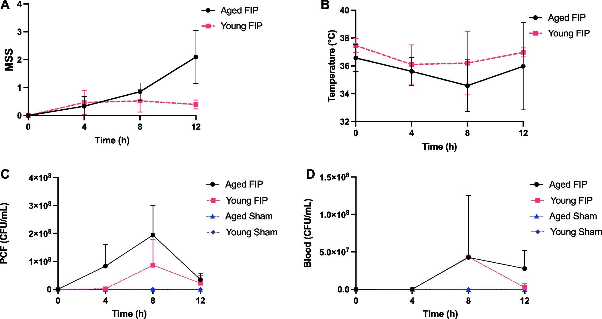
Modified murine sepsis score (MSS) was used to assess sepsis severity [16, 22]. The modified MSS involves observing posture, respiration quality, responsiveness, activity, and appearance [16, 22]. The MSS component scores were standardized to a four-point scale ranging from 0 (healthy) to 3 (sick) (Additional file 1: Table S1). Mice were humanely euthanized if their average MSS was ≥ 1.75, if any component of MSS was equal to 3, or if they reached the end of the study.
Temperature monitoring
We measured body temperature using contactless temperature chips that were subcutaneously inserted beneath the dorsal skin in the neck (United Information Devices, Lake Villa, Illinois, USA).
12 h and 72 h FIP studies
To investigate time-dependent changes in disease severity during the early phase of sepsis, young and aged FIP and sham mice were monitored and culled at the following timepoints: 4 h, 8 h, and 12 h (outlined in Additional file 1: Table S2). In a separate cohort of mice (FIP, sham, and naive), the animals were monitored for 72 h and were euthanized at humane endpoint or at study endpoint (outlined in Additional file 1: Table S2). Mice in all cohorts were randomly selected from their cages and randomly allocated to the experimental groups. Fluids, antibiotics, and analgesia were administered to the mice, consistent with standard practice for the treatment of human sepsis and current recommendations [14, 16]. For both studies, the mice were injected with 0.05 mg/kg of buprenorphine (subcutaneous) at 4 h. For the 72 h study, the mice received antibiotics (25 mg/kg of imipenem IP), Ringer’s lactate (15 mL/kg subcutaneous), and 0.05 mg/kg of buprenorphine at 12 h and every 12 h until study endpoint.
Collection of blood and peritoneal cavity fluid
At humane or experimental endpoint, mice were anesthetized with isoflurane and oxygen inhalation. Phosphate buffered saline (PBS) was injected into the peritoneal cavity and peritoneal cavity fluid (PCF) was collected. Blood was collected via the inferior vena cava into a one tenth volume of 3.2% buffered citrate. Plasma was prepared by centrifugation at 5000 × g for 10 min (twice), aliquoted, and stored at − 80 °C.
Quantification of bacterial cultures
At humane or experimental endpoint, bacterial loads were assessed in the PCF and blood. Ten-fold serial dilutions of PCF and blood in phosphate buffered saline were prepared until 10,000X dilution. Ten μL of each dilution starting from 10X were spotted in triplicate on 5% blood agar plates. Agar plates were placed at 37 °C overnight and colonies from the highest dilution were counted.
Quantification of lung myeloperoxidase, interleukin-6, interleukin-10, thrombin-antithrombin complexes, cell-free DNA, monocyte chemoattract protein (MCP-1)/CCL2, and ADAMTS13 activity
Levels of lung MPO were quantified using a mouse MPO DuoSet (R&D Systems, Minneapolis, Minnesota, USA). Plasma levels of Interleukin (IL)-6, IL-10, and monocyte chemoattract protein (MCP-1)/CCL2 were measured using the mouse IL-6 Duoset, the mouse IL-10 quantikine ELISA, and the Mouse CCL2/JE/MCP-1 DuoSet respectively, (R&D Systems, Minneapolis, Minnesota, USA). Plasma levels of thrombin antithrombin (TAT) complexes were quantified using the matched-pair antibody set (Affinity Biologicals, Ancaster, Ontario, Canada) according to the manufacturer’s protocol. CFDNA was quantified as per the manufacturer’s instructions using their Quant-iT™ PicoGreen™ assay (ThermoFisher Scientific, Waltham, Massachusetts, USA). ADAMTS13 activity was measured using FRETS-VWF73 (Anaspec Inc, Fremont, California, USA).
Organ histology
Histology was performed on the lung, liver, and kidneys. Organ sections were stained with hematoxylin and eosin (H&E). The sections were scored in a blinded fashion by two individuals, on a scale of 0 to 5 (0 = normal, 5 = severe) based on inflammation, thrombosis, and organ morphology. A composite score (organ damage) was calculated as a sum of the 3 categories (described in Additional file 1: Table S3).
Statistical analysis
Statistical analyses were performed using GraphPad Prism version 9.1.1 for macOS (GraphPad Software, San Diego, California, USA, www.graphpad.com). Values were expressed as mean ± standard deviation (SD) and p-values < 0.05 were considered significant. Significant differences between groups were determined using an ordinary one-way analysis of variance (ANOVA) or mixed-effects analysis. For the 72 h study, since there was only one mouse that survived in the aged FIP group, the aged FIP survivor group (n = 1) was not included in the statistical analysis. Survival curves were analyzed using a Log-rank (Mantel-Cox) test.


留言 (0)