Animal study and reagent
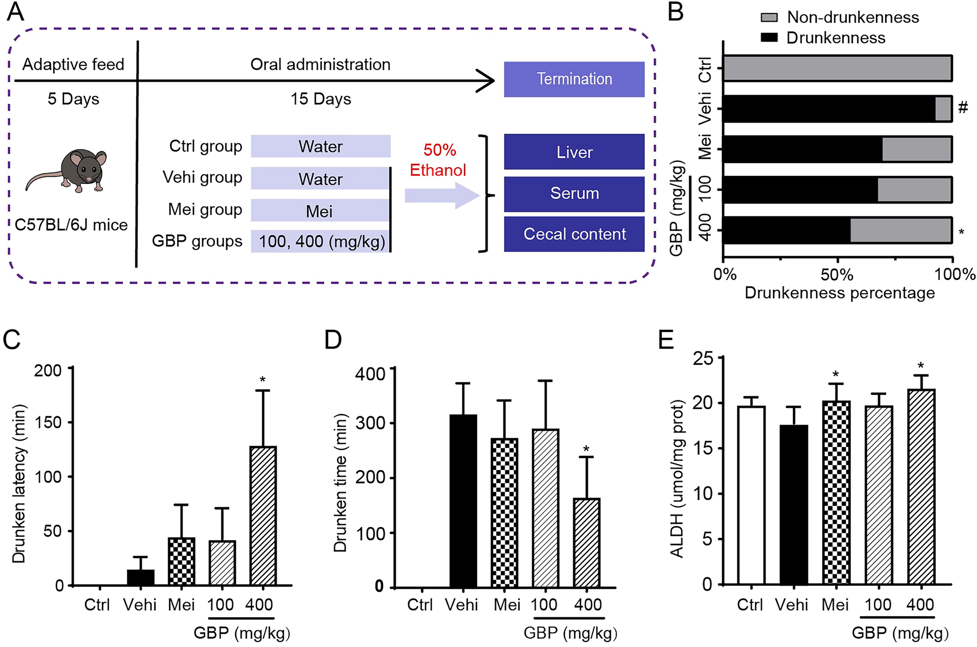
Male Sprague Dawley rats weighing 260–280 g were sourced from Zhejiang Vital River Laboratory Animal Technology. These rats were accommodated in a specific pathogen-free-grade animal cabin, ensuring unrestricted access to food and water. The housing conditions were meticulously controlled, maintaining a 12-h light/dark cycle, 60–80% humidity, and a temperature of 22 ± 1 °C. After acclimatization, OA was induced using the destabilization of the medial meniscus (DMM) method [15]. After anesthesia and disinfection, a surgical incision in the medial parapatellar skin revealed the knee joint. Then, the medial meniscotibial ligament was carefully transected. The joint was closed, and the rats were allowed to recover. Starting at the 7th day after surgery, intra-articular injections of either 100 µg or 200 µg TCG (CAS: 34209-69-3, Purity ≥ 98%, Tianzhi Biotechnology, Wuhan, China) or an equivalent volume of vehicle were administered twice a week for 8 weeks. The rats were sacrificed for further study. The animal experiments were conducted in compliance with the Guide for the Care and Use of Laboratory Animals (National Institutes of Health publication 85–23, revised 1996) and were approved by the Shanghai Jiao Tong University School of Medicine Animal Study Committee (2018-0027).
Histological analysis
The isolated joints were fixed in 4% paraformaldehyde (PFA) for 7 days, then decalcified, paraffin embedded, and sliced. The sections underwent staining using safranin O and fast green (S–O/FG), hematoxylin–eosin (HE), and immunohistochemical staining with anti-Collagen II antibody (Col2a1). OARSI cartilage histopathology assessment system was applied to evaluate the extent of articular cartilage damage [16]. The overall OARSI score, ranging from 0 to 24, was determined by multiplying OA grade (ranging from 0 to 6) and OA stage (ranging from 0 to 4).
Von Frey test
To assess pain sensitivity in rats, Von Frey hairs (Semmes–Weinstein Monofilaments, Galesville, USA) were used as previously reported [17]. Rats were acclimated to the testing environment for 20 min. Filaments with different forces were applied to the hind paw, starting from 0.4 g. Responses were recorded as "O" for negative and "X" for positive. The formula for calculating 50% paw withdrawal threshold (PWT) is “50% PWT = 10[Xf+kδ]/104”, in which Xf represent the value (in log units) of the last filament used, k is related to the response pattern (modified from Dixon), and δ is a constant (in log units) determined by standard deviation (SD) of serial force.
Micro-CT analysis
The isolated joints were fixed in 4% PFA for 7 days. Subsequently, a high-resolution micro-CT scanner (SkyScan-1176, Bruker, Belgium) was used to conduct quantitative analysis of knee joints. Morphometric parameters were calculated through 3D analysis using CTAn software (Bruker, Belgium). The visual 3D reconstruction was created by CTvox software (Bruker, Belgium). More importantly, all analyses were carried out in a blinded manner.
Isolation of mouse primary cells and reagent treatment
Bone marrow-derived macrophages (BMMs) were isolated from male C57/BL6 mice at the age of 4 to 6 weeks by collecting bone marrow cells from the femurs and tibias as previous reported [18]. The cells were incubated with α-modified minimal essential medium (α-MEM) complete medium with 1% penicillin/streptomycin (PS) and 10% fetal bovine serum (FBS). BMMs were induced into osteoclasts under the stimulation of 30 ng/mL macrophage-stimulating factor (M-CSF) and 50 ng/mL nuclear factor kappa-B ligand (RANKL).
Primary mouse chondrocytes (PMCs) were isolated from neonatal C57BL/6 mice knee cartilage within 5 days after birth as previous reported [19]. The harvested cells were incubated in Dulbecco’s Modified Eagle Medium/Nutrient Mixture F-12 (DMEM F12) with 1% PS and 10% FBS at 37 °C in a normal oxygen incubator with 20% oxygen or in a low oxygen incubator with 1% oxygen (Esco Lifesciences, Singapore).
8-pCPT-2′-O-Me-cAMP-AM (CAS: 1152197-23-3) used in this article to activate Ras association proximate 1, Rap1) were purchased from APExBIO (Huston, USA). 2-Deoxy-D-glucose (CAS: 154-17-6, 2-DG) and LW6 (CAS: 934593-90-5) used to treat PMCs were obtained from MedChemExpress (Monmouth Junction, USA).
TRAP staining and Phalloidin staining
BMMs were induced to form osteoclasts while being treated with indicated doses of biotin, TCG, or TCG-biotin. Osteoclasts were stained using the leukocyte acid phosphatase (TRAP) staining kit (Sigma-Aldrich Inc., St. Louis, USA) following the manufacturer's instructions. Under the microscope, cells exhibiting a burgundy color and possessing multiple nuclei (three or more) were considered as osteoclasts for inclusion in the analysis. To visualize F-actin, the cells were incubated with rhodamine phalloidin (2 units/mL) for 30 min away from light. 4,6-diamidino-2-phenylindole (DAPI, 1 μg/mL) was used to stain nuclei. The F-actin ring was observed as red ring under fluorescence microscope.
Bone resorption assay
To investigate the impact of TCG on the osteoclast bone resorption function, BMMs were induced into osteoclasts in a collagen-coated dish. Subsequently, the cells were gently transferred onto bone slices and treated with a specified dose of TCG for 48 h. As previously described, bone resorption pits were assessed using a confocal laser scanning microscope [20].
CCK-8 assay
BMMs and PMCs were exposed to indicated doses of TCG for a specified period of time. Following treatment with CCK-8 reagent, cell viability was assessed by measuring the optical density at 450 nm using the Infinite F200 PRO absorbance microplate reader. The calculated cell viability was normalized to the control.
qRT-PCR analysis
RNA extraction reagent (Vazyme Biotech, Nanjing, China) was used to extract the total RNA from the sample of interest. The concentration and purity of RNA were measured using a NanoDrop spectrophotometer. Then, PrimeScript Reverse TranscriptMasterMix (TaKaRa, Otsu, Japan) was employed to conduct reverse transcription. Subsequently, QuantStudio Real-Time PCR system (Thermo Fisher, Waltham, USA) was utilized to perform qRT-PCR. Details of Primer sequences are listed in Additional file 2: Table S1. The relative mRNA expression was assessed using the ΔΔCT method.
Western blot and immunofluorescence assay
Total protein lysates were collected. Lysates were then subjected to SDS-PAGE and subsequently transferred to polyvinylidene fluoride membranes. After 1-h of membrane blocking, the membrane was then incubated overnight at 4 °C with primary antibodies (Additional file 2: Table S2). Next day, after a 1-h incubation with secondary antibodies at room temperature, chemiluminescence reagents and eBlot Touch Imager (eBlot, Shanghai, China) were utilized to detect protein bands.
For immunofluorescence assays, cells were fixed, permeabilized, and blocked. Subsequently, the cells were incubated overnight at 4 °C with p65 antibodies (1:400 dilution). Following this, cells were incubated for 1 h at room temperature with Alexa Fluor 488-conjugated antibodies. Images were captured using a Zeiss LSM880 confocal microscope.
RNA sequencing analysis
The total RNA from each sample was prepared and subjected to quality control using the Agilent bioanalyzer 2100 (Agilent, Santa Clara, USA). Subsequently, the RNA is converted into a cDNA library using NEBNext Ultra RNA Library Prep Kit (Illumina, San Diego, USA). High-throughput sequencing was performed on an Illumina HiSeq platform (Illumina, San Diego, USA). Bioinformatics analysis including read trimming, filtering, and mapping to a reference genome was then conducted on the raw data by using HISAT2 tools. Further functional annotation and pathway analysis, utilizing resources like Kyoto Encyclopedia of Genes, Genomes (KEGG) and Gene Ontology (GO) and gene set enrichment analysis (GSEA), provide a comprehensive understanding of the biological significance of the identified genes. Differentially expressed genes were identified by setting a threshold at log2FC ≥ 1, FDR < 0.01 in TCG-treated BMMs, or log2FC ≥ 0.5, p-value < 0.05 in TCG-treated PMCs.
Active Rap1 detection assay
To isolate active GTP-bound Rap1 (Rap1-GTP), we utilized the Rap1 Activation Assay Kit (Cell Biolabs, San Diego, USA) in accordance with the manufacturer's guidelines. Subsequently, SDS-PAGE analysis was performed on both pulldown and input samples to identify the presence of Rap1-GTP.
Small molecule pull-down
The chemical synthesis process of biotin-linked TCG TCG-biotin) is shown in Additional file 2: Table S3. A pull-down experiment was performed coupled with mass spectrometry (MS) analysis to identify TCG-bound proteins. Briefly, lysates from BMMs were incubated with either biotin beads (Thermo Fisher, Waltham, USA) or TCG-biotin beads for 4 h at 4 °C. The proteins attached to the beads were subsequently isolated through SDS-PAGE. The gel's protein-containing band was meticulously excised, and in-gel digestion was performed. The resulting peptides underwent liquid chromatography-tandem mass spectrometry (LC–MS/MS) analysis for the identification of proteins.
Virtual docking
Crystal structure of human Rap1b (PDB ID: 3X1W, chain A) was used for molecular docking study. The chain A of 3X1W was preprocessed by Protein Preparation Wizard. The ligand TCG was prepared by LigPrep and then docked into the protein by Induced Fit Docking. The resulting 3D structures of the ligand–protein complexes and the corresponding 2D ligand–protein interaction diagrams were visualized using Maestro.
EdU assay
Cells were exposed to EdU for a 2-h duration at 37 °C. After fixed and permeabilized, a click reaction solution was applied for a 30-min staining period. Subsequently, cells were counterstained with Hoechst 33342 and observed using fluorescence microscopy to quantify EdU-positive cells and determine the percentage of proliferating cells.
Flow cytometry
Flow cytometry was employed to detect PMCs apoptosis. PMCs were washed and centrifuged to achieve a single-cell suspension. This suspension was incubated with Annexin V to identify phosphatidylserine externalization. Propidium iodide, a nuclear dye, was added to distinguish apoptotic cells from necrotic cells. The samples were analyzed using a Thermo Fisher flow cytometer (Waltham, USA), and the fluorescence signals intensity and distribution were measured to quantitatively assess the extent of apoptosis.
Measurement of extracellular acidification rate (ECAR)
For Seahorse extracellular flux analysis, the measurement of the ECAR was carried out as follows: Cells were seeded onto Seahorse assay plates and allowed to adhere. Then, the assay plates were loaded into the Seahorse XF Analyzer (Seahorse Biosciences, North Billerica, USA). During the experiment, a sequence of reagents was sequentially loaded including 10 mM glucose, 2 μM oligomycin, and 0.1 M 2-DG. The Seahorse XF Analyzer continuously monitors the changes in ECAR in real-time, allowing for the dynamic observation of cellular responses to metabolic perturbations.
Lactate measurements
Lactate levels were quantified following the manufacturer's guidelines. Cells were seeded in 96-well plates and exposed to TCG for 24 h. Lactate levels in the culture medium were determined using the L-lactate assay kit (Eton Biosciences, Union, USA). The measurements were normalized to the total protein content in each well.
Statistical analysis
The results were presented as mean ± SD. Statistical evaluations included independent t-tests and one-way ANOVA for datasets conforming to a normal distribution. A significance threshold of p < 0.05 was deemed statistically significant. Statistical analyses were conducted using SPSS 20.0 (SPSS, Inc., Chicago, USA) and GraphPad Prism 9.0 (GraphPad Software Inc., La Jolla, USA).


留言 (0)