
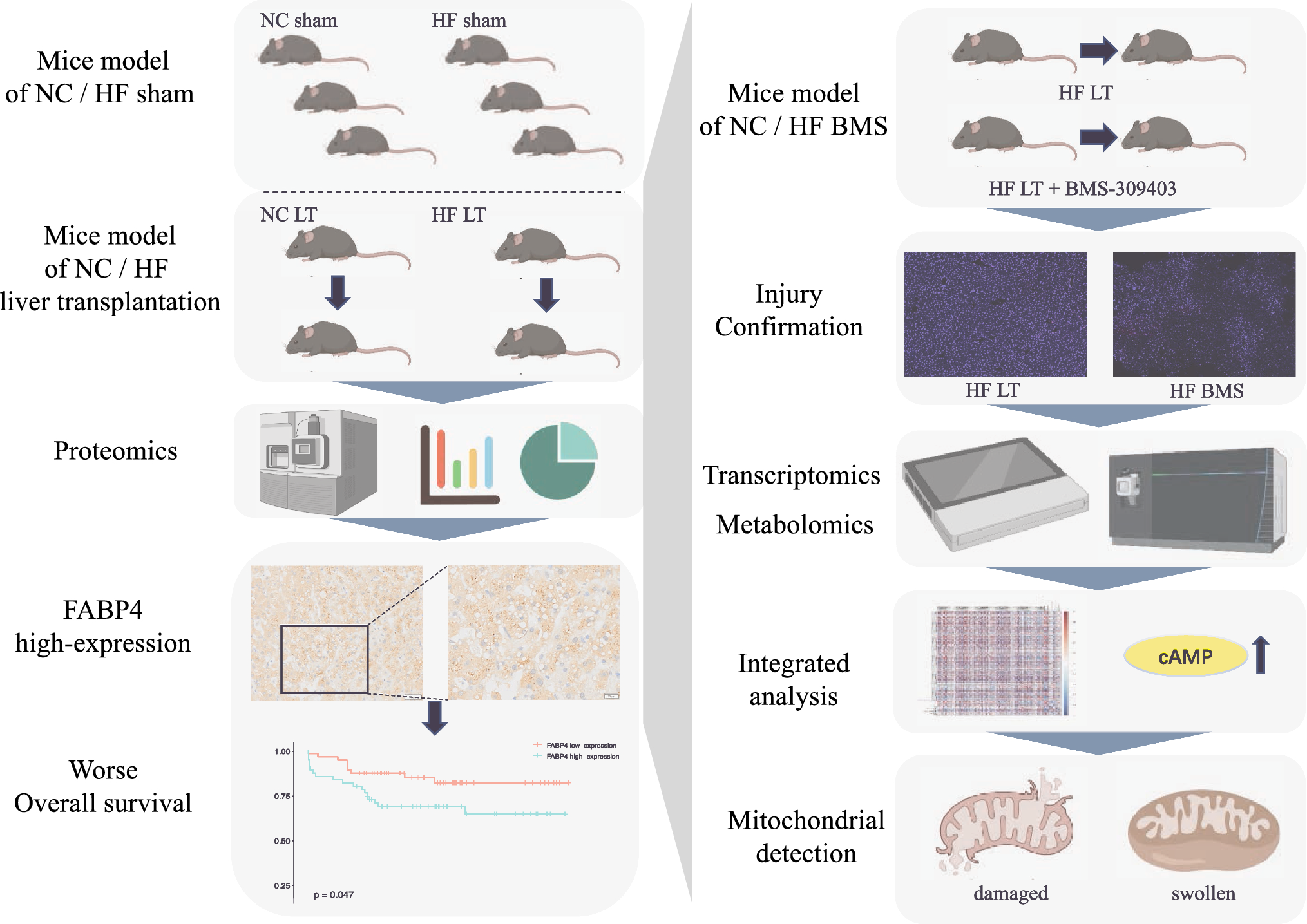
記住我
Donor mice were fed different diets for eight weeks and transplanted into normal mice. Oil Red O staining was used to identify the success of fatty liver development (Fig. 1A). Hepatic steatosis in the HF LT group was 30–60% under the microscope, and no hepatic steatosis was observed in the NC LT group. H&E staining displayed considerable inflammatory cell infiltration and fat vacuoles in the HF LT group (Fig. 1B). TUNEL staining was performed, and more apoptotic liver cells were found in the HF LT group than in the NC LT group (Fig. 1C and D).
Fig. 1
Establishment and validation of mouse liver transplantation model. A Oil Red O staining of mouse liver transplantation model. B H&E staining. C, D TUNEL staining. E–H Liver function detection of ALT, AST, LDH, and TB. I–L Relative expression of Bax, cleaved Caspase-3, and cleaved PARP. ns, not significant; *P < 0.05; **P < 0.01; ***P < 0.001; ****P < 0.001
Liver function analysis demonstrated that serum ALT, AST, and LDH levels were significantly higher in the HF LT than in the NC LT group (1970 ± 550 vs. 1420 ± 221 U/L, P < 0.05; 1849 ± 349 vs. 1235 ± 194 U/L, P < 0.01; 6796 ± 1739 vs. 4918 ± 1029 U/L, P < 0.05; Fig. 1E–G). The relative expression of Bax, cleaved Caspase-3, and cleaved PARP was significantly higher in the HF LT than in the NC LT group (P < 0.01, P < 0.05, P < 0.001, respectively; Fig. 1J–L). All above results demonstrated severe hepatocellular damage in the HF LT group.
The corresponding NC and HF SHAM groups detection was conducted, including Oil Red O, H&E, and TUNEL staining (Supplementary Fig. 1).
Proteomic profile revealing the change of FABP4 during liver transplantationProteomics was adopted among the four groups, including NC SHAM, HF SHAM, NC LT, and HF LT. A total of 5131 quantifiable proteins were identified, and the differentially expressed proteins were revealed in a heatmap (Fig. 2A). Principal component analysis (PCA) depicted that duplicate samples were statistically consistent. The clustering degree between samples was quite dense, and circles representing the NC LT and HF LT groups were far from the SHAM circles and each other (Fig. 2B). The fold change and P-values were considered while screening for differentially expressed proteins. After screening, 168 proteins were identified between NC SHAM and HF SHAM groups (Fig. 2C), 297 proteins were identified between NC LT and HF LT groups (Fig. 2D), 2,241 proteins were identified between NC SHAM and NC LT groups, and 1994 proteins were identified between HF SHAM and HF LT groups. Shared and discrete proteins were identified among these groups (Fig. 2E).
Fig. 2
Proteomic profile revealing the change during liver transplantation. A Heatmap of the differentially expressed proteins. Red rectangles mean that proteins are upregulated, and green ones mean that they are downregulated. B Principal component analysis of the duplicate samples, in which the degree of aggregation among samples represents statistical consistency. C Volcano plot of the differentially expressed proteins between NC SHAM and HF SHAM groups. Gray dots represent genes that are not differentially expressed; red dots and blue dots represent genes that are upregulated and downregulated significantly. D Volcano plot of the differentially expressed proteins between NC LT and HF LT groups. E Venn diagram demonstrating shared and discrete proteins in each of these four groups. F Expression patterns of these differentially expressed proteins based on membership and expression. G Protein–protein interaction network of differentially expressed proteins with FABP4
The molecular function of the GO pathway indicated that fatty acid elongase activity, fatty acid synthase activity, and oxidoreductase activity were separated by enrichment analysis between NC SHAM and HF SHAM groups. In contrast, the KEGG pathway analysis revealed that ferroptosis and fatty acid elongation were screened. Compared with NC LT and HF LT groups, cellular components revealed that the mitochondrial protein complex, mitochondrial inner membrane, inner mitochondrial membrane protein complex, and endoplasmic reticulum were screened. KEGG pathway analysis revealed that the inflammatory mediator regulation of TRP channels was screened. Comparing HF SHAM and HF LT groups, biological processes found that the apoptotic signaling pathway, extrinsic apoptotic signaling pathway, and mitochondrial transmembrane transport were analyzed. The cellular components revealed that ribosomes, mitochondrial respirasomes, respiratory chain complexes, and mitochondrial protein complexes were screened. KEGG pathway analysis revealed autophagy and apoptosis in both groups. Clustering was used to assess the expression patterns of differentially expressed proteins based on their membership and expression (Fig. 2F). Clustering analysis of the four groups revealed significant changes in the expression profiles of the ribosomes, proteasomes, endocytosis, and oxidative phosphorylation pathways (Supplementary Fig. 2).
After analyzing the differently expressed proteins among all comparison groups, fatty acid binding protein 4 (FABP4), which also interacts with fatty acid metabolism and synthesis, was screened for changes in each pair of these four groups. The differential protein–protein interaction relationship was obtained using the String database, and the protein–protein interaction network was visually displayed (Fig. 2G).
Relationship with FABP4 expression and liver transplantation recipients’ overall survivalA total of 110 LT liver tissue microarrays of donors were sectioned, and FABP4 IHC staining was performed. The clinical characteristics of donors and recipients, including prognostic follow-up, were collected. The histochemistry score was calculated for each sample. FABP4’s high expression (Fig. 3A) and low expression (Fig. 3B) were determined based on the median histochemistry score. A total of 55 FABP4 high- and 55 FABP4 low-expression cases were enrolled.
Fig. 3
Relationship with FABP4 expression and liver transplantation recipients’ overall survival. A FABP4 is a high expression of donors’ liver tissue microarray IHC staining. B FABP4 low expression of IHC staining. C Comparison of overall survival rate between FABP4 high- and low-expression groups. D Comparison of the incidence of early allograft dysfunction. E Comparison of the risk of liver steatosis
A survival curve was plotted based on FABP4 expression. The 1-year overall survival rate of the FABP4 high-expression group was 68.5%, which was significantly worse than that of the FABP4 low-expression group (68.5 vs. 87.3%, P < 0.05, Fig. 3C). The incidence of early allograft dysfunction (EAD) was relatively higher in recipients in the FABP4 high-expression group (54.5 vs. 25.5%, P < 0.01, Fig. 3D). In addition, donor grafts in the FABP4 high-expression group were often accompanied by liver steatosis (27.3 vs. 9.1%, P < 0.05, Fig. 3E).
Influence of FABP4 inhibitor on mouse high fatty liver transplantation modelOne hour before donor liver procurement, the FABP4 inhibitor BMS-309403 was administered. H&E staining displayed that the liver had many fat vacuoles, a moderate decrease in inflammatory cell infiltration, and a considerable decrease in apoptotic hepatocytes in HF BMS compared with HF LT (Fig. 4A). A small amount of hepatocyte rupture and nuclear lysis was observed in the NC BMS group than in the NC LT group. TUNEL staining revealed a significant decrease in the number of apoptotic hepatocytes after BMS-309403 treatment (Fig. 4B).
Fig. 4
Influence of FABP4 inhibitor on mouse high fatty liver transplantation model. A H&E staining of mouse liver transplantation model after using FABP4 inhibitor. B TUNEL staining. C–F Liver function detection of ALT, AST, LDH, and TB. G–I Oxidative stress injury assay of GSH, MDA, and SOD. J–N Relative expression of FABP4, Bax, cleaved Caspase-3, and cleaved PARP. ns, not significant; *P < 0.05; **P < 0.01; ***P < 0.001; ****P < 0.001
Liver function detection revealed that serum ALT, AST, and LDH were significantly higher in the HF LT group than in the NC LT group (P < 0.01, P < 0.001, P < 0.05, respectively; Fig. 4C–E). These three indicators and TB decreased significantly after the adoption of BMS-309403 compared to the HF BMS and HF LT groups (P < 0.05, P < 0.05, P < 0.05, and P < 0.0001). However, this trend was not significant compared with NC BMS and NC LT groups (P > 0.05, P < 0.05, P > 0.05, and P > 0.05). The adoption of BMS-309403 could improve the liver function indicators in the HF BMS group compared to those in the NC LT group (P > 0.05, P > 0.05, P > 0.05, P < 0.05).
After BMS-309403 was used, the oxidative stress damage tests demonstrated substantial variations. The values of GSH and SOD decreased, and MDA levels increased in the HF BMS group compared with those in the HF LT group (P < 0.01, P < 0.01, P < 0.01, respectively; Fig. 4G–I). This trend was also observed when comparing NC BMS and NC LT groups (P < 0.05, P < 0.01, and P < 0.05). The relative expression of FABP4, Bax, cleaved Caspase-3, and cleaved PARP has decreased significantly in the HF BMS group compared to the HF LT group (P < 0.05, P < 0.01, P < 0.01, and P < 0.01, Fig. 4K–N). Correspondingly, the relative expression of these proteins was also lower in the NC BMS group than in the NC LT group (P < 0.01, P < 0.01, P < 0.01, and P < 0.05).
Transcriptomic profiles revealing the influence of FABP4 inhibitorTranscriptomics was used in HF LT and HF BMS groups. As displayed in Fig. 5A, all genes were moderately expressed. PCA revealed that duplicate samples conformed to statistical consistency (Fig. 5B). In total, 289 quantifiable genes were identified. The differentially expressed genes are shown in a heatmap (Fig. 5C), where 153 upregulated and 136 downregulated genes were identified and are depicted in a volcano map (Fig. 5D).
Fig. 5
Transcriptomic profiles revealing the influence of FABP4 inhibitor. A Violin diagram of expression patterns of all genes. B Principal component analysis of the duplicate samples. C Heatmap of the differentially expressed genes. D Volcano plot of the differentially expressed genes between HF LT and HF BMS groups. E Gene ontology pathway enrichment for differentially expressed proteins. The circle sizes represent the number of genes enriched in pathways, and the circle’s color means significance. F KEGG pathway enrichment for differentially expressed proteins
Immune response, defensive response, and stimulus regulation were screened by enrichment analysis of biological processes (Fig. 5E). Cellular components such as phagocytic vesicles, early phagosomes, and endoplasmic reticulum membranes were screened. Molecular function analysis revealed that ion binding and cation binding were screened. KEGG pathway analysis revealed Fc γ R-mediated phagocytosis, and phagosomes were screened (Fig. 5F). The pathway enrichment analysis is shown in Supplementary Fig. 3.
Metabolomic profiles revealing the influence of FABP4 inhibitorMetabolomics was also adopted to compare HF LT and HF BMS groups. The total ion chromatogram spectral overlap comparison displayed overlapping response intensities and retention times (Fig. 6A). PCA revealed that close clustering of the quality control samples indicated good repeatability of the experiment (Fig. 6B). A total of 159 upregulated and 105 downregulated metabolites were identified, as demonstrated in the volcano map (Fig. 6C). Cluster analysis of the differential metabolites demonstrated similar expression patterns for metabolites in the same cluster (Fig. 6D).
Fig. 6
Metabolomic profiles revealing the influence of FABP4 inhibitor. A Total ion chromatogram of spectral overlap comparison, with response intensity and retention time overlapping. B Principal component analysis of the duplicate samples. C Volcano plot of the differentially expressed metabolites between HF LT and HF BMS groups. D Heatmap of the differentially expressed metabolites. E Correlation analysis between metabolites and visualized in the form of correlation heatmaps. F Revealing the co-regulatory relationships between various metabolites by chord diagrams
Metabolic proximities were analyzed to further understand the mutual regulatory relationships between metabolites during the biological state (Fig. 6E). A chord diagram of metabolic proximities also revealed a correlation between the various metabolites (Fig. 6F). The lipids and lipid-like compounds were tested for organic acids and their derivatives. Only the VEGF and cAMP signaling pathways were evaluated between the two groups by the KEGG pathway.
Integrated transcriptomic and metabolomic analysisVisualizing gene and metabolite expression is important for interpreting high-throughput omics experiments to understand how IR injury in steatotic LT is initiated. Gene–metabolite interaction analysis was used to study and depict differential gene–metabolite interactions. Transcriptomics screened 216 pathways, and metabolomics screened 24 pathways together (Fig. 7A); 12 pathways were found to be involved in both genomes (Fig. 7B). Aldosterone synthesis and secretion, steroid hormone biosynthesis, and cAMP signaling pathways have more molecules annotated in the two groups for the biological pathway.
Fig. 7
Integrated transcriptomic and metabolomic analysis. A KEGG pathways that transcriptomic and metabolomic respectively enriched. B KEGG pathways that transcriptomic and metabolomic simultaneously enriched. C Spearman’s correlation hierarchical clustering analysis of differences in the expression patterns. D Correlation network analysis of significant differences in key node locations
Spearman correlation hierarchical clustering was conducted on genes and metabolites with substantial differences to intuitively represent expression patterns (Fig. 7C). Different genes or metabolites that appear in the same cluster have similar expression patterns. Correlation network analysis was performed to screen for genes and metabolites with significant differences in key node locations (Fig. 7D). The metabolite 6-hydroxycoumarin was associated with six genes, two positively and four negatively correlated.
Influence of FABP4 inhibitor on cAMP signaling pathwayTranscriptomic profiles revealed that 6 genes were differently expressed in cAMP signaling pathway, including 5 upregulated (Hhip, Adrb2, Rac2, Adcy7, and Rapgef4) and 1 downregulated (Grin3b). The metabolomic profiles revealed 2 upregulated metabolites (Prostaglandin i2 and N-oleoylethanolamine). Western blot assay revealed that the relative expression of Hhip, Adrb2, Rac2, and protein kinase A (PKA) was significantly higher in the HF BMS than in the HF LT group (P < 0.05, P < 0.001, P < 0.05, P < 0.05, respectively; Fig. 8B–E). The content of metabolites of two groups was also validated by ELISA. Prostaglandin i2 and N-oleoylethanolamine increased more than twofold after FABP4 inhibitors treatment (P < 0.01, P < 0.05; Fig. 8F, G). Moreover, the mRNA expression of Hhip, Adrb2, Rac2, and Adcy7 was elevated in HF BMS group, indicating activation of the cAMP signaling pathway (P < 0.01, P < 0.05, P < 0.01, P < 0.05, respectively; Fig. 8H–K). The adoption of FABP4 inhibitors might activate the cAMP signaling pathway.
Fig. 8
Influence of FABP4 inhibitor on cAMP signaling pathway. A–E Relative expression of HHIP, ADRB2, RAC2, and PKA. F, G The content of Prostaglandin i2 and N-oleoylethanolamine validated by ELISA. H–K The mRNA expression of Hhip, Adrb2, Rac2, and Adcy7 of HF LT and HF BMS groups. ns, not significant; *P < 0.05; **P < 0.01; ***P < 0.001; ****P < 0.001
Influence of FABP4 inhibitor on the high fatty liver mitochondrionTEM was used to observe the mitochondrial morphology of the mouse liver. There was no edema in the hepatocytes, and intracellular organelles were abundant in the HF SHAM group (Fig. 8A). The bile duct structure was in good condition. The nucleus was round, and the nuclear membrane was complete. Mitochondria are ovoid, shallow, and without bulging, fractured, or shortened cristae. Lipid droplets were scattered throughout the distribution. There were more autophagosomes and autolysosomes in the HF LT group (Fig. 8B), mitochondrial swelling, unclear structure, and partial disintegration of the ridge. The HF BMS group had an obvious mitochondrial structure, and the injured and healthy mitochondria were lytic, suggesting autophagy (Fig. 8C).
Mitochondrial membrane potential was detected using multiplex immunofluorescence staining (Fig. 8D–F). The staining results displayed a larger area of mitochondrial membrane potential loss in the HF LT group than in the HF SHAM group regarding mitochondrial membrane homeostasis. Membrane depolarization still existed but depicted a significant easing trend after adopting BMS-309403 in the HF BMS group. Compared to the HF LT group, the TUNEL fluorescence intensity in the HF BMS group was also weakened.
Compared to HF LT, DRP1 expression decreased considerably in HF BMS (P < 0.01, Fig. 8H). The relative expression of MFN-1 in the HF BMS group displayed an increasing trend but was insignificant (Fig. 9).
Fig. 9
Influence of FABP4 inhibitor on high fatty liver mitochondrion. A–C Transmission electron microscopy image of mouse liver in each group. A The mitochondrial structure was clear, and no autophagosomes were presented in the HF SHAM group. B Autophagosomes and autolysosomes were found in the HF LT group with mitochondrial swelling. C The mitochondrial structure of the HF BMS group was clear, and damaged parts of mitochondria were lytic. D–F Multiplex immunofluorescence staining of rhodamine reagent for detecting mitochondrial membrane potential. Green fluorescence represents mitochondrial membranous potential, red fluorescence represents apoptotic hepatocytes, and blue fluorescence represents nuclear staining. G–I Relative expression of DRP1 and MFN-1. ns, not significant; *P < 0.05; **P < 0.01; ***P < 0.001; ****P < 0.001. AP, autophagosomes; ASS, autolysosome; LD, lipid droplets; M, mitochondria; RER, rough endoplasmic reticulum
Influence of FABP4 siRNA on in vitro hypoxia / reoxygenation modelFlow cytometry was performed to detect the hypoxic injury-induced apoptosis of AML12 cells after the cells were stained with Annexin V / propidium iodide. As shown in Fig. 10, the apoptosis rate from the HF group (Fig. 10C) increased significantly compared with that of the NC group (Fig. 10A), while the HF siRNA group (Fig. 10D) had a lower apoptosis rate than the HF group. The supernatant of each group was collected for detecting ALT, AST, LDH, and TB. The levels of ALT, AST, and LDH were significantly higher in the HF group than in the NC group, which were significantly increased in HF siRNA group (P < 0.05, P < 0.01, P < 0.05, respectively; Fig. 4E–G). Thus, adoption of FABP4 siRNA in steatotic murine AML12 hepatocytes could protect from the hypoxia/reoxygenation injury.
Fig. 10
Influence of FABP4 siRNA on in vitro hypoxia / reoxygenation model. A–D Flow cytometry was performed to detect the hypoxic injury-induced apoptosis of AML12 cells. E–H Liver function detection of ALT, AST, LDH, and TB using the supernatant of each group. I–P Relative expression of FABP4, PKA, RAC2, HHIP, ADRB, DRP1, and MFN-1. ns, not significant; *P < 0.05; **P < 0.01; ***P < 0.001; ****P < 0.001
Western blot assays were applied to demonstrate the activation of cAMP signaling pathway. The relative expression of PKA, Rac2, and Hhip was significantly higher in the HF siRNA group than in the HF group (P < 0.05, P < 0.05, P < 0.05, respectively; Response Fig. 4K–M). The relative expression of Adrb2 was also higher but not significant in the HF siRNA group than in the HF group (P > 0.05, Fig. 4N). Compared to HF group, DRP1 expression decreased considerably in HF siRNA group (P < 0.05, Response Fig. 4O). The relative expression of MFN-1 in the HF siRNA group displayed an increasing trend (P < 0.05, Fig. 4P), indicating the modulating mitochondrial membrane homeostasis role of FABP4 siRNA, which probably owing to the activation of cAMP signaling pathway.
留言 (0)