
Remember me
The initial search identified 1251 publications. Twenty-three of these met the eligibility criteria for the pooled analysis, including 1153 patients treated by distraction osteogenesis [8, 12, 14, 15, 22,23,24,25,26,27,28, 31,32,33,34,35,36,37,38,39,40,41,42]. Details of each study are shown in Table 1. Extracted data from each study and pooled analysis are shown in by docking site intervention group in Table 2 (planned intervention) [12, 14, 15, 23, 25,26,27,28, 31,32,33] and Table 3 (observed) [8, 24, 33, 38,39,40,41,42]. Where patients from a single study had different docking site protocols, these were included in the relevant group. In total, there were 407 patients in the planned intervention group and 746 in the observed group. In the planned intervention group, 368 of 407 (90%) docking sites went on to union without further intervention, compared to 497 of 746 (66%) in the observed group. This result was statistically significant (chi-squared p < 0.0001). Included within these failures at the docking site were 7 refractures in the planned intervention group [12, 28, 32, 33] compared with 21 in the observation group [23, 24, 27, 32,33,34, 41] (not significant, p = 0.28). For those studies reporting the outcome, more surgical procedures overall (including planned and unplanned) were required to achieve union in the planned intervention group than the observation group (mean 3.8 in 160 patients from four studies vs. mean 2.2 in 333 patients from 10 studies). The proportion of patients in whom union was ultimately achieved was similar between the two groups, being 402 of 407 (99%) in the planned procedure group vs. 737 of 746 (99%) in the observed group (chi-squared p =0.612). Overall treatment time is difficult to estimate between groups without access to the raw data. Calculating an average based upon the data as reported in each paper, where this was available, and the number of patients treated reveals that this is similar at 383 days (275 patients) in the planned intervention group and 393 days (642 patients) in the observed group. These results were highly variable between studies due to their heterogenous nature, and the methodology of pooled analysis is weak; these results should be viewed with caution.
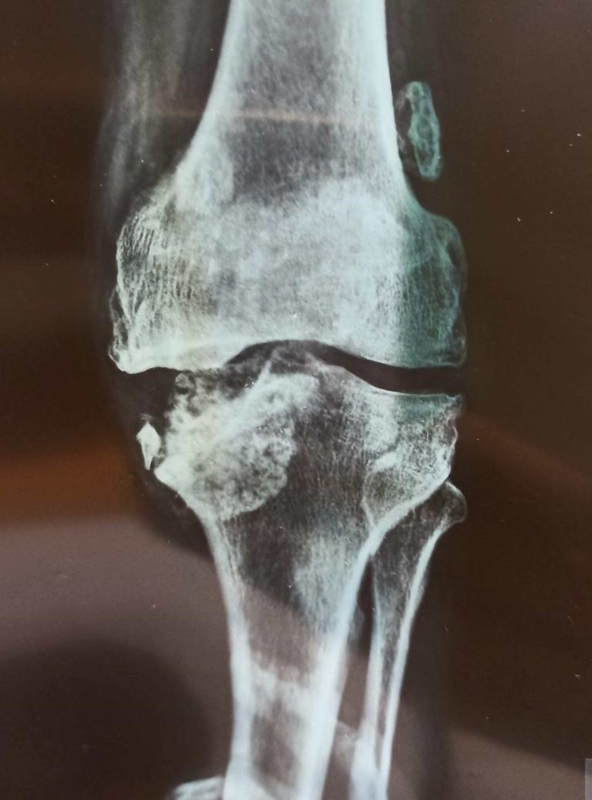
Table 1 Summary of papers included in the pooled analysisTable 2 Patient outcome and pooled analysis for those treated by protocols including routine docking site procedures (planned intervention group)Table 3 Patient outcome and pooled analysis for those treated by protocols including initial period of observation with intervention only if spontaneous docking site union did not occur (observed group)Other factors associated with docking site outcomeThe heterogenous nature of the studies and problems with reporting made it difficult to draw conclusions regarding what type of docking procedure is most effective. All studies in the planned procedure group included refreshing the bone ends in their operative protocol for the docking site intervention, though some undertook this using minimally invasive techniques [25, 27]. Many used autologous bone graft, though this was used selectively within studies, making any attempt to understand the effectiveness of this intervention compared to others prone to selection bias. In several studies, it was not possible to determine the outcome of different patients based upon which intervention was applied, as this was not part of the main research question and therefore not reported. Considering studies where it was possible to reliably extract relevant data, in patients where the bone graft was applied, union occurred without further intervention in 90 of 105 cases (86%) compared with 58 of 59 cases (98%) where this was not used (Fisher’s exact test p < 0.01). For the reasons stated above, this should be interpreted with caution. We present a case of docking site non-union in a patient treated for infected non-union of the femur managed by bone grafting in Fig. 3.
Fig. 3
Docking site non-union in patient treated for infected non-union of femur managed by bone grafting. A Initial radiographs following removal of femoral nail and debridement (left). A monolateral external fixator is applied for bifocal bone transport with partial shortening (right). B Standing alignment films show transport and re-lengthening is complete. C Distraction segment has consolidated but docking site appears to only have tenuous union at best. Simple compression has been applied follow by compression and distraction (accordian technique). CT confirms docking site non-union. D Patient undergoes docking site procedure with freshening of the bone ends and application of autologous bone graft along with bone marrow aspirate concentrate and bioactive glass graft expander. E At 4 months post grafting, the docking site appears to have united, and the fixator is removed. F Radiographs at 12 months post fixator removal show union and remodelling of the docking site
Due to the nature of the intervention, all patients in the planned intervention group were treated by bone transport, whereas some in the observed group underwent acute shortening and re-lengthening (ASR) [33, 35, 37, 38, 41]. Outcomes were therefore compared only for those patients treated without routine docking site intervention (observed group) between those managed by ASR or bone transport. In papers, where it was possible to reliably extract data, the uncomplicated docking site union rate in patients treated with ASR was 96 of 128 (75%) compared to 420 of 618 (68%) managed by bone transport. This difference was not statistically significant (chi-squared p = 0.11).
Some studies managed patients by bifocal and some by trifocal bone transport. In several studies, a mix of these techniques was used, and in several, it was not possible to determine outcomes by this factor due to the nature of reporting. In the remaining papers, docking site union was compared by this factor in the observed group, where it likely has the most impact. The rate of uncomplicated docking site union was 209 of 280 in those managed by bifocal transport (75%) compared to 31 of 37 managed by trifocal transport (84%). This result was not statistically significant (Fisher’s exact p = 0.31), though due to the issues stated this is difficult to interpret and the numbers small.
Two studies in the observed group reported the use of either poly-methyl methacrylate or calcium sulphate bone cement within the defect (54 patients) [22, 37]. The rationale for this is to induce a biologically active membrane to assist union and regenerate formation. These patients had a higher spontaneous docking site union rate when compared with the remaining 692 patients from the observed group (45 of 54 (83%) vs. 420 of 692 (61%), chi-squared test p = 0.0009). Again, given the heterogenous nature of the studies, this result should be viewed with caution.
Results of narrative reviewDocking site union is likely to occur by a combination of endochondral and intramembranous ossification, and the exact mechanism remains unclear [7]. As for bone healing in other situations, the interaction of host biology, local and systemic and mechanics will influence this. The following factors, relevant to the surgical technique, are thought likely to be particularly important in docking site union [7, 9, 24, 29].
(1)Bone contact: Greater contact area between bone surfaces will increase the probability of union and decrease rates of refracture. Bone contact is influenced by the geometry of the bone cuts at debridement and the accuracy of the bone transport device in maintaining alignment. We present a case of docking site refracture managed non-operatively in Fig. 4.
(2)Infection: Unresolved infection at a docking site will affect local biology and is likely to be detrimental to union. Appropriate debridement and antimicrobial strategies likely influence this.
(3)Vascularity: Sufficient blood supply is critical to bone healing. Thorough debridement of necrotic bone is likely to be important in achieving vascularized bony surfaces to facilitate healing.
(4)Mechanics: Alignment and stability are important in generating appropriate mechanical environments for bony healing. This factor is affected by the stabilisation technique employed for the docking site and will also be influenced by the degree of contact.
Fig. 4
Docking site refracture managed non-operatively. A Patient has apparently successfully completed treatment for a Gustilo and Anderson IIIB open tibial fracture with bone loss. Note consolidated proximal transport segment and mid-diaphyseal docking site which appears united after simple compression. B Radiographs early after frame removal appear to show a united tibia. C Patient stumbles and suffers a low energy fracture. Radiographs reveal a minimally displaced refracture at the docking site. D A plaster cast is applied and wedged to restore alignment. E Spontaneous union of the refracture occurs over 4 months. Note that the docking site has better union than prior to the refracture
Docking site healing is substantially different from fracture healing. In all circumstances, the local biology at the docking site will have been significantly damaged by trauma, surgery and in some cases infection. This will result in decreased local blood supply, deleterious to healing. The haematoma and inflammation, which constitute the first stage of secondary bone healing, are missing in cases of bone transport because docking occurs several weeks after the injury [43]. This results in the formation of a fibrous connective cap, again believed to have an adverse effect on bone healing. Though the bone ends are fashioned so that there should be good coaptation on contact, this is difficult to achieve, and during the transport process, bone resorption often occurs, worsening contact further [17]. The lack of fracture haematoma at docking will exacerbate this problem, potentially making gap healing less likely to occur. Even in cases of acute shortening, surgical intervention removes this haematoma to a significant extent, and the damage to the local blood supply may still result in bone resorption and problems with bone contact, with similar results [44].
Docking site operations are not without the potential for complication. The most commonly reported are local infections, soft tissue problems, and donor site morbidity [15, 25, 26]. Undertaking open surgical procedures within the confines of a circular frame can be challenging and raises issues around the maintenance of a sterile field. This leads to concerns over deep infection threatening the ultimate outcome. Docking procedures also do not guarantee union. The studies considered in the pooled analysis report rates of further docking site problems requiring intervention 0 to 32% (10% overall) in the routine intervention group. This should be considered when consenting patients for these procedures.
Location of the defectThe majority of included studies deal with tibial bone loss [8, 12, 14, 15, 22,23,24,25,26,27,28, 31,32,33,34,35, 37,38,
Comments (0)