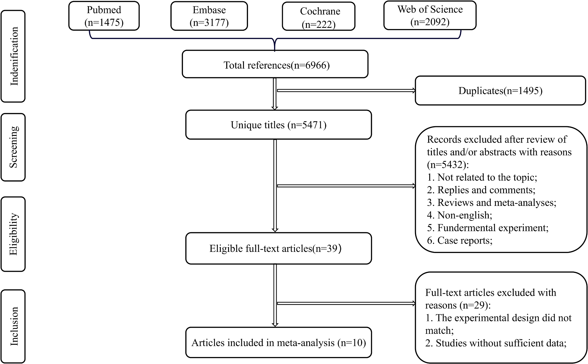
In this cross-sectional study, male patients presented more back pain and leg pain compared to female patients in the clinical presentation and presented smaller Cobb’s angle, less apex vertebrae rotation, larger LL, smaller PI-LL and lower paravertebral muscle FIR when compared to female cohorts in radiological characteristics, and we speculate that the difference of these parameters between male and female patients may be of potential etiological significance for the DLS.
A large body of the literature clearly demonstrates that men and women respond differently to pain, with increased pain sensitivity and risk for clinical pain commonly being observed among women [10]. In the present study, male DLS patients, however, showed more severe low back pain and leg pain than female patients. Based on the findings of Nakamae et al [11]. that low back pain due to DLS is not directly related to the patient’s age or the severity of scoliosis, we believe that a study by Robinson and colleagues [12] gives a more plausible explanation. Since women are more sensitive to pain than men, men are more reluctant to report pain. Therefore, in the present cross-sectional study, unlike female patients, male patients with DLS opted for hospitalization only when they experienced more severe pain.
The natural progression of degenerative scoliosis with age is inevitable. A study of 51 patients with early degenerative lumbar scoliosis followed for 13.7 years found that the patients’ scoliosis curve progressed by an average of 0.4° to 1.4 °per year [13]. The mean curve progression over 5 years was reported to average 3 degrees per year in 73% of patients, Grade 3 apical rotation, a Cobb angle ≥30 degrees, lateral vertebral translation ≥6 mm and prominence of L5 in relation to the intercrest line were important factors for predicting curve progression [14]. In the current cross-sectional study, however, male DLS patients were relatively older than female patients but had a smaller Cobb angle compared to the female cohort. We propose that the effect of gender differences on scoliosis progression is much greater than the effect of natural progression. As a cross-sectional study, we strictly followed the inclusion and exclusion criteria to screen patients within 10 years of our institutional case base and still obtained results that male patients were on average older than female patients. One possible reason for this is that the prevalence of DLS is greater in female than in male patients, and the male sample size results in an unavoidable selection bias. However, another more plausible explanation is that the age of onset of DLS may be greater in male patients than in females. According to Xu et al.’s study [5], osteopenia, gender of female, and aged older than 65 years could contribute to the presence of DLS. In this study, the BMD (L1 vertebral HU value) of male subjects was not significantly different from female subjects and the mean value was smaller in female subjects. This interpretation is also supported by the fact that the Cobb angle was significantly smaller in male subjects than in female subjects. And another possible explanation for the larger Cobb angle in female subjects than male subjects is that gender differences in muscle atrophy play a role. One possible explanation is that gender differences in muscle atrophy play a role. Studies have shown that aging affects muscle function more in women than in men, and that skeletal muscle attachment quality is lower in older women compared to men [15]. It is generally accepted that the paraspinal muscles are the dynamic stabilizers of the spine column [16]. Research by both Crawford et al. [17] and Urrutia et al. [18] showed that fat infiltration of the paraspinal muscles was greater in females than in males in all age groups. A study by Yagi et al. [19] concluded that there is a causal relationship between paraspinal muscle degeneration and the development of DLS. In the present study, the paraspinal muscles of male patients were significantly less degenerated than those of female patients. We believe that this may be one of the reasons for the different severity of scoliosis in male and female DLS patients. The third possible reason is that the dramatic decrease in estrogen production in women after menopause also plays a role. It is now generally accepted that disk degeneration is the initiating factor in DLS [20]. Our team has long been engaged in research on the correlation between estrogen and intervertebral disk degeneration and found that estrogen has an excellent ability to inhibit the apoptosis of intervertebral disk cells and revealed the related mechanism of action [21, 22]. The female patients in this cross-sectional study were of postmenopausal age, and the rapid decline in estrogen secretion led to the loss of estrogen protection of the disk cells, which accelerated the degeneration of the disks and thus the progression of DLS, which may be another reason for the severity of scoliosis in the female patients compared with the male patients.
Different studies have reached varied conclusions regarding the correlation between apex vertebrae rotation and DLS progression. Pritchett et al. [14] and Kohno et al. [23] suggest that apex vertebrae rotation is a predictor of curve progression. In contrast, Park et al.’s research [13] found no significant correlation between apex vertebrae rotation and scoliotic angle in the early stages of DLS. In this study, we found a significant correlation between apical vertebrae rotation and coronal Cobb angle in both males and females. Combined with previous studies, we believe that the apex vertebrae rotation does not precede the appearance of scoliosis, but occurs with the progression of scoliosis.
Pelvic incidence (PI), first described by Duval Beaupere, is unique for skeletal mature individuals and is not affected by posture [24]. Since PI is the sum of sacral slope (SS) and pelvic tilt (PT), it is a valid descriptor of the global shape of the pelvis and the position of the sacrum in the pelvic unit [25]. Pelvic incidence minus lumbar lordosis (PI-LL) was used to quantify the mismatch between pelvic morphology and lumbar curve [26, 27]. In this study, there was no difference in PI values between male and female DLS patients, LL was greater in male patients than in female patients, and PI-LL was less in male patients than in female patients according to geometric calculations. In addition, the PI-LL values of male and female patients were positively correlated with their Cobb angles. Han et al. [28] found significantly lower LL values in DLS patients compared to normal subjects in a comparative study. A prospective study by Jimbo et al. [29] concluded that the decrease in LL is a result of the progression of lumbar degeneration and leads to further deformities. These studies all support our conclusion that it is precisely because female patients exhibit a more severe scoliosis angle than male patients, which in turn results in smaller LL values, and larger PI-LL values. Furthermore, the PI-LL mismatch was higher in female than male, and ODI showed no statistical difference according to sex. This findings suggested that sex-based difference can impact manifestation of DLS. It also strengthens our conclusion that low back pain was more pronounced in male patients and scoliosis was more severe in female patients.
In previous research on the pathogenesis of DLS patients, male and female patients were usually analyzed as a whole. Our current research shows that the clinical manifestations of male and female DLS patients are not identical. We believe that the influence of gender should be considered in future research on the pathogenesis of DLS patients.
We recognize that the current study still has limitations. As it was a retrospective study, we could not provide strong evidence of causality for the various differences between male and female DLS patients. However, as a cross-sectional study, we can still demonstrate that gender factors play a role in the development of scoliosis in DLS. In addition, our study was a single-center study, and the patients were all from the north of China, and whether the findings can be generalized to other ethnic groups still needs to be confirmed by further multi-center large-sample studies.


Comments (0)