
記住我
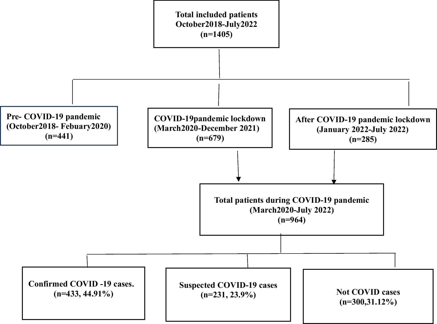


Fifty-nine patients (31 women), aged 75.7 ± 12.0 years, were enrolled. Twenty-nine were still alive 12 months after ICH. As far as non-survivors are concerned, 28/30 (93.3%) succumbed within the first 30 days after ICH. In detail, the leading causes of death were arrhythmia (11/30; 36.7%), infection/sepsis (8/30; 26.7%), and cardiovascular disease (7/30; 23.3%), followed by respiratory failure (2/30; 6.7%) and recurrent hemorrhage (2/30; 6.7%). A flow diagram is provided as Fig. 1.
Fig. 1
Flow diagram of the study
Characteristics of patients and detailed comparisons between 12-month survivors and non-survivors are presented in Table 1. Of note, 4/59 patients (6.8%) presented thrombocytopenia due to infection/sepsis (1 case), liver cirrhosis (1 case), myelodysplastic syndrome (1 case), and bone marrow infiltration from metastatic lung cancer (1 case)". No patient received platelets, fresh frozen plasma, or any other treatment for thrombocytopenia.
Table 1 Patients’ characteristics and univariate analysis based on 12-month survival statusUnivariate analysis demonstrated that younger age (P = 0.001), absence of diabetes mellitus (P = 0.013), absence of arterial hypertension (P = 0.019), elevated Hb (P = 0.019), elevated glucose (P = 0.021), and increased sPO2 at admission (P = 0.023) were correlated with survival (Table 1). Moreover, increased PMI2 (P = 0.020) and PLT at day 2 (P = 0.017) were correlated with survival (Table 2).
Table 2 Platelet number and indices (measured at admission, day 2, and day 7), as well as univariate and multivariate analysis based on 12-month survival statusAiming to elucidate whether PMI values might be used as early predictors of survival, repeated measures General Linear Model was used. In that model, PMI1 and PMI2 were independently correlated with survival (P = 0.048 and P = 0.004, respectively), while PMI7 was not (P = 0.332), after adjustment for age, diabetes mellitus, arterial hypertension, Hb, and sPO2 at admission (Table 2, Fig. 2). Glucose was initially excluded from adjustment due to collinearity issues attributed to diabetes mellitus.
Fig. 2
Estimated Marginal Means of 12-month survival at mean of covariates (age, hypertension, diabetes, Hb, and sPO2) for PMI1 (day 1; admission), PMI2 (day 2), and PMI7 (day 7) using Repeated Measures General Linear Model
To further investigate the contribution of additional potential confounders to within-samples variability concerning the consecutive measurements of PLT and PMI, Repeated Measures GLM multivariate models based on 12-month survival status adjusted for age, diabetes, hypertension, Hb, and sPO2 were performed (Table 3). These models suggested that hyperlipidemia, glucose, lactates, and temperature, but not gender, history of coronary artery disease (CAD), antiplatelets, and anticoagulants, might constitute true confounders. PMI2 were still independently correlated with survival (P = 0.012) after adjustment for these additional confounders (Table 2).
Table 3 PLT and PMI (measured at admission, day 2, and day 7) multivariate analysis based on 12-month survival status additionally adjusted for age, diabetes, hypertension, Hb, and sPO2 using Repeated Measures GLM: Contribution of potential confounders to within-samples variabilityBinary discretization of PMI2, after adjustment for age, diabetes mellitus, arterial hypertension, Hb, and sPO2 at admission, suggested 2,400 fL/μL as cut-off (Fig. 3). Using binary regression, PMI2 ≥ 2,400 fL/μL was independently correlated with survival (OR 0.123; 95% CI: 0.022–0.694; P = 0.018), after adjustment for age (OR 1.872 per decade; 95% CI: 0.959–3.655; P = 0.066), diabetes mellitus (OR 3.527; 95% CI: 0.231–53.910; P = 0.365), arterial hypertension (OR 9.837; 95% CI: 1.318–73.426; P = 0.026), Hb (OR 0.395 per g/dL; 95% CI: 0.187–0.834; P = 0.015), and sPO2 at admission (OR 0.664 per %; 95% CI: 0.434–1.016; P = 0.059) (Fig. 4).
Fig. 3
Ridge paths determining the best cut-off for PMI2 (2,400 fL/μL)
Fig. 4
Forest plot depicting binary regression model for PLT2 ≥ 2,400 fL/μL; OR < 1 favors 12-month survival
PLT values, when measured at day 2, were also independently correlated with survival (P = 0.017); however, PLT values, when measured at admission and day 7, failed to demonstrate prognostic value (P = 0.069 and P = 0.460, respectively). Moreover, MPV, PDW, and P-LCR had no prognostic value (Table 2).
Of note, PLT2 (r = − 0.312; P = 0.022), and PMI2 (r = − 0.285; P = 0.038) were negatively correlated with mRS. Moreover, these parameters presented a weaker, yet significant negative correlation with ICH score (PLT2: r = − 0.278; P = 0.042), and PMI2: r = − 0.275; P = 0.046) (Fig. 5A–D).
Fig. 5
Scatterplots depicting correlations between A PLT2 with MRs (left column; upper row), B PMI2 with MRs (right column; upper row), C PLT2 with ICH score (left column; lower row), and D PMI2 with ICH score (right column; lower row)
留言 (0)