Bone of dermal origin is considered to be phylogenetically older than bone that forms the endoskeleton as no mineralized tissue forming the endoskeleton has been identified in early vertebrates. (Donoghue and Sansom, 2002, Sire et al., 2009). The earliest evidence of osteocytic bone in an early vertebrate appears concurrently with anosteocytic bone in the fossil record (Smith and Sansom, 1997, Witten et al., 2004), but subsequent to the earliest reported anosteocytic bone (Smith et al., 1996). Furthermore, the presence of acellular bone, dentine and enameloid has been shown to be present in the dermal skeleton of the earliest verterbrates (Bystrow, 1959, Denison, 1967, Donoghue et al., 2000, Keating et al., 2015, Keating et al., 2018, Moss, 1968a, Moss, 1968b, Ørvig, 1989, Sansom et al., 2005, Schaeffer et al., 1977). A close association between the evolution of bone and dentin is likely as they are found together in most dermal skeleton elements of early vertebrates (Donoghue et al., 2000, Sansom et al., 2005, Sweet and Donoghue, 2001). Over evolutionary time, in most extant lineages, bone, dentin and enameloid, which were components of various dermal skeletons of early vertebrates, was secondarily reduced or lost (Sire and Kawasaki, 2012). These are fascinating insights into the early evolution of vertebrate mineralized tissues.
These observations, together with many more such observations of the dermal tissues from modern (Lees et al., 2012, Leprevost et al., 2017, Meunier and Brito, 2004, Sire and Huysseune, 1993, Yang et al., 2014), and fossil fish (Giles et al., 2013, Keating et al., 2015, Meunier and Brito, 2004, Sansom et al., 2005, Smith et al., 1996), are however almost entirely based on two-dimensional (2D) observations using light and electron microscopy to examine thin sections. For the last almost two decades it has been possible to obtain 3-dimensional (3D) data at very high resolution (around 10nms) and in volumes of thousands of microns cubed, using a focused ion beam-scanning electron microscope (FIB-SEM). FIB-SEM provides consecutive slices with a thickness of 8–10 nm which form a volume of continuous data. At this resolution the characteristic repeating structure of individual collagen fibrils with their characteristic 67 nm D-banding, can be visualized. This in turn makes it possible to reconstruct the organization of the basic building block of vertebrate mineralized tissues, namely the mineralized collagen fibril bundle. In this study we use mainly structural information obtained from FIB-SEM to revisit aspects of the plasticity of mineralized tissues that possibly contributed to their evolution by studying the dermal scales of the sturgeon.
The approach we use is to integrate the very detailed high-resolution structural information from the FIB-SEM digital image volumes into the tissue as a whole by showing the broader contexts from which the volumes were obtained. Analyzing the 3D organization of the mineralized collagen fibril bundles (CFBs) within the FIB-SEM digital image volumes is also challenging. Here we use not only observations of the characteristic collagen banding patterns, but we also take advantage of the fact that in almost all vertebrate mineralized tissues, the extent of mineralization of the CFBs is incomplete. This results in the fibril bundles having foci of well mineralized ellipsoids (now termed tesselles (McKee et al., 2022), that are surrounded by less mineralized fibrils. Less mineralized fibrils can be identified digitally in FIB-SEM images using a so-called eigenvalue approach based on the characteristic 50–70 nm diameter of a fibril (Ibrahim et al., 2023). In this approach the less mineralized fibrils are segmented, and their orientation characterized by an ellipsoid which yields three eigenvectors. The largest eigenvector shows the primary orientation of the less mineralized fibrils, and the orientation of these fibrils is extrapolated to define the orientation of the entire fibril bundle. Interpretation of CFBs within the image volume is achieved by generating an eigenvector 3D volume by mapping the largest eigenvectors in 3D, and by superimposing the eigenvector 3D volume on the FIB-SEM digital image volume (Ibrahim et al., 2023). We note that FIB-SEM has been used to study the 3D structural organizational motifs within lamellar bone (Raguin et al., 2021, Reznikov et al., 2014, Reznikov et al., 2013), fibrolamellar bone (Magal et al., 2014, Raguin et al., 2021), woven bone (Ibrahim et al., 2023), and in several mineralized tissues from fish (Atkins et al., 2015, Raguin et al., 2020). The latter do indeed expand our understanding of the diversity of mineralized tissue structures that are known mostly from mammals. At present no studies have been performed on non-teleost ray-finned fishes, and future studies looking at other hard tissues of fish, such as isopedin, are required for proper comparison.
Sturgeon is the common name for 29 species of fish in the family Acipenseridae, which diverged from the line leading to present day fish about 385–200 million years ago (Dillman and Hilton, 2015, Near et al., 2012, Patterson, 1982). Sturgeons have a well-developed dermal skeleton with 5 distinct rows of scutes, the largest of the calcified elements in the skin, running the length of the body. The small structures embedded in the skin between the rows of scutes have been called scales or denticles (Jollie, 1980, Sewertzoff, 2005, Sire et al., 2009, Weisel, 1978), however, the criteria used to name these structures is unclear. Scales of the sturgeon have been divided into 5 groups (including scutes) based on anatomical location, with the smallest scales of the body described as the fundamental unit due to their ability to group together to form larger scales (Findeis, 1993).
Scales are mineralized elements embedded in the skin of fish (the integumentary skeleton)(Kresja, 1979, Moss, 1972, Zylberberg et al., 1992) which together with bones of the skull, pectoral girdle, pectoral fin, teeth, and tooth-like elements of the oral and pharyngeal cavities, form the dermal skeleton (Sire et al., 2009). The dermal skeleton of fish thus encompasses a large variety of structures (Francillon-Vieillot et al., 1989, Huysseune and Sire, 1998, Sire and Huysseune, 2003, Zylberberg et al., 1992), formed from a wide array of mineralized tissues which differ in their basic structure (Meunier and Huysseune, 1992, Moss, 1960, Moss, 1961, Moss, 1963, Moyle and Cech, 2004, Weiss and Watabe, 1979). In general, it is thought that scales are composed of bone with a scale-specific covering such as cosmine in cosmoid scales and ganoine in ganoid scales (Benthon, 2004, Moyle and Cech, 2004, Zhu et al., 2012). However, the basal bone and scale-specific material may be highly reduced or absent. Fish scales have been classified into cosmoid scales, elasmoid scales, ganoid scales, leptoid scales (cycloid and ctenoid), and placoid scales, however, structures of scales are diverse and complex and at present there is no consensus as to their nomenclature and classification (Donoghue, 2002, Schultze, 2018, Sire et al., 2009). A detailed understanding of the structures of scales, especially in 3D, may provide important insights into the plasticity of mineralized tissues that possibly contributed to the evolution of vertebrate mineralized tissues.
From an evolutionary perspective sturgeons are an early branching lineage of ray-finned fishes which preserve some primitive characteristics, and belong to the Order Acipensiforms, a distinctive group of fish (Dillman and Hilton, 2015, Near et al., 2012, Patterson, 1982), having undergone very little morphological, molecular and genetic change over time (Du et al., 2020, Gardiner, 1984, Krieger and Fuerst, 2002). It is noteworthy that scales distributed between rows of scutes have been described in the skin of fossilized sturgeons (Grande and Hilton, 2006, Hilton and Grande, 2023, Sato et al., 2018). While the scales of fossilized sturgeons show some superficial similarities to extant sturgeons, nothing is known about the material from which they are formed. It is therefore with these interesting considerations in mind, that we chose to study the structure of a modern mineralized dermal tissue, the scale of the sturgeon.
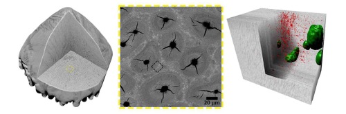
Isolated scales of the body of the sturgeon vary in size from 0.4 mm − 2.0 mm. Scales develop from the front to the back of the body after the development of the scutes, and while they cover a large part of the body, each is surrounded by skin (Sewertzoff, 2005). The scales on the dorsal part of the body are larger and have a higher density with smaller spaces between individual scales compared to the scales on ventral part of the body. Scales on the ventral part of the body are rhomboid and aligned in oblique rows, with bigger spaces between them (Weisel, 1978). Fifteen shapes of scales have been described. however, a round or spade shaped basal plate supporting 1–5 caudally directed protrusions or points is common to all (Sewertzoff, 2005). The base is not visible as it is buried beneath the surface of the skin. However, the points penetrate and extend above the surface of the skin where they are visible (Jollie, 1980, Sewertzoff, 2005, Weisel, 1978).
The aim of this study is to characterize the 3D structures comprising the bulk material(s) of sturgeon scales at the CFB level in order to determine if this material contains organizational motifs that are homologous to motifs in other known vertebrate mineralized tissues. We also characterize the scales at lower resolution in order to better understand their tissue level organization.


留言 (0)