Bacterial isolation and identification
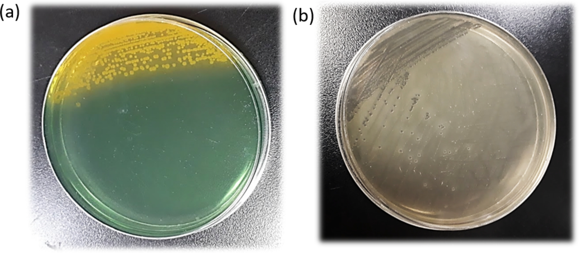
V. vulnificus was isolated from the patient’s blood sample using a blood culture on modified cellobiose-polymyxin B-colistin (mCPC) agar medium and purified on blood plate agar medium. V. vulnificus was identified using Matrix-Assisted Laser Desorption/Ionization-Time Of Flight Mass Spectrometry (MALDI-TOF MS) and COMPACT VITEK2 and named vv16015.
Ethics statement
All animal experiments were approved by the Clinical and Research Ethics Committee of Shenzhen Centre for Diease Control and Prevention.
Antimicrobial susceptibility testing (AST)
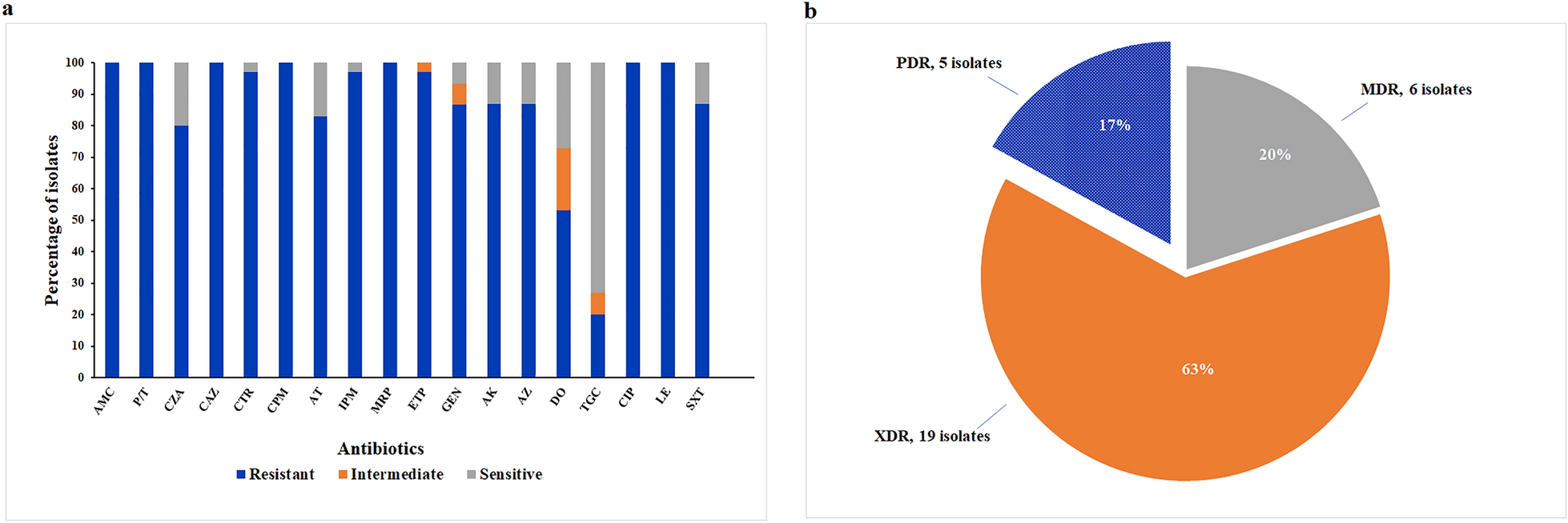
Susceptibility of vv16015 to 21 antibiotics studied using the VITEK 2 AST-GN09 antimicrobial susceptibility board: amikacin, ampicillin, ampicillin/sulbactam, aztreonam, cefazolin, cefepime, cefotetan, ceftazidime, ceftriaxone, cefuroxime, ciprofloxacin, gentamicin, imipenem, levofloxacin, meropenem, nitrofurantoin, piperacillin, piperacillin/tazobactam, tobramycin, trimethoprim/sulfamethox, and tetracycline. The results were automatically interpreted by the VITEK 2-COMPACT fully automated microbiological analyser, using E. coli ATCC25922 as the quality control strain, according to the recommendations of the Clinical and Laboratory Standards Institute document M100-S30 [14].
Genomic DNA extraction and bioinformatics analysis
Genomic DNA was extracted using the QIAamp DNA Mini Kit and whole genome sequencing was performed by BGI (Shenzhen, China). Raw data were filtered using Trimmomatic v0.39 [15], SPAdes v3.9.1 [16] was used to perform de-novo genome assembly. Kraken2 (https://ccb.jhu.edu/software/kraken2/) was used to identify V. vulnificus, Prokka 1.14.6 [17] was used for gene annotation. The vv16015 carries virulence genes identified through the VFDB online database (http://www.mgc.ac.cn/VFs/) and resistance genes identified through CARD online database (https://card.mcmaster.ca/).
From the National Center for Biotechnology Information (NCBI), we downloaded the genome sequences of 133 V. vulnificus strains isolated from eight countries between 1993 and 2022. To show the evolutionary relationships between vv16015 and the 133 other V. vulnificus strains, a maximum-likelihood phylogeny was constructed, with CMCP6 as the reference genome as it was considered to be the most complete and accurate of the published V. vulnificus clinical strain genomes. Snippy Pipeline v4.6.0 was used to identify core genomic single nucleotide polymorphisms (core-SNPs), then Maximum-likelihood phylogeny was constructed using fasttree and visualised and annotated in iTOL.
Galleria mellonella virulence assay
The Galleria mellonella larvae model is widely used to study the pathogenic potential of Vibrio as it is easily survivable, simple, inexpensive and more ethically acceptable than mammalian infection models [18]. We established a G. mellonella larvae model to study the virulence of V. vulnificus vv16015, vv15018 and vv220015 were selected for experimental comparison and sourced from the Shenzhen Centre for Disease Control and Prevention strain bank. Vv15018 was isolated from a clinical patient with no specific disease history and discharged from hospital in good condition after an acute gastroenteritis caused by a poor diet. Vv22015 was isolated from an oysters and represents an environmental strain that was used as the experimental control because its virulence genes are approximately the same as those of vv16015 and vv15018.
Sixth instar G. mellonella larvae were 2-3 cm long with a mean weight of 0.33 g (Tianjin Huiyude Biotechnology Company Limited) and selected for vigour and absence of black spots. There were seven groups each with ten larvae. One group remained untreated controls (group C1) and six groups were injected with 10µL of control or bacterial suspension on the last right-side pair of hind feet. Group C2 received 0.9% sterile saline, T1 received 1 × 10⁴ CFU/mL, T2 1 × 10⁵ CFU/mL, T3 1 × 10⁶ CFU/mL, T4 1 × 10⁷ CFU/mL and T5 1 × 10⁸ CFU/mL. After injection G. mellonella larvae were placed in separate sterile disposable petri dishes and incubated in a constant temperature incubator at 37 °C for 48 h and survival curves were plotted for G. mellonella larvae. Survivorship was recorded every 6 h.
Statistical analysis
All statistical analyses were performed using GraphPad Prism 9.5.0 software. Larval survival were plotted by the Kaplan-Meier method. Log-rank (Mantel-Cox) test was used to compare the significance of differences in larval survival rates between groups. P < 0.05 was considered to indicate significant differences between groups.


Comments (0)