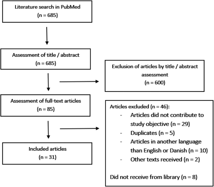
Yurac R, Matamala JM, Zamorano JJ, Harrop JS, Davies BM, Nouri A. et al. Degenerative cervical myelopathy. Rev Med Chil. 2022;150:339–52.
Article
PubMed
Google Scholar
Nouri A, Tessitore E, Molliqaj G, Meling T, Schaller K, Nakashima H, et al. Degenerative cervical myelopathy: development and natural history [AO Spine RECODE-DCM Research Priority Number 2]. Global Spine J. 2022;12:39S.
Badhiwala JH, Ahuja CS, Akbar MA, Witiw CD, Nassiri F, Furlan JC. et al. Degenerative cervical myelopathy - update and future directions. Nat Rev Neurol. 2020;16:108–24.
Article
PubMed
Google Scholar
Nouri A, Martin AR, Mikulis D, Fehlings MG Magnetic resonance imaging assessment of degenerative cervical myelopathy: a review of structural changes and measurement techniques. Neurosurg Focus. 2016 [cited 2022 Nov 29];40. Available from: https://pubmed.ncbi.nlm.nih.gov/27246488/.
Kim TH, Ha Y, Shin JJ, Cho YE, Lee JH, Cho WH Signal intensity ratio on magnetic resonance imaging as a prognostic factor in patients with cervical compressive myelopathy. Medicine (United States). 2016 [cited 2022 Dec 7];95. Available from: https://journals.lww.com/md-journal/Fulltext/2016/09270/Signal_intensity_ratio_on_magnetic_resonance.6.aspx.
Ponticorvo S, Manara R, Russillo MC, Erro R, Picillo M, Di Salle G, et al. Magnetic resonance T1w/T2w ratio and voxel-based morphometry in multiple system atrophy. Sci Rep. 2021 Dec 1 [cited 2022 Nov 29];11.
Sasiadek MJ, Szewczyk P, Bladowska J Application of diffusion tensor imaging (DTI) in pathological changes of the spinal cord. Med Sci Monit [Internet]. 2012 [cited 2022 Nov 29];18. Available from: https://pubmed.ncbi.nlm.nih.gov/22648262/.
Liu H, MacMillian EL, Jutzeler CR, Ljungberg E, MacKay AL, Kolind SH, et al. Assessing structure and function of myelin in cervical spondylotic myelopathy: evidence of demyelination. Neurology. 2017;89:602.
Article
CAS
PubMed
PubMed Central
Google Scholar
Martin AR, De Leener B, Cohen-Adad J, Cadotte DW, Kalsi-Ryan S, Lange SF. et al. A Novel MRI Biomarker of Spinal Cord White Matter Injury: T2*-weighted white matter to gray matter signal intensity ratio. AJNR Am J Neuroradiol. 2017;38:1266–73.
Article
CAS
PubMed
PubMed Central
Google Scholar
He B, Sheldrick K, Das A, Diwan A. Clinical and research MRI techniques for assessing spinal cord integrity in degenerative cervical myelopathy: a scoping review. Biomedicines. 2022;10:2621.
Article
CAS
PubMed
PubMed Central
Google Scholar
Yang HE, Kim WT, Kim DH, Kim SW, Yoo WK, Yang HE. et al. Utility of diffusion and magnetization transfer MRI in cervical spondylotic myelopathy: a pilot study. Diagnostics. 2022;12:2090.
Article
PubMed
PubMed Central
Google Scholar
Glasser MF, van Essen DC. Mapping human cortical areas in vivo based on myelin content as revealed by T1- and T2-weighted MRI. J Neurosci. 2011;31:11597.
Article
CAS
PubMed
PubMed Central
Google Scholar
De Leener B, Lévy S, Dupont SM, Fonov VS, Stikov N, Louis Collins D, et al. SCT: Spinal Cord Toolbox, an open-source software for processing spinal cord MRI data. Neuroimage. 2017;145:24–43.
Article
PubMed
Google Scholar
Ganzetti M, Wenderoth N, Mantini D. Whole brain myelin mapping using T1- and T2-weighted MR imaging data. Front Hum Neurosci. 2014;8:671.
Article
PubMed
PubMed Central
Google Scholar
Hannoun S, Kocevar G, Codjia P, Barile B, Cotton F, Durand-Dubief F. et al. T1/T2 ratio: a quantitative sensitive marker of brain tissue integrity in multiple sclerosis. J Neuroimag. 2022;32:328–36.
Article
Google Scholar
Pareto D, Garcia-Vidal A, Alberich M, Auger C, Montalban X, Tintoré M. et al. Ratio of T1-weighted to T2-weighted signal intensity as a measure of tissue integrity: comparison with magnetization transfer ratio in patients with multiple sclerosis. Am J Neuroradiol. 2020;41:461–3.
Article
CAS
PubMed
PubMed Central
Google Scholar
Fischl B. FreeSurfer. Neuroimage. 2012;62:774.
Article
PubMed
Google Scholar
Martin AR, de Leener B, Cohen-Adad J, Cadotte DW, Kalsi-Ryan S, Lange SF, et al. Clinically feasible microstructural MRI to quantify cervical spinal cord tissue injury using DTI, MT, and T2*-weighted imaging: assessment of normative data and reliability. AJNR Am J Neuroradiol. 2017;38:1257–65.
Article
CAS
PubMed
PubMed Central
Google Scholar
Cohen-Adad J, Alonso-Ortiz E, Abramovic M, Arneitz C, Atcheson N, Barlow L, et al. Open-access quantitative MRI data of the spinal cord and reproducibility across participants, sites and manufacturers. Scientific Data 2021 8:1 [Internet]. 2021 Aug 16 [cited 2022 Dec 1];8:1–17. Available from: https://www.nature.com/articles/s41597-021-00941-8.
Teraguchi M, Yamada H, Yoshida M, Nakayama Y, Kondo T, Ito H. et al. Contrast enrichment of spinal cord MR imaging using a ratio of T1-weighted and T2-weighted signals. J Magn Reson Imaging. 2014;40:1199–207.
Article
PubMed
Google Scholar
Bansal R, Hao X, Liu F, Xu D, Liu J, Peterson BS. The effects of changing water content, relaxation times, and tissue contrast on tissue segmentation and measures of cortical anatomy in MR images. Magn Reson Imaging [Internet]. 2013;31:1709.
Article
CAS
PubMed
Google Scholar
Boaventura M, Sastre-Garriga J, Garcia-Vidal A, Vidal-Jordana A, Quartana D, Carvajal R, et al. T1/T2-weighted ratio in multiple sclerosis: a longitudinal study with clinical associations. 2022.


留言 (0)