This pilot study was observational and prospective. The protocol was approved by the Ethics Committee : Comité de Protection des Personnes (CPP) Est IV (ID 2018-A00008-47).
Patients
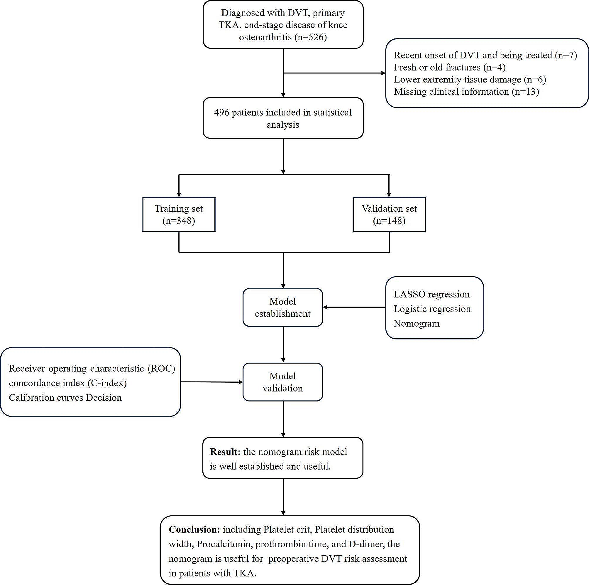
We screened a total of 201 patients with HIT suspicion between September 2018 and September 2020. Patients were included if their 4T score was > 3, including a platelet count decrease equal or greater than 30% from the initial count and heparin treatment administered for at least 5 days [1].
Of those 201 patients, 83 were not included because they did not meet the inclusion criteria and 13 because not enough plasma was stored after the initial work up. The decision to stop heparin and switch to an alternative anticoagulant is made by the haematologist and the attending clinician, based on the careful examination of the case presentation leading, but not restricted to the 4T score and the results of the first available biological tests results. Sometimes the alternative treatment is promptly started (before blood sampling) indeed ; those cases were not included in the study to avoid possible interference in the three techniques evaluated.
For each included patient, the following clinical and laboratory information was recorded: age, gender, hospital unit (medical, surgical or ICU), nature of anticoagulant treatment and corresponding anti-Xa level, 4T score, platelet count, HIT immunoassay results, heparin-dependent platelet activation results (Table 1).
Laboratory diagnosis of HIT relied on a positive platelet activation test with light transmission aggregometry [11] and/or positive ELISA with an optical density (OD) > 2 (Asserachrom® HPIA Diagnostica Stago, Asnières sur Seine France).
In order to increase the number HIT + patients we added six diagnosed until July 2021. Among the 19 patients diagnosed with HIT (13 over the study period + 6 supplementary patients), ten suffered from a thrombotic event (HITT + patients) present at the diagnosis or during the first week, documented with relevant imaging, such as Doppler ultrasound (US) and computer tomography (CT-scan) for venous thromboembolism. Of note, in case of confirmed HIT diagnosis, systematic Doppler US was systematically performed.
Diagnosis of HIT was based on 4T score, laboratory testing (ELISA anti-FP4 antibodies; platelet activation test with normal PRP and light transmission aggregometry), thrombotic complication, correction of platelet count after shifting to non-heparin anticoagulant treatment (danaparoid or argatroban).
Table 1 Characteristics of the study patients
Blood collection and preparation
Blood samples were collected at the time of HIT suspicion (initial sampling), into citrate 3.2% tubes (BD Vacutainer, France), 9 volumes of blood for 1 volume of citrate solution. Blood samples were centrifuged at 2500 g for 15 min to obtain platelet poor plasma. Plasma was then aliquoted (1mL) and kept frozen at -80 °C until further analysis. The only anticoagulant treatment potentially present in samples was heparin (unfractionated heparin or low molecular weight heparin).
Procoagulant phospholipids of circulating extracellular vesicles
Procoagulant phospholipids (ProcoagPPL) associated with extracellular vesicles (phosphatidylserine exposure), formerly known as microparticles, were assessed in plasma samples with a commercially available assay (STA®-Procoag PPL, Diagnostica Stago, France). The assay measures the clotting time initiated with added factor Xa and calcium to the plasma sample mixed with a normal, procoagulant phospholipid-depleted, plasma. The shorter the clotting times, the greater the procoagulant phospholipids plasma levels.
STA®-Procoag PPL is considered to be insensitive to heparin levels up to 1.5 IU anti-Xa/mL (unfractionated heparin) or 2.0 IU anti-Xa/mL (low molecular weight heparin). The kit was used according to manufacturer’s instructions on STA R® Max analyzer (Diagnostica Stago, France).
Fibrin monomers
Fibrin monomers (FM), also known as soluble fibrin, result from thrombin action on fibrinogen, when fibrinopeptides A are released and fibrin monomers are not polymerized to such an extent that they are no longer soluble [12]. FM levels were measured using an immunoturbidimetric assay (STA®- Liatest® FM, Diagnostica Stago, France) on STA R® Max analyzer according to manufacturer’s instructions. The assay principle is based on immunocapture of FM by a mouse monoclonal anti-human FM antibody coated onto latex microparticles and OD is converted into FM concentration using a dedicated calibration (STA®-Liatest® FM Calibrator, Diagnostica Stago, France).
Thrombin generation
Thrombin generation (TG) monitors the course of active thrombin over time in a clotting plasma sample [13]. The principle of this assay was already described, with a fluorometric detection of thrombin over time [14,15,16]. All devices and reagents were from Diagnostica Stago. We studied TG on ST Genesia analyzer using the STG-BleedScreen reagent. The choice of the initiating reagent, the one with the lowest concentration of tissue factor (TF), was aimed at being as sensitive as possible to the expected hypercoagulable states secondary to procoagulant microparticles in HIT. We performed runs of samples from 12 patients, along with one calibration curve (with STG-ThrombiCal), two quality controls measurements (low and normal TG level). The calibration curve was run in parallel with STG-FluoSet enabling calculation of a correction factor for the optical characteristics of each plasma sample as described earlier [20]. Four parameters from the time course of active thrombin i.e. thrombogram were used for further analysis: Lag Time, which corresponds to the time elapsed before the first quantities of thrombin appear (this quantity being equal to 1/6th of the thrombin level at Peak Height); Peak Height, which is the maximum concentration of thrombin; Time to Peak, corresponding to the time to reach the Peak Height; and Endogenous Thrombin Potential (ETP), which corresponds to the area under the TG curve and represents the total thrombin potential of the sample. Results were kept for statistical analysis, as soon as ST Genesia analyzer delivered results with a thrombogram (exclusion if no value was delivered with the message “TG too low”).
TG results were recorded as absolute and normalized against reference plasma STG-RefPlasma BLS [17, 18]. The reference plasma is a lyophilized standard sample to which a correction factor, provided by the manufacturer, is applied to scale results to normal (or 100%) [16]. This normalization step of TG results is meant to smooth for inter-runs and inter-batches variability [19, 20]. However, as our study was run in a single site, using a single batch of reagent and within few runs of tests, we chose to only represent absolute results here. Normalized results, expressed as ratios for lag time and time to peak and as percentages for peak height and ETP, are available as supplementary material.
Heparin neutralization
Samples sent to the laboratory for HIT diagnosis work up often contained heparin because blood was collected early, as soon as HIT was suspected and TG is very sensitive to presence of heparin in the plasma sample [20, 21]. Consequently, we had to perform the assay after neutralization of heparin. For this, we considered two reagents: polybrene (hexadimethrine bromide, Sigma-Aldrich, France) at concentrations described elsewhere [22] and heparinase (Dade Hepzyme®, Siemens Healthcare Diagnostics, Marburg Germany), using one vial of product for 1mL of plasma, according to manufacturer’s instructions. In preliminary experiments, plasmas from two healthy controls and five non-HIT patients under unfractionated heparin (UFH) and low molecular weight heparin (LMWH) were incubated with polybrene or Dade Hepzyme® at room temperature for 15 min before checking neutralization efficiency as follows. Coagulation tests (APTT, PT) and chromogenic anti-Xa assay (STA®-Liquid Anti-Xa, Diagnostica Stago, France) were performed with a STAR® Max analyzer before and after heparin neutralization. As some residual anti-Xa levels (0.10–0.20 IU/mL) were observed in polybrene-treated samples, we performed the study treating each individual patient’s sample with Dade Hepzyme® before TG study.
Statistical analysis
Statistical analyses were performed using MedCalc Statistical Software version 17.4.4 (MedCalc Software bvba, Ostend, Belgium). Results of PPL, FM, and TG were compared (i) among HIT + patients, those with or without thrombosis (HITT + and HITT- respectively) primary objective; (ii) between HIT + and HIT- patients.
Statistical significances of differences for each group pair were explored with Mann-Whitney test (cut-off p-value set at 0.05).


留言 (0)