Our study showed that NASH mice induced by a high-fat, high-sugar diet with trans-fat as the main fat for 24 weeks with an increased ratio of cholesterol and bile salts exhibited advanced liver inflammation, hepatic steatosis, and fibrosis. After the HFHCCC diet model, significant changes were observed in the levels of innate immune cells and their released cytokines in the liver of mice, while the related changes in adaptive immune cells were not obvious. This model is more inclined to trigger an innate immune response than an adaptive immune response.
After 24 weeks of HFHCCC diet induction, the content of TG, as the main indicator of liver fat content, in hepatic mice had significantly elevated. Mice showed significant hyperglycemia and insulin resistance. Pathologically, NASH in mice is characterized by hepatic steatosis, infiltration of neutrophils and macrophages. The stage of liver fibrosis in mice is mostly F1-2. The primary clinical manifestation of NASH includes dyslipidemia, steatosis, and liver cell damage, inflammation, and varying degrees of fibrosis [11]. In this study, the main features of the HFHCCC-induced NASH model were very consistent with the main clinicopathological features of NASH patients.
Theoretically, the "ideal" NASH model should reflect the full spectrum of biochemistry and histology of clinical human liver disease, as well as the characteristics of the associated metabolic syndrome, and should not be too long (e.g., more than one year) [12]. At present, commonly used NASH models include methionine choline deficiency diet (MCD) model, carbon tetrachloride diet (CCL4) model, high-fat diet (HFD) model, high-fat and high-sugar diet (HFHC) model, etc. Although a MCD or CCL4 can induce fibrosis, but it cause weight loss that does not exhibit pathological characteristics similar to that of humans, and lack of insulin resistance or promote fibrosis without hepatic steatosis. Diet-induced obesity models are more similar to the physiology of human NASH patients. However, significant liver fibrosis couldn’t be developed by a high-fat diet alone in most rodents [13]. C57BL/6J mice fed a traditional high-fat diet (60% of energy comes from fat) showed obesity and disorders of glycolipid metabolism after 10 weeks, steatosis and inflammation at 16 weeks, and took 50 weeks to induce mild fibrosis [14]. After 30 weeks of feeding C57BL/6 mice a high-fat and high-sugar diet, they showed NASH characteristics such as balloon-like change, glucose and lipid metabolism disorders, liver damage, and inflammation, but the degree of fibrosis was very mild [7]. Compared with these conventional high-fat diet, high-fat and high-sugar diet models, models with added cholesterol and bile salts can better induce disease characteristics similar to those in humans. Mels et al. [15] found that C57BL/6J mice modeled with a high-trans-fat, high-sucrose and high-cholesterol diet not only developed steteaosis, lobular inflammation, hepatocyte balloon-like transformation, but also showed fibrosis. Tu et al. [16] induced NASH with high-fat and high-cholesterol bile salt diet modeling, in which 37.1% of the energy came from fat, the cholesterol content was 1.25%, and the sodium cholate content was 0.5%, the pathological results showed that the mice showed steatosis and inflammation after the diet modeling, but the degree of fibrosis was very low, our study increased the cholesterol content (2%) and increased high-sugar drinking water based on the research of Lan N. Tu et al. further accelerating the progress of NASH. More pronounced disorders of glycolipid metabolism were induced, and fibrosis occurred. In summary, the HFHCCC diet was more capable of replicating NASH models similar to human metabolic and histological characteristics than other methods-induced NASH models.
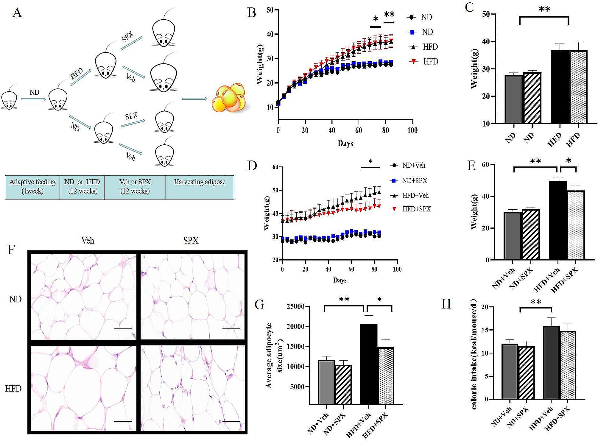
However, it was worth noting that although the HFHCCC diet-treated mice showed significantly increased liver weight and liver body ratio compared to control diet-treated mice, yet mice in the HFHCCC group exhibited significant decreased body weight by approximately 9% at 24 weeks compared to that in the control group. A possible reason is that sodium cholate can participate in the metabolism of bile acids and promote the metabolism and absorption of cholesterol, activate G protein-coupled receptor 5, induce the activity of type 2 deiodinase, promote the conversion of tetraiodothyronine to triiodothyronine, and finally promote the energy expenditure, resulting in weight loss [17]. In conclusion, this model could simulate the entire disease course of human NASH patients, while exhibiting the characteristics of NASH fibrosis, showing characteristics of patients with NASH, especially non-obese patients with NASH liver fibrosis.
More and more evidences show the role of immune response in the pathogenesis of NASH. The innate immune cells of liver include Kupffer cells (KCs), neutrophils, dendritic cells (DC) and natural killer cells (NK). In order to further analyze the immune disorder of the model, we detected the expression of immune cells and related inflammatory factors in liver tissue. The results showed that HFHCCC model induced the activation of KCs, neutrophils, DCs and other innate immune cells in the liver of mice, accompanied by increased release of cytokines; However, most of the adaptive immune-related cells in the mouse liver did not change, and the cytokine released by the adaptive immune cells did not change significantly. The results suggested that HFHCCC diet was more likely to activate the innate immunity of mice.
Clinical studies had found that immune cells were activated at all stages of human NAFLD, including T cells, B cells, macrophages and neutrophils [18]. The recruitment of KCs and the expression of pro-inflammatory cytokines such as CCL2, TNF and IL-1βcould be observed in the liver tissue of patients with NASH [19]. After being activated, KCs could secrete pro-inflammatory cytokines such as CXCL2, CXCL1 and IL-8, promoted the recruitment and differentiation of other immune cells, such as neutrophils and dendritic cells, to regulate the immune response under metabolic stress. Some studies found that NASH tended to activate innate immunity before fibrosis F0-1, and adaptive immune response during fibrosis F2-4 [20], macrophages were the first immune cells detected in patients with NASH, and could be detected at the stage of steatosis. However, the increase of CD3+ T cells, CD4+ T cells, and CD8+ T cells in the liver or peripheral blood could not be observed until NASH fibrosis F2-4. In this study, HFHCCC diet was used to induce NASH liver fibrosis, with the stage of F1-2, and KCs cells were observed in the liver tissue of mice at 24 weeks The significant recruitment of neutrophils and the recruitment and release of pro-inflammatory cytokines, chemokines and colony-stimulating factors, but CD4+ T cells, CD8+ T and other adaptive immune-related cells and cytokines did not change significantly before and after the HFHCCC diet modeling, which is also basically consistent with the results of human research.
Many NASH models have been used in the related research of NASH immune response, including MCD diet-induced NASH mouse model [21], HFD diet-induced NASH mouse model [22], HFHC diet-induced NASH mouse model [23], ob/ob mouse model [24], etc. Studies have found that MCD diet can induce both innate and adaptive immunity. However, the current studies on the immune response of NASH induced by MCD diet mostly focus on KCs, NKT cells, Inflammasome and other innate immune responses. After using NASH induced by MCD diet, it was found that KCs [22],NKT cells [21], DC cells [25] and NK cells [26] were significantly activated, and CD4T lymphocytes were significantly recruited [27], cytokines such as IFN-γ, IL1α, IL1β, IL12 (p40), GM-CSF, and CCL3 secreted by these cells are also higher. Similarly, the liver tissue of NASH mice induced by HFD also showed significant activation of KCs [22], neutrophil infiltration [28] and large increase of DC cells [29], increased release of pro-inflammatory cytokines such as TNF-α, IL-6, MCP-1, etc. released by innate immune cells. Kind et al. [30]. found that the activation of innate immune cells such as NK cells and DC cells appeared in the liver tissue and adipose tissue of mice for 15 weeks, while there were no significant changes in the effector T cell subsets. We observed macrophage and neutrophil infiltration and TNF in the liver tissue of mice 24 weeks after modeling with HFHCCC diet and found macrophage and neutrophil infiltration and release of other related inflammatory factors such as TNF-α, IL-6, IL-1α, IL-1β and in mouse liver tissues 24 weeks after modeling with HFHCC diet. Higher levels of NKT cells and DC cells were detected, but no changes in adaptive immune-related cells and cytokines such as CD4+ T cells and CD8+ T cells were found. To some extent, it is proved that the immune characteristics of NASH model induced by MCD diet and HFD diet are similar to those of our study.
It is worth noting that there are some immune cells whose role in NASH is complex and controversial. Different studies on NKT cells have shown different conclusions. Zheng et al. [31] constructed two models, high-fat and high-sugar diet model and MCD model, to induce fibrosis in NASH mice, and found that the liver NKT cells of mice fed a high-fat and high-sugar diet were significantly less than those in the normal diet group, while the abundance of liver NKT cells in mice fed the MCD diet did not change significantly compared with the normal diet group. A human immune-related study found NKT cell aggregation in the fibrous septum in patients with NASH stage 3–4 liver fibrosis [32]. Our findings show significant recruitment of NKT cells after dietary induction of HFHCC, which is closer to the human findings. The results of different studies vary, which may be related to the different stages of disease development caused by different modeling methods.
This study had some potential limitations. Inconsistent with the disease characteristics of most patients with NASH, the weight of the model mice was lower than that of the normal group, which was mainly related to the involvement of bile salts in bile acid metabolism affecting energy metabolism; on the other hand, this model also provided a reference for the study of non-obese NASH. In addition, due to the limitation of the number of experimental channels for flow analysis, the study only used immunohistochemistry for the analysis of liver macrophages, and lacked the results of flow analysis; in terms of adaptive immunity, Th1, Th2, Th17, and Treg have not been further detected and analyzed, which is also the direction for further research in the later stage.
In conclusion, we established an HFHCCC diet-induced NASH model that is stable and reproducible, showing the emergence of NASH pathological features such as inflammation, steatosis, and fibrosis. This model is likely to trigger innate immunity. This could serve as a suitable experimental model for drug testing and for understanding the pathogenesis of innate immunity in NASH.


留言 (0)