Cell culture
GBM U87 cells were bought from the Institute of Basic Medicine’s Cell Center at Peking Union Medical College (China), and they were grown in high-glucose DMEM (Gibco, USA) with 10% fetal bovine serum (Gibco). At 37 degrees Celsius, the cells were incubated with saturated humidity, 5% carbon dioxide, and oxygen. The ratio of the added medium that could be used for a subculture and the total number of cells was approximately 1:3.
Cell counting kit 8 assay for cell viability assay
100-μl cell suspensions were prepared in 96-well plates, 10,000 cells were added to each well, five compound holes were set, and the plate was precultured in the incubator for 24 h. A medium containing hederagenin (PureChem-Standard Co., Ltd., Chengdu, China) was put on the culture plate in place of the fresh media. Twenty-four hours were spent incubating the plate. Cell Counting Kit 8 (CCK-8) (Biosharp, Hefei, China) solution was added to each well after the medium had been changed. For 1-4 hours, the dish was incubated. A microplate analyzer was used to test the absorbance at 450 nm. Cell viability was calculated as [(ODHED − ODblank)/(OD0 − ODblank)] × 100%, in which OD is optical density and HED is hederagenin. Furthermore, ODHED represents the absorbance of holes with cells, solutions, and drug solutions; ODblank represents the absorbance of holes with culture media and solutions but no cells, and OD0 represents the absorbance of holes with cells and solutions but no drug solutions. The quantity of hederagenin digested in U87 cells was also determined using the equation’s results, using the approximate treatment concentration as the 50% inhibitory concentration (IC50).
Preparation of protein extraction
U87 cells were treated with 40 μg/ml (according to IC50 result) of hederagenin for 24 h on the petri dish and gently cleared twice with low-temperature phosphate-buffered saline; the supernatant was discarded, and an appropriate amount of sodium lauryl sulfonate and Tris-HCl was added for lysis. The cells were rapidly scraped, placed in a boiling water bath for 15 minutes, and then ultrasonically centrifuged at 14000 g for 15 minutes to get the supernatant. It was harvested from the filtrate. Protein quantification was carried out using the bicinchoninic acid technique.
The samples were separated and stored at − 20 °C. A total of 20 μg of protein was added to the 6X loading buffer for each sample, and then for 5 min, the samples were submerged in boiling water. Then, 12% sodium lauryl sulfonate-polyacrylamide gel electrophoresis (constant pressure of 250 V, 40 min) was performed for Coomassie bright blue staining. The filter-aided proteome preparation (FASP) technique was then used to prepare the protein solution [10].
TMT labeling
Thermo TMT Labeling kit (Thermo Fisher Scientific, Waltham, USA) instructions were followed for labeling 100 g of the peptide from each sample for TMT. Each group’s labeled peptides were combined and graded using an Agilent 1260 Infinity II HPLC system (Agilent Technologies Inc., CA USA). 10 mM of HCOONH4 and 5% acetonitrile made up buffer A, whereas 10 mM of HCOONH4 and 85% acetonitrile made up buffer B. (pH 10.0). An automated sampler inserted the samples onto the chromatographic column at a flow rate of 1 ml/min after the column had been balanced with liquid A. The liquid phase’s gradient was as follows: Between 0 and 25 minutes, liquid B was at 0%; between 25 and 30 minutes, it had a linear gradient of 0 to 7%; between 30 and 65 minutes, it had a linear ladder of 7 to 40%; between 65 and 70 minutes, it had a linear ladder of 40 to 100%; and between 70 and 85 minutes, it remained at 100%. The components were collected every minute throughout the elution, and the absorbance at 214 nm was measured. Forty elution components were collected in total. After being freeze-dried, the samples were divided into 10 pieces and redissolved in 0.1% formic acid.
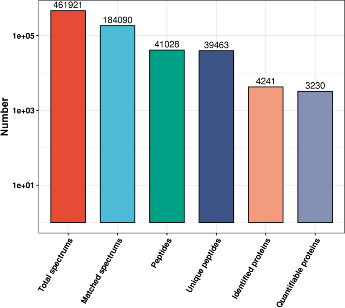
Mass spectrometry analysis
Each sample was separated using an Easy nLC system with a nanoliter flow rate (Thermo Fisher Scientific, USA). Chromatography was used to separate the materials, and a Q Exactive Plus mass spectrometer was used for analysis (Thermo Fisher Scientific, USA). Using a high-resolution mass spectrometer called the Q Exactive Plus, quantitative proteome analysis of TMT was carried out. The original graph files (.raw files) were converted into. mgF files by Q Exactive Plus using Thermo Fisher Scientific’s Proteome Discoverer 2.2 program. The MASCOT2.6 server received the files for database retrieval. The data were then filtered by Proteome Discoverer 2.2 using the standard of a false discovery rate of less than 0.01 after the library file (.dat file) created on the MASCOT server was delivered back to the software. DEPs were indicated by a fold change of more than 1.2 and a P value (Student’s t-test) of less than 0.05. Data from protein mass spectrometry were qualitatively analyzed using the Uniprot HomoSapiens _20367 20,200,226 (http://www.uniprot.org). For the mass spectrometry analysis, raw data were employed. Mascot 2.6 and Proteome Discoverer 2.2 were employed for database identification and quantitative analysis.
Bioinformatics analysis
The target protein set’s alignment sequences with the highest bit scores were retrieved using the Blast2GO Command-Line and the Blast retention findings to produce the Gene Ontology (GO) items (www.geneontology.org). The target protein sequence was mapped to the GO entries obtained throughout the mapping process using the Blast2GO Command-Line. The KEGG Orthology (KO) and Links Annotation, or KOALA, program compared the KEGG genes database using Kyoto Encyclopedia of Genes and Genomes (KEGG) pathway annotations, and KO categorized the target protein sequence. The target protein sequence’s pathway information was automatically derived based on KO categorization. The distribution of each GO classification or KEGG pathway in the target protein and the overall protein was compared by Fisher’s exact test in the GO enrichment annotation or KEGG pathway annotation on the target protein set to assess the level of significance of protein enrichment in a particular GO term or KEGG pathway. Projections were positioned to account for variations in protein analysis using the WOLF PSORT (https://wolfpsort.hgc.jp/) program. We examined the functional domain annotation of various proteins using the InterPro database. The significance level of the enrichment degree in a functional domain was assessed by comparing the distribution of various proteins in the total protein using Fisher’s exact test. The target protein’s quantitative data were adjusted for cluster analysis. A hierarchical clustering heat map was produced using Matplotlib software while classifying samples and protein expression in two dimensions.
Identification by Western blotting
Choose the hub protein from DEPs for WB confirmation, as the TMT data suggest. In the logarithmic stage, U87 cells were divided into two groups: the control group and the 40 g/mL hederagenin group. After 24 h, U87 cell proteins were collected and eliminated using a RIPA lysis cushion containing 1 mM PMSF from Beijing Applygen Technologies Co., Ltd. (Beijing Applygen Technologies Co., Ltd.). The BCA (Beijing Elabscience Co., Ltd.) technique did not wholly lock down protein. Then, each collection of 20 ng protein samples was deposited onto a PVDF layer using SDS-PAGE and electrophoresis (Millipore, USA). The layer was inhibited with 5% skim milk for 1 hour and short-term, at 4 degrees Celsius, brooded with necessary antibodies. The layer was treated with TBST (Beijing Applygen Technologies Co., Ltd.) several times the next day before being brooded for 1 h at room temperature with optional antibodies.
TBST was used to repeatedly wash the layer before ECL was used to identify the goal groups (Thermo, USA). To normalize protein levels, −actin (Beijing zsbio, Inc.) was used as the internal control. KIF7 is one of the crucial antibodies used in this investigation (Proteintech Group, Inc. USA). Additional antibodies came from Beijing zsbio.


留言 (0)