
Remember me


Sporadic cerebral small vessel disease (SVD) is a common finding in the ageing brain, defined by neuroimaging features [1] and a range of histological lesions [2, 3]. While critical to providing a uniform structure to future research, where histopathological definitions are defined, particularly Skrobot et al. [2], there is no attempt at gradation, and they are defined in the context of a single clinical outcome, cognitive decline. Clinically significant, SVD causes 25% of ischaemic strokes [3] and 85% of intracerebral haemorrhage [4]. It is the major cause of vascular dementia (VaD) [5] and is associated with a range of other cognitive [6, 7] and physical problems [8, 9].
Small vessel disease is best visualised in vivo using magnetic resonance imaging (MRI). An international working group from the Centres of Excellence in Neurodegeneration developed definitions and imaging standards for markers and consequences of SVD identifying white matter hyperintensities (WMH), lacunes, enlarged perivascular spaces (PVS), microbleeds and at high field, microinfarcts [1, 10].
Currently, there is no comparable consensus document regarding histological SVD lesions [11]. Variation between MRI appearances and histopathology may reflect this lack of consistency in definitions or may be a consequence of the scarcity of studies detailing the pathology of imaging-detected lesions [12]. Additionally, early stage disease may be under-reported and most autopsy-identified lesions may be end-stage ‘scars’ [13]. Genetic abnormalities and molecular pathways [14, 15] have been identified, reflecting the heterogeneous underlying pathophysiology [16]. Potential mechanisms for vascular and tissue damage include blood–brain barrier (BBB) dysfunction [17, 18], para- and peri-vascular space abnormalities [19, 20] and abnormal perfusion [21, 22]. However, precise mechanisms at different stages and in different lesions remain unclear.
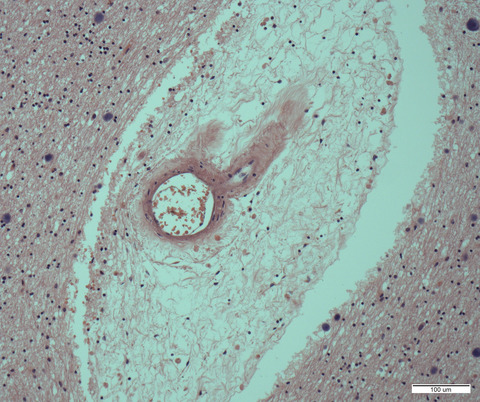
Some lesions are clearly identified by both MRI and histology, such as lacunes [2], PVS [23] and small infarcts [24]. Others, like early stage WMH, are easily observed on MRI but difficult to detect histologically, contributing to apparent discrepancies about lesion composition, notably water content and axon degeneration when assessed by histology and MRI respectively. Damaged micro-vessels (i.e. arteriolosclerosis, lipohyalinosis, fibrinoid necrosis and cerebral amyloid angiopathy [CAA]) are below current imaging resolution thresholds [2, 25, 26], but are key descriptors in histological assessment.
Detailed imaging-histological correlations, particularly of individual lesions and subtypes, are required. We aimed to summarise current knowledge of precise MRI-histological correlations in common SVD lesions, to assess their reliability and generalisability and identify where greater consistency in methodology and definition is needed.
METHODSIn this systematic review, we identified all studies correlating ex vivo medical imaging with histopathology of SVD lesions. Given the dynamic nature of SVD during life, we focused on studies that used ex vivo imaging to avoid extended timelines between imaging and histology.
Search termsWe searched for the terms ‘small vessel disease’, ‘SVD’ and all relevant commonly used neuroradiological [1] and histopathological terms (Table 1), in the Medline online database from its inception in 1966 to the 9 September 2020. The search terms were refined through several iterations to ensure all relevant SVD lesions on imaging and histology were included, and that all potential lesion types were included (Table 1). We hand-searched reference lists in textbooks, review papers and two relevant journals (Stroke and Neuropathology and Applied Neurobiology) and for relevant primary publications.
TABLE 1. Final search strategy Results Search 1 exp Brain/pa, pp [Pathology, Physiopathology] 2 *Cerebrovascular Disorders/pa [Pathology] 3 *Stroke, Lacunar/et, pa, pp [Etiology, Pathology, Physiopathology] 4 (cerebrovascular disease or cerebrovascular lesions or cerebrovascular pathology).ti,ab. 5 SVD.ti,ab. 6 small vessel disease.ti,ab. 7 lipohyalinosis.ti,ab. 8 arteriolosclerosis.ti,ab. 9 (atherosclerosis adj3 small*).ti,ab. 10 CAA.ti,ab. 11 cerebral amyloid angiopathy.ti,ab. 12 (WMH or white matter hyperintensit$).ti,ab. 13 (WML or white matter lesion$).ti,ab. 14 ((lacune or lacunar) adj2 (stroke or infarct$)).ti,ab. 15 microbleed.ti,ab. 16 perivascular space.ti,ab. 17 leukoaraiosis.ti,ab. 18 small deep infarct.ti,ab. 19 fibrinoid necrosis.ti,ab. 20 (histopatholog$ or histolog$ or patholog$).ti,ab. 21 (IHC or immunohistochemi$).ti,ab. 22 (ICC or immunocytochemi$).ti,ab. 23 1 or 2 or 3 24 4 or 5 or 6 or 7 or 8 or 9 or 10 or 11 or 12 or 13 or 14 or 15 or 16 or 17 or 18 or 19 25 20 or 21 or 22 26 23 and 24 and 25 27 limit 26 to humans Inclusion/exclusion criteriaWe included all studies published in full which carried out post-mortem computerised tomography (CT) or MR imaging and histological examination of adult human brain tissue to study sporadic SVD (Figure 1). We excluded: studies reporting only biopsy material; studies performing post-mortem imaging but no histopathology or vice versa; studies not reporting on human sporadic SVD; non-English publications; and studies of haemorrhagic SVD lesions only as these were reviewed recently [27, 28]. We excluded all duplicate publications, editorials and conference abstracts. Where studies came from the same groups and it was not clear which, if any, cases were included more than once, we included only the largest study for subject characteristics and results, and only distinct individual findings from the other papers.

Summary of a literature evaluation. PVS perivascular spaces; SVD, small vessel disease
Quality of reportingTo assess methodology quality, we adapted the Standards for the Reporting of Diagnostic Accuracy studies (STARD) criteria [29] for observational studies to give a score out of 25. Information was extracted regarding the authors expertise, aims, the study population, study dates, data collection, participant recruitment, pathological and radiological methods, blinding, analysis, results reporting and discussion of findings (Table 2). Each individual feature could gain one point, or part thereof if only part of the criteria was met. The authors discussed and resolved uncertainties regarding inclusion, exclusion and scoring when required.
TABLE 2. Quality assessment Title Neuropathologist and neuroradiologist in the authors Introduction State the research questions or study aims Methods ParticipantsDescribe the study population: inclusion and exclusion criteria, setting and locations where the data were collected
Report when the study was done, including beginning and ending dates of recruitment
Describe participant recruitment
Describe data collection: prospective or retrospective
PathologyDescribe the pathological methods: rationale, technical specifications (and/or cite references)
Define pathological lesions and criteria used for assessments
Describe the number and expertise of the persons executing the macroscopic and microscopic neuropathological assessment
Were the individuals carrying out the macroscopic and microscopic assessment blinded to the results of other assessments and any other available information?
Assess PM tissue quality and/or report post-mortem interval
ImagingDescribe the radiological methods: rationale, technical specifications (and/or cite references)
Define radiological lesions and criteria used for assessments
Describe the number and expertise of the persons executing the radiological assessment
Were the assessors blinded to the results of other assessments and any other available information?
Describe measures taken to minimise artefact on MRI
Statistics Describe statistical methods for calculating or comparing measures of diagnostic accuracy, adjusting for variables, and the statistical methods used to quantify uncertainty (e.g. 95% confidence intervals) Results ParticipantsReport clinical and demographic characteristics of the study population (e.g. age, sex, race, clinical info etc.)
Report characteristics for individual subjects
Report clinical features associated with SVD
ResultsReport the distribution of disease severity
Report a cross-tabulation (including indeterminate and missing results)
DiscussionDiscuss the clinical applicability and/or use of the findings
Discuss the scientific importance use of the findings
Discuss the future use of the findings
Note Adapted from the Standards for the Reporting of Diagnostic Accuracy studies checklist. Each feature assessed could gain 1 point, or a part thereof if the only part of the data was reported up to a total score of 25. Abbreviations: MRI, magnetic resonance imaging; SVD, small vessel disease. Data extractionData were extracted from study characteristics including study design and dates, prospective or retrospective recruitment, data collected, sample size, blinding and analysis methods. Subject data included demographic and clinical data including cognitive status, in vivo investigations, age, gender and ethnicity. Radiological specifics included scanner field strength, sequence resolution, tissue scanned, measures to avoid artefact and involvement of neuroradiologists. We collected details of pathological processing, assessment of tissue quality, macroscopic examination, staining, grading systems used, terminology and definitions and involvement of neuropathologists. All three authors read the papers and had regular meetings to agree a consistent approach and achieved consensus through discussion.
Various pathological terminologies were used in the papers, not always consistently, hence we condensed the information into manageable categories based on the terminologies in each paper. For reference, we provide a full list of the terms encountered and similar expressions in Table S1. We recorded the method used to relate tissue locations on MRI/CT to that on histology, the analyses used and the neuroradiological–neuropathological findings.
RESULTSThe initial search yielded 1627 articles: 16 duplicates were removed, and we excluded 1403 papers including 37 studying monogenic SVD (reasons detailed in Figure 1). For interest, the excluded papers that studied monogenic SVD are listed in the supplement, two of which compared lesion appearances on post-mortem imaging and histology [30, 31]. Searching reference lists of 183 review papers provided nine additional relevant papers, and two further papers were identified from hand-searching journals. This resulted in 38 relevant papers, that reported imaging of at least 1146 individual patients in total, 342 of whom had SVD lesions on imaging. Studies were published between 1986 and September 2020, all using ex vivo MRI (Table 3). Of these we created three groups; 29 primarily focused on imaging-identified white matter lesions (WML), six studying microinfarcts and three on PVS and lacunes.
TABLE 3. Summary of papers studying non-haemorrhagic SVD Paper (lead author, year) Cohort origin Number of cases with SVD (total scanned) Number of female subjects in those with SVD (in total) Mean age (range) (unless otherwise stated) Data collection MRI field strength Tissue scanned (medium) Quality score WML Awad (1986) [51] Arizona, US 7 (7) 2 (2) 72 (61–84) Not stated 1.5T Whole brain (2 fresh in isotonic saline, 8 fixed in formaldehyde solution) 16.75 Braffman (1988) [32] Philadelphia, USX
Duplicated subjects used in Braffman 1988 (33)
X 67 (60–78) Not stated 1.5T Whole brain (water) 10.50 Marshall (1988) [54] California, US 6 (18) — (—) >60 (—) Not stated 0.35T Whole brain (—) 7.75 Revesz (1989) [47] UK 4 (6) 2 (—) 52 (40–68) Retrospective 0.5T Slices (—) 12.00 Fazekas (1991) [52] Austria 4 (4) 0 (0) 59 (52–63) Not stated 1.5T Whole brain (—) 11.00 Grafton (1991) [46] Seattle, US 5 (7) — (—) 77 (69–88) Retrospective 0.5T Whole brain (air) 15.75 Van Swieten (1991) [50] Netherlands 10 (40) — (19) 76 (—) Retrospective 1.5T Whole brain (formalin) 12.50 Chimowitz (1992) [45] Ohio, US 7 (7) 4 (4) 68 (64–74) Retrospective 1.5T Whole brain (water) 12.25 Munoz (1993) [57] Canada 13 (15) — (11) 69 (47–87) Not stated 1.5T Whole brain (air) 14.00 Scarpelli (1994) [48] Italy 17 (21) 7 (9) 74 (65–94) Retrospective 1T Whole brain, slices scanned if MR lesions but macroscopically normal (water) 15.25 Scheltens (1995) [49] Netherlands — (15) — (9) 73 (60–83) Retrospective 0.6T Whole brain (—) 15.75 Fazekas (1999) [53] Austria 9 (11) 4 (5) 72 (45–90) Not stated 1.5T Slices (—) 17.00 Smith (2000) [34] Kentucky, USX
Can't be sure subjects not same as those in Smith 2000 (63)
X — (83–99) Prospective — Whole brain (—) 15.50 Smith (2000) [65] Kentucky, US 52 (54) 52 (54) 89 (SD 4.8) Prospective — — (—) 13.50 Bronge (2002) [44] Sweden 6 (6) 3 (3) 87 (81–101) Retrospective 1.5T Whole brain (air) 13.25 Fernando (2004) [59] UK CFASX
Can't be sure subjects not same as those in Fernando 2006 (58)
X 85 (median) (70–97) Prospective 1T Slices (in plastic bag) 19.00 Moody (2004) [56] N Carolina, US 12 (21) 6 (9) 72 (58–90) Not stated 1.5T Slices (between cooled plastic sheets) 14.25 Fernando (2006) [60] UK CFAS — (456)a — (—) Over 77% >80 years (—) Prospective 1T Slices (sealed in polythene) 12.75 Matsusue (2006) [66] Japan — (—) — (—) — (—) None given 1.5T Whole brain (—) 6.00 Simpson (2007) [63] UK CFASX
Can't be sure subjects not same as those in Fernando 2006 (58)
X Median ages of cohorts range 84–87 (68–95) Prospective 1T Slices (plastic bag) 16.00 Simpson (2007) [64] UK CFASX
Can't be sure subjects not same as those in Fernando 2006 (58)
X Median ages of cohorts range 80–87 (68–100) Prospective 1T Slices (plastic bag) 14.50 Young (2008) [66] Australia 20 (20) 8 (8) 76 (61–94) Prospective 3T Whole brain (agar) 18.50 Polvikoski (2010) [58] Finland 132 (132) 114 (114) 91b (85–105) Not stated 1.5T Whole brain (0.1mM MnCl2) 16.00 Auriel (2011) [43] Hungary — — 64 (—) Retrospective 1.5TWhole brain (—)
Ex vivo scanning only of brains with poor quality in vivo MRI and control cases
15.50 Murray (2012) [62] Florida, US — (4) — (2) 85 (74–90) Prospective 3T Hemisphere (distilled water) 14.75 McAleese (2013) [55] UK NBTR — (40) — (25) 83 (68–98) Not stated 4.7T Hemisphere (air) 14.25 Hainsworth (2017) [61] UK CFASX
Can't be sure subjects not same as those in Fernando 2006 (58)
X 86 (SD 7.7) Prospective 1T Slices (in polythene bag) 14.75 Van Veluw (2019) [69] Massachusetts, US 9 (11) 2 (3) 75 (64–95) Not stated 3T Hemisphere (plastic bag filled with periodate-lysine-paraformaldehyde fixative) 18.75 Waller (2019) [67] UK CFASX
Can't be sure subjects not same as those in Fernando 2006 (58)
X — (71–101) Prospective 1T Slices (in plastic bag) 15 PVS and lacunes Bokura (1998) [41] Japan — (12) — (2) 76 (61–90) Retrospective 1.5T Whole brain (—) 12.25 Braffman (1998) [33] Philidelphia, US 9 (36) 5 (18) 66 (37–78) Not stated 1.5T Whole brain (water with very dilute amount of formalin) 10.50 Van veluw (2016) [42] Netherlands1 (1)
4 additional cases appear to be used in van Veluw 2015 (37)
1 (1) 76 (68–84) Retrospective 7T Slices (10% formalin) 14.50 Microinfarcts Van Veluw (2013) [38] Netherlands
Comments (0)