* Prof. Dr. Guillermo Stok, **Dra. Carolina López Sanabria.
*Prof. Cátedra de ORL, Facultad de Medicina, Universidad Nacional de Tucumán, Argentina. Médico staff
Hospital Centro de Salud Zenón Santillán, Tucumán, Argentina.
** Médico de staff Hospital San Bernardo, Salta, Argentina.
Introduction
The verrucous carcinoma of Ackerman is a rare and well differentiated tumor of
the squamous carcinoma, that can manifest in the vocal cords as in the oral
cavity.
Verrucous carcinoma was first described as a distinct clinicopathological entity
by Dr. Ackerman in 1948. Although found primarily in the oral cavity, the larynx
is the second most affected site, accounting for almost all cases.1
The incidence of verrucous carcinoma of the larynx is 1% to 2% of all malignant
neoplasms that affect this organ, mostly between the ages of 40 and 69 years.2
There is a clear association between tobacco consumption and verrucous
carcinoma, as well as the presence of papilloma virus as a possible etiological
factor, especially genotypes 16 and 18.3
It is a well-differentiated, slow-growing verrucous tumor with invasion of local
structures, without distant metastases, and local metastases are uncommon.5
Macroscopically, both appear as warty masses, with notable papillary
projections of the epithelial surface. The histological appearance of the
verrucous carcinoma is of a well differentiated hyperplastic epithelial lesion. A
densely keratinized surface and strongly circumscribed deep margin, often
described as a “pushing border”, is what characterizes these tumors.6, 7
Long-term dysphonia is the most common symptom, since the glottic region
was the most common site of verrucous carcinoma.
Local recurrences are common when treatment is inadequate. But its benign
behavior allows conservative treatment mostly by endoscopic laser route, and
selective lymph node dissection is not indicated.8
Clinical case
Obese patient, 50 years old, with permanent dyspnea, worse for walking and
supine position. Supraclavicular and suprasternal chimney flow. Progressive
dysphonia.
Background of larynx microsurgery in the year 2013 due to papillomatous right
vocal cord lesion. Biopsy of the year 2013 with report of vegetative
papillomatous lesion with abundant blood vessels.
In December of 2016 he consulted for dyspnea and dysphonia.
Fibrolaryngoscopy shows a lesion that occupies the entire glottis, vegetative,
keratinized, papillomatous type.
In laryngeal microsurgery with C02 laser, lesion of the right vocal cord and the
mucosa was resected. There wasn ?t invasion of the vocal ligament. The airway
was permeabilized, and the patient was discharged at 5 hours.
At the control month there wasn ?t lesion with excellent voice and breathing
quality
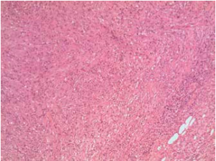
Biopsy reports tumor regions with papillomatous appearance and presence of
verrucous carcinoma.
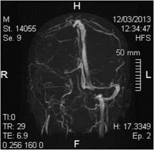
Fig. 1, 2,3: Right vocal with cord verrucous lesion.
Fig. 4: Control at the month of surgery.
Discussion
Verrucous carcinoma is a form of squamous cell carcinoma with specific
clinical, morphological and cytokinetic characteristics. The term verrucous
carcinoma refers to those exophytic or cutaneous squamous mucous tumors
that are stacked on the epithelial surface with a papillary micronodular
appearance.2
Tumors originated most frequently in the oral cavity (55.9%) and larynx (35.2%).
Although the majority of patients are male (60.0%), tumors of the oral cavity are
more common in older women.3
Although verrucous carcinoma is related to tobacco consumption and the
presence of human papillomavirus, its etiology is not yet defined.4
The tumor most frequently in the larynx originates in the true vocal cords. The
incidence of verrucous carcinoma of the larynx is 1% to 2% of all malignant
neoplasms that affect this organ, with the peak incidence between 40 and 69
years.2
The most representative symptoms of verrucous carcinoma of the larynx are
dysphonia and dyspnea that increase gradually.9
Diagnosis Verrucous carcinoma can only be performed by biopsy, and in some
cases additional biopsies are required to assess the relationship between the
epithelium and the underlying stroma. In laryngoscopy, these lesions are warty
and exophytic, often resulting in superficial biopsies.10, 11
The histopathological features of verrucous carcinomas are described as well
differentiated squamous lesions with local invasion through descending growth
intrastromal invaginations containing fine fibrous vascular cords.12, 13
Verrucous carcinoma exists within the histologic continuum ranging from benign
squamous hyperplastic lesions to invasive squamous cell carcinoma.
Distinguishing verrucous carcinoma from these benign and malignant
processes can be difficult. By definition, all verrucous carcinomas are classified
as well differentiated (or Grade 1)
In order to differentiate verrucous carcinoma of keratosis, verruca vulgaris or
squamous carcinoma with verrucous appearance, it is necessary to obtain deep
margins of the lesion, for the detection of microscopic foci of invasive squamous
carcinoma inside or adjacent to the keratosis of the lesion.
Clinically, the lesions appear as grayish, exophytic and verrucous growths that
tend to grow slowly and are not associated with metastases to the regional
cervical lymph nodes.1
The clinical behavior of verrucous carcinoma can be destructive despite its
deceptively benign microscopic appearance. These lesions can grow very large
and can infiltrate extensively to destroy adjacent tissues including cartilage and
bone. The presence of cervical lymphadenopathy is mostly due to reactive
lymph node hyperplasia secondary to the inflammatory reaction at the stromal
tumor interface.4
In 94.9% of the cases reported in the literature, the extent of the disease was
only a local invasion, with little invasion of adjacent structures (4.2%), and
regional metastases in only one case (0.8%). Distant metastasis was not
reported in any of these cases at presentation.14
Despite their locally aggressive nature, the metastases of verrucous carcinoma
are extremely rare.15
Surgery is the mainstay of treatment, achieving local control rates ranging from
77% to 100% .5, 10
The use of radiotherapy as a treatment is discussed, without reaching a
concomitant due to the scarce collection of clinical cases. In addition to having a
partial response to treatment with radiotherapy, some papers support the
possibility of anaplastic carcinoma after radiotherapy due to the presence of the
human papilloma virus.15
Conclusion
Ackerman verrucous carcinomas are rare tumors. In the presence of suspected
presence, the total resection of the lesion should be performed and not a
superficial biopsy, to avoid a misdiagnosis of the disease. The treatment of
choice is endoscopic surgery or laryngotomies, since treatment with
radiotherapy would provide an adequate response and the possible
transformation to anaplastic carcinoma
Reference
1- Ackerman LV: Verrucous carcinoma of the oral cavity. Surgery
1948;23:670-678.
2- Koch BB, Trask DK, Hoffman HT, et al. National survey of head and neck
verrucous carcinoma: patterns of presentation, care, and outcome.
Cancer. 2001;92:110-120.
3- Ishiyama A, Eversole LR, Ross DA, Raz Y, Kerner MM, Fu YS, et
al. Papillary squamous neoplasms of the head and
neck. Laryngoscope 1994; 104: 1446–52.
4- Gissmann L, zur Hausen H: Human papilloma viruses: Physical mapping
and genetic heterogeneity. Proc Natl Acad Sci USA 1976;73:1310\x=req-
\ 1313.
5- Varshney S, Singh J, Saxena RK, Kaushal A, Pathak VP. Verrucous
carcinoma of larynx. Indian J Otolaryngol Head Neck Surg. 2004;56:54-
56.
6- Mounts, P., Shah, K. V., and Kashima, H. Viral etiology of juvenile-onset
and adult-onset squamous papilloma of the larynx. Proc. NatI. Acad. Sci.
USA, 79: 5425—5429,1982.
7- Steinberg, B. M., Topp, W. C., Schneider, P. S., and Abramson, A. L.
Laryngeal papillomavirus infection during clinical remission. N. Engl. J.
Med.,308:1261—1264, 1983.
8- Dubal PM, Svider PF, Kam D, Dutta R, Baredes S, Eloy JA. Laryngeal
verrucous carcinoma: a population-based analysis. Otolaryngol Head
Neck Surg. 2015;153:799-805.
9- Barnes L, Eveson JW, Reichart P, Sidransky D. Pathology and Genetics
of Head and Neck Tumours. Lyon, France: IARC Press; 2005.
10- Ferlito A, Recher G. Ackerman’s tumor (verrucous carcinoma) of the
larynx: a clinicopathologic study of 77 cases. Cancer. 1980;46:1617-
1630.
11- Damm M, Ecke HE, Schneider D, Arnold G. CO2 laser surgery for
verrucous carcinoma of the larynx. Lasers Surg Med. 1997;21:117-123.
12- Hyams VJ, Batsakis JG, Michaels L. Tumors of the upper respiratory
tract and ear. Atlas of Tumor Pathology. Armed Forces Institute of
Pathology. Fascicle 1986;25: 72–6.
13- Rosai J. Ackerman’s surgical pathology, 8th ed. vol. 1. St. Louis:
Mosby, 1996: 223–55.
14- Batsakis JG, Hybels R, Crissman JD, Rice DH. The pathology of head
and neck tumors: verrucous carcinoma. Part 15. Head Neck
Surg 1982;5: 29–38.
15- Van Nostrand AWP, Olofsson J: Verrucous carcinoma of the larynx: A
clinical and pathologic study of ten cases. Cancer 1972;30:691-702.


Comments (0)