
Remember me
Noninvasive ventilation (NIV) is one of the interventions that have been used to aid weaning process and prevent extubation failure. Previous studies demonstrated the benefit of NIV in reduction of in-hospital mortality, ventilator-associated pneumonia, and intensive care unit (ICU) and hospital length of stay.1–3 NIV had been used efficiently in chronic obstructive pulmonary disease exacerbation,4 cardiogenic pulmonary edema,5 and post-operative patients.6 For post-extubation, the benefit of NIV in reduction of reintubation rate is still restricted to high-risk patients.3,7,8 And their benefit in medical ICU population has been conflicting.7,9–11
Dual control modes of ventilation are auto-regulated pressure- controlled modes of mechanical ventilation with a user-selected tidal volume target. The ventilator adjusts the pressure limit of the next breath as necessary according to the previous breath's measured exhaled tidal volume. Peak airway pressure varies from breath to breath according to changes in the patient's airway resistance and lung compliance. Volume Support Ventilation (VSV) is a pressure-limited mode that uses a target tidal volume and minute ventilation for feedback control. Thus, the level of pressure support is continuously adjusted to deliver the preset tidal volume (Vt).12,13
The use of dual control modes post-extubation has been studied in few trials and still no evidence to support its role in preventing reintubation.14
2 Materials and methods 2.1 Study design and patient selectionThis parallel group randomized controlled non-blinded trial was conducted from June 2017 to June 2018 in the Critical Care Department, Cairo university (80 beds adult medical ICU). The study protocol was revised and approved by the local department ethics committee and board review of the department. Written consent was obtained from each patient or the patient next of kin. The study was conducted according to the Declaration of Helsinki.
Inclusion criteria were patients 18 years or older; on invasive mechanical ventilation for a period longer than 72 hours due to acute respiratory failure; and planned for weaning from invasive mechanical ventilation and ready for extubation.
Exclusion criteria were patients with facial abnormality or trauma or recent facial surgery; previous intubation in this current admission; tracheostomized patients; and the presence of hemodynamic instability.
Randomization was conducted using a computer-generated random-number sequence in 1:1 sequence. The attending physicians randomly assigned patients to both groups.
2.2 Treatment protocolThe patients were ready for extubation after being weaned from invasive ventilation then randomized into 2 groups:
2.2.1 VSV groupPatients assigned to VSV after extubation, used a proper size full-face silicone mask which was fitted for each patient and adjusted to confirm and ensure patients’ comfort. Volume Support mode started. The device was set at a delivered TV of 6 mL/kg of ideal body weight. Positive end-expiratory pressure was set at 5 cm H20 and pressure alarm limit of 25 cm H2O (Drager, V300 machines were used in the study, Drager Inc., Germany). After 24 hours of usage, NIV was replaced by a nasal oxygen cannula.
2.2.2 Oxygen mask groupPatients assigned to oxygen mask group administered oxygen via simple oxygen mask with humidification at fixed oxygen flow of 6 lpm immediately after extubation. After 24 hours of usage, the oxygen mask was replaced by an oxygen nasal cannula.
2.3 MonitoringAll patients in the study received standard nursing care in medical ICU and were monitored for vital signs (heart rate, respiratory rate, and blood pressure) and oxygen saturation. Blood gases were withdrawn from an arterial line or radial artery at baseline, 30 minutes, 2 hours, and 24 hours post-extubation. Patients were followed up until discharge from ICU or 28 days after extubation (Fig. 1).
 Figure 1:
 Study design.
Extubation failure and reintubation criteria were:
Lack of improvement in the partial pressure of carbon dioxide and worsening of respiratory acidosis. Decrease in the oxygen saturation to less than 85%, despite the use of a high fraction of inspired oxygen. Changes in mental status. Signs of persistent respiratory-muscle fatigue. Hypotension or marked arrhythmia (newly developed). Copious secretions. Gastrointestinal bleeding. 2.4 Data collection and definitionsThe baseline demographic and clinical characteristics that were collected were age, gender, comorbidities, APACHE II score on admission, reason for ventilation, days of invasive ventilation, and reintubation rate specifically at 2 and 7 days post-extubation.
2.5 Statistical analysisThe data collected were verified, coded and entered, and analyzed using IBM Statistical Package for Social Science Statistics 22. Values are expressed as mean ± standard deviation. For qualitative data, bivariate associations were examined using chi-square tests and Student's t tests, as appropriate. For continuous variable that was normally distributed, independent sample t test was used to compare the two groups. All P-values with P < .05 were considered statistically significant.
3 ResultsDuring the treatment period, 73 consecutive patients on invasive mechanical ventilation and ready for weaning and extubation were evaluated for eligibility to participate in the study. Of these, 23 were excluded because of refusal to participate (11 patients) or the presence of exclusion criteria (12 patients) (Fig. 2). The remaining 50 patients were randomized into 2 equal groups: VSV group (25 patients) and oxygen mask group (25 patients).
 Figure 2:
 Study flow chart.
3.1 Baseline characteristics of the treated and control groupsThe baseline characteristics of the 2 groups are shown in Table 1. Both groups were similar in terms of baseline characteristics and demographic data.
Table 1 - Baseline characteristics of the study groups Variable VSV (n = 25) Oxygen mask (n = 25) P value (a) Gender (M/F) 17/8 16/9 .76 Age (yrs), mean (SD) 60.5 (7.1) 61 (8.1) .84 CNS, number (%) 3 (12) 3 (12) 1 COPD, number (%) 4 (16) 2 (8) .66 Renal imp., number (%) 2 (8) 4 (16) .66 DM, number (%) 9 (36) 8 (32) .76 HTN, number (%) 5 (20) 5 (20) 1 Days of invasive ventilation, mean (SD) 10.3 (5) 9.8 (4.2) .73 APACHE II, mean (SD) 20.9 (4.8) 20.7 (4) .89 PaO2 pre-extubation, mean (SD) 122.2 (5.4) 121.8 (5.7) .98 PaCO2 pre-extubation, mean (SD) 36.7 (6.3) 36.3 (5.5) .81Significant values with significant levels of 0.05. APACHE II, Acute Physiology and Chronic Health Evaluation; CNS, central nervous system affection (stroke); COPD, chronic obstructive pulmonary disease; VSV, Volume Support Ventilation.
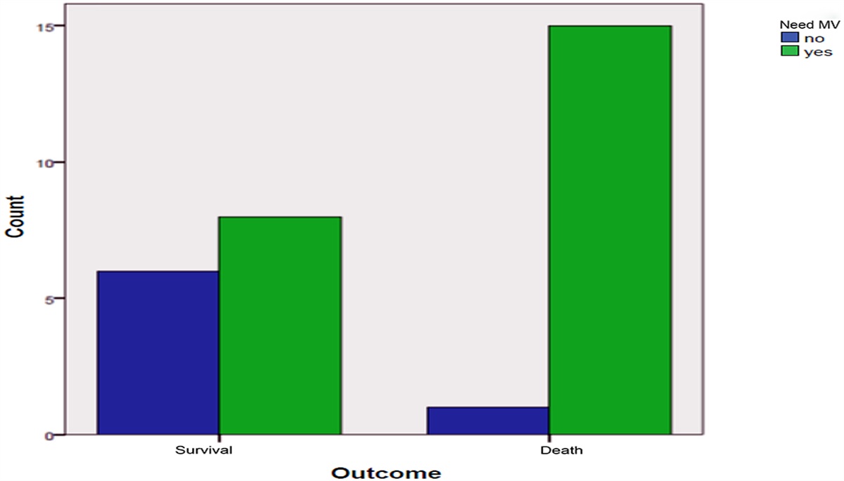
There was a significant lower rate or reintubation on the second day post-extubation in the VSV group compared to the oxygen mask group [1 pt (4%) versus 10 pts (40%) with P value = .005]. This was also noted on the seventh day post-extubation, 3 patients (12%) were reintubated in the VSV group versus 12 patients (48%) in the oxygen mask group (P = .012) (Table 2). The causes of reintubation in our study groups are shown in Table 3.
Table 2 - Outcome of the study groups Outcome VSV (n = 25) Oxygen mask (n = 25) P value (a) Reintubation day 2, number (%) 1 (4) 10 (40) .005 Reintubation day 7, number (%) 3 (12) 12 (48) .012 In-hospital mortality, number (%) 3 (12) 8 (32) .17 VAP, number (%) 2 (8) 6 (24) .24 LOS, mean (SD) 15.2 (5.9) 16.2 (5.7) .75Significant values with significant levels of 0.05. LOS, length of stay in ICU; VAP, ventilator-associated pneumonia; VSV, Volume Support Ventilation.
VSV, Volume Support Ventilation.
There was no difference between the VSV and oxygen mask groups in terms of in-hospital mortality, 3 of the 25 patients (12%) died in-hospital in the VSV group compared to 8 out of 25 patients (32%) in the oxygen mask group (P = .12) (Table 2). The incidence of VAP showed no significant difference between both groups (Table 2).
4 DiscussionTo the best of our knowledge, the present study is the first randomized controlled trial to perform prophylactic VS-NIV (volume target mode breath by breath) in patients post-extubation to prevent reintubation. In our study, we assessed the impact of volume support mode with a noninvasive interface on patients outcome after planned extubation. The implementation of a prophylactic Volume Support Noninvasive Ventilation protocol significantly reduced the rate of reintubation in our patients.
Many studies have suggested that post-extubation NIV could reduce the risk of reintubation, particularly in hypercapnic patients.15–17 In these studies, the rate of reintubation assessed within the 48 to 72 hours following extubation ranged from 19% to 24% with standard oxygen and from 8% to 11% with prophylactic NIV.15–17
On concordance to our study, some studies have found a significant reduction in the rate of reintubation.3,9,17,18 In the study by Nava and colleagues, prophylactic NIV applied at least 8 hours per day during the first 48 hours following extubation significantly reduced the rate of reintubation from 24% to 8% (P = .027). Another study observed a decrease in reintubation rate from 39% to only 5% in the group receiving NIV (P = .016).9
On the other side, Ferrer and colleagues stated that prophylactic NIV helped to reduce the risk of post-extubation respiratory failure and to decrease mortality, even though the reintubation rate in their study was not significantly decreased.19 Indeed, as NIV could be used as rescue therapy in case of respiratory distress, it enabled a number of patients from the control group receiving standard oxygen to avoid reintubation.
Thanthitaweewat et al. used target volume ventilation in comparison to oxygen mask and found no significant difference in the reintubation rate between the 2 groups this might be due to utilizing other rescue interventions besides reintubation when extubation failure developed, as stated in their study. Yet their study showed a significant decrease in the extubation failure and 2 days reintubation. Their study was one of the few studies using dual controlled modes post-extubation like ours.14
Conventional pressure support NIV was not tested against volume support NIV in our study. However, we hypothesized that the volume support NIV might be more convenient than conventional pressure support NIV due to steadier Vt (that was supported by unpublished data comparing both modes done in our department).
5 ConclusionProphylactic VSV used immediately post-extubation reduces the reintubation rate in medical ICU patients compared to oxygen mask, yet there is no evidence on its impact on mortality or VAP reduction.
6 Study limitationOur study had some limitations, which must be acknowledged. Being a single centre trial, multicentre study with larger sample size is warrant to evaluate the impact of VSV on mortality and ICU length of stay, beside we did not study the impact of longer duration VSV post-extubation, also comparing different NIV modes to VSV would have emphasized more of its potential benefit yet this was tried already in a non-published work by our researcher. For practical reasons, our investigator was not blinded to the administered mode. This unblinded design may have affected their assessment of respiratory symptoms and discomfort.
References [1]. Antonelli M, Bello G. Noninvasive mechanical ventilation during the weaning process: facilitative, curative, or preventive? Crit Care 2008;12(2):136. [2]. Ferrer M, Valencia M, Nicolas JM, et al. Early noninvasive ventilation averts extubation failure in patients at risk: a randomized trial. Am J Respir Crit Care Med 2006;173:164–170. [3]. Nava S, Gregoretti C, Fanfulla F, et al. Noninvasive ventilation to prevent respiratory failure after extubation in high-risk patients. Crit Care Med 2005;33:2465–2470. [4]. Brochard L, Mancebo J, Wysocki M, et al. Noninvasive ventilation for acute exacerbations of chronic obstructive pulmonary disease. N Engl J Med 1995;333:817–822. [5]. Gray A, Goodacre S, Newby DE, et al. Noninvasive ventilation in acute cardiogenic pulmonary edema. N Engl J Med 2008;359:142–151. [6]. Squadrone V, Coha M, Cerutti E, et al. Continuous positive airway pressure for treatment of postoperative hypoxemia: a randomized controlled trial. JAMA 2005;293:589–595. [7]. Bajaj A, Rathor P, Sehgal V, Shetty A. Efficacy of noninvasive ventilation after planned extubation: a systematic review and meta-analysis of randomized controlled trials. Heart Lung 2015;44:150–157. [8]. Thille AW, Boissier F, Ben-Ghezala H, et al. Easily identified at-risk patients for extubation failure may benefit from noninvasive ventilation: a prospective before-after study. Crit Care 2016;20:48. [9]. Ornico SR, Lobo SM, Sanches HS, et al. Noninvasive ventilation immediately after extubation improves weaning outcome after acute respiratory failure: a randomized controlled trial. Crit Care 2013;17(2):R39. [10]. Figueroa-Casas JB. Preventive use of noninvasive ventilation after planned extubation. Respir Care 2012;57:318–320. [11]. Su CL, Chiang LL, Yang SH, et al. Preventive use of noninvasive ventilation after extubation: a prospective, multicenter randomized controlled trial. Respir Care 2012;57:204–210. [12]. Branson RD, Davis K Jr. Dual control modes: combining volume and pressure breaths. Respir Care Clin N Am 2001;7:397–408. [13]. Jaber S, Delay J, Matecki S, Sebbane M, Eledjam J, Brochard L. Volume- guaranteed pressure-support ventilation facing acute changes in ventilatory demand. Intensive Care Med 2005;31(9):1181–1188. [14]. Chirakalwasan N, Thanthitaweewat V, Muntham D. Targeted-volume noninvasive ventilation reduces extubation failure in postextubated medical Intensive Care Unit patients: a randomized controlled trial. Ind J Crit Care Med 2018;22(9):639–645. [15]. Keenan SP, Sinuff T, Burns KE, et al. Canadian Critical Care Trials Group/Canadian Critical Care Society Noninvasive Ventilation Guidelines GroupClinical practice guidelines for the use of noninvasive positive-pressure ventilation and noninvasive continuous positive airway pressure in the acute care setting. CMAJ 2011;183:E195–E214. [16]. Epstein SK, Ciubotaru RL, Wong JB. Effect of failed extubation on the outcome of mechanical ventilation. Chest 1997;112:186–192. [17]. Epstein SK, Ciubotaru RL. Independent effects of etiology of failure and time to reintubation on outcome for patients failing extubation. Am J Respir Crit Care Med 1998;158:489–493. [18]. Ferrer M, Valencia M, Nicolas JM, Bernadich O, Badia JR, Torres A. Early noninvasive ventilation averts extubation failure in patients at risk: a randomized trial. Am J Respir Crit Care Med 2006;173(2):164–170. [19]. Ferrer M, Sellares J, Valencia M, et al. Noninvasive ventilation after extubation in hypercapnic patients with chronic respiratory disorders: randomised controlled trial. Lancet 2009;374(9695):1082–1088.
Comments (0)