Patient cell lines
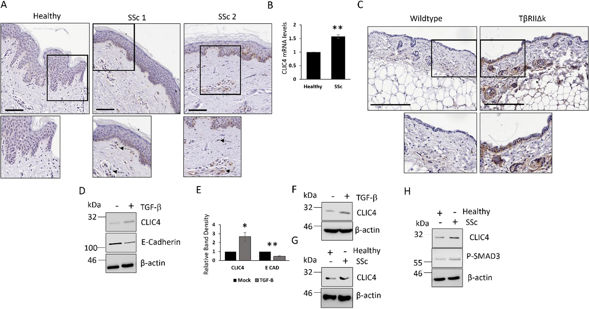
Full thickness skin biopsies were surgically obtained from the forearms of four adult healthy controls and four adult patients with recent onset SSc, defined as a disease duration of less than 18 months from the appearance of clinically detectable skin induration. All patients satisfied the 2013 ACR/EULAR criteria for the classification of SSc as defined by LeRoy et al. [13]. All participants provided written informed consent to participate in the study. Informed consent procedures were approved by NRES-011NE to FDG. Fibroblasts and keratinocytes were isolated and established as previously described [14]. Primary cells were immortalized using human telomerase reverse transcriptase (hTERT) to produce healthy control hTERT (N = 4) and SSc hTERT (N = 4).
Cell culture
hTERT patient fibroblasts (300,000 cells seeded per experimental condition) and the keratinocyte cell line HaCaT (600,000 cells seeded per experimental condition) (purchased from ATCC) were maintained in Dulbecco’s modified Eagle medium (DMEM) (Gibco) supplemented with 10% FBS (Sigma) and penicillin–streptomycin (Sigma). Primary SSc keratinocytes were maintained in keratinocyte growth media (Promocell). Human umbilical vein endothelial cells (HUVEC) (300,000 cells seeded per experimental condition) were maintained in ECGM2 media. Cells were treated with the chloride channel inhibitors (NPPB (25μM), IAA-94 (50μM)) or JAK1 inhibitor (Tofacitinib (2μM)) or TGF-β receptor/ALK5 inhibitor (SD-208 (1μM)) for 48h.
Trans-well co-culture experiments
Healthy and SSc fibroblasts were seeded onto 0.4-micron pore polyethylene terephthalate (PET) transmembranes (Corning). The transmembrane was inserted in wells containing an equal number of HaCaT. After 48 h, the well was removed and the HaCaTs were harvested for protein.
Immune agonist stimulation
Healthy dermal fibroblasts were serum starved for 24 h in DMEM containing 0.5% FBS and stimulated with Poly I:C (10 μg/ml), Poly dAdT (50 ng/ml), IFN-α2 (2 ng/ml), ODN2216 (1 μM) for 48h.
siRNA transfections
A pool of four siRNAs specific for different regions of CLIC4 (70nM final concentration) or a negative control scrambled siRNA (Qiagen) were transfected into HaCaTs or healthy and SSc patient fibroblasts cells using Lipofectamine 2000 (Thermo Fisher). Briefly the 4 siRNAs were combined in 300μl of Opti-MEM. 1μg/ml Lipofectamine was added to the transfection mixture and incubated for 20 min. The transfection mixture was added to the cells were and incubated for 48 h prior to harvesting.
Western blotting
Total proteins were extracted from fibroblasts in RIPA buffer and resolved by SDS-PAGE (10–15% Tris–Glycine). Proteins were transferred onto Hybond nitrocellulose membranes (Amersham biosciences) and probed with antibodies specific for α-smooth muscle actin (Abcam AB7817, 1/2000), CLIC4 (Santa Cruz sc135739, 1/2000), phosphorylated and total STAT1 (Cell signalling 9167, 9171, 1/1000), CTGF (Abcam AB209730 1/1000), phosphorylated IRF3 (Abcam AB75493, 1/1000), β-catenin (cell signalling 8480, 1/1000), phosho-SMAD3 (S423/S425) (Abcam AB52903, 1/1000), total SMAD3 (Cell signalling 9523, 1/1000), E-Cadherin (Santa Cruz sc8426, 1/1000), CXCL10 (Abcam AB9807, 1/1000), STING (Cell signalling 13,647, 1/1000) and β-Actin (Sigma A5441, 1/5000). Immunoblots were visualized with species-specific HRP conjugated secondary antibodies (Sigma) and ECL (Thermo/Pierce) on a Biorad chemiDoc imaging system. Western blot quantification was performed using Image J software.
Quantitative real time PCR
RNA was extracted from cells using commercial RNA extraction kits (Zymo Research). RNA (1ug) was reverse transcribed using cDNA synthesis kits (Thermo). QRT-PCRs were performed using SyBr Green PCR kits on a Thermocycler with primers specific for MX1 (Forward: CGACACGAGTTCCACAAATG Reverse: AAGCCTGGCAGCTCTCTACC), CXCL10 (Forward; GGTGAGAAGAGATGTCTGAATCC Reverse; GTCCATCCTTGGAAGCACTGCA), CXCL11 (Forward; TCCCCCATGTTCAAAAGAGGAC Reverse; ATATCTGCCACTTTCACTGCTTTTAC), IFIT1 (Forward; GACTGGCAGAAGCCCAGACT Reverse; GCGGAAGGGATTTGAAAGCT), CLIC4 (Forward: CATCCGTTTTGACTTCAGTGTTG; Reverse: AGGAGTTGTATTTAGTGTGACGA) and GAPDH (Forward; ACCCACTCCTCCACCTTTGA Reverse; CTGTTGCTGTAGCCAAATTCGT). Data were analysed using the ΔΔ Ct method. GAPDH served as a housekeeping gene. The experiments were run on the Quantstudio 5 Real-Time PCR system (Thermo Fisher).
TβRIIΔk transgenic mice experiment
All mice were housed in a clean conventional animal facility. Tissue samples were obtained from adult (age 12–18 week) sex matched littermate pairs (n = 3) from transgenic and non-transgenic mice genotypes by transgene specific PCR assay. The mouse strain genetic background in C57BL/6. Detailed description of the derivation and characterisation of the TβRIIΔk-fib strain [15].
Compresstome precision Microtome
3 mm skin biopsies were embedded in low melting point agarose. These tissue sections were sliced into 300 μm sections using the compresstome vibrating microtome (Precisionary). The sections were grown in DMEM (2% FBS) and stimulated with TGF-β (5ng/ml, Sigma Aldrich) plus NPPB for 48h. The sections were harvested for RNA.
Immunohistochemistry
Immunohistochemistry was performed as previously described [1]. Sections were stained with CLIC4 (1/200) antibody, visualised using an HRP conjugated mouse secondary and DAB substrate. The sections were counterstained with haematoxylin. Skin sections from age matched wildtype and TβRIIΔK-fib transgenic mouse model of human SSc were also stained for CLIC4 expression in additional control experiments.
SSc fibroblast conditioned media stimulation
Sub-confluent healthy and SSc dermal fibroblasts were grown in sera depleted DMEM for 48 h. The media was collected and centrifuged at 2000 g for 30 min. The media was added to HaCaT cells neat for 48 h or to HUVECs cells neat for 24 h.
IRF-Lucia Luciferase reporter assay
Thp1 dual cells (Invivogen thpd-nfis) were stimulated with conditioned media from healthy and SSc dermal fibroblasts for 48 h. The media was collected from the Thp1 cells after 48 h and IRF reporter activity was measured using the QUANTI-Luc™ 4 Lucia/Gaussia, a Lucia and Gaussia luciferase detection reagent.
Immunofluorescence
HaCaT were seeded onto coverslips. After stimulation/treatment, the cells were fixed in 4% paraformaldehyde and permeabilised with 0.1% trition- × 20 for 10 min. The cells were stained with an CLIC4 antibody and visualised with a secondary antibody conjugated to alexa-594. Nuclei were visualised by DAPI contained within the mounting media. Scale bars represent 20 μm.
Exosome isolation and stimulation
Exosomes were isolated following the protocol previously described [16]. HaCaT were stimulated with 1% total volume of exosomes for 48 h.
Analysis of single cell RNA sequencing dataset
The public scRNA-seq dataset GSE138699 [17], consisting of 12 systemic sclerosis (SSc) and 10 healthy control samples, was obtained from the Gene Expression Omnibus. Data processing was performed using the Seurat package v4 in R, including quality control (minGene = 200 maxGene = 5000 pctMT = 30), multi-sample integration using harmony package, normalization, and dimensional reduction (PCA and UMAP). A nearest neighbour graph was constructed using FindNeighbors, followed by clustering with FindClusters at a resolution of 0.8. Clusters were manually annotated based on top marker genes. CLIC4 expression was visualized across clusters, and statistical differences in expression levels between SSc and healthy controls in keratinocyte clusters were assessed using the Wilcoxon rank-sum test in the rstatix package.
Statistical analysis
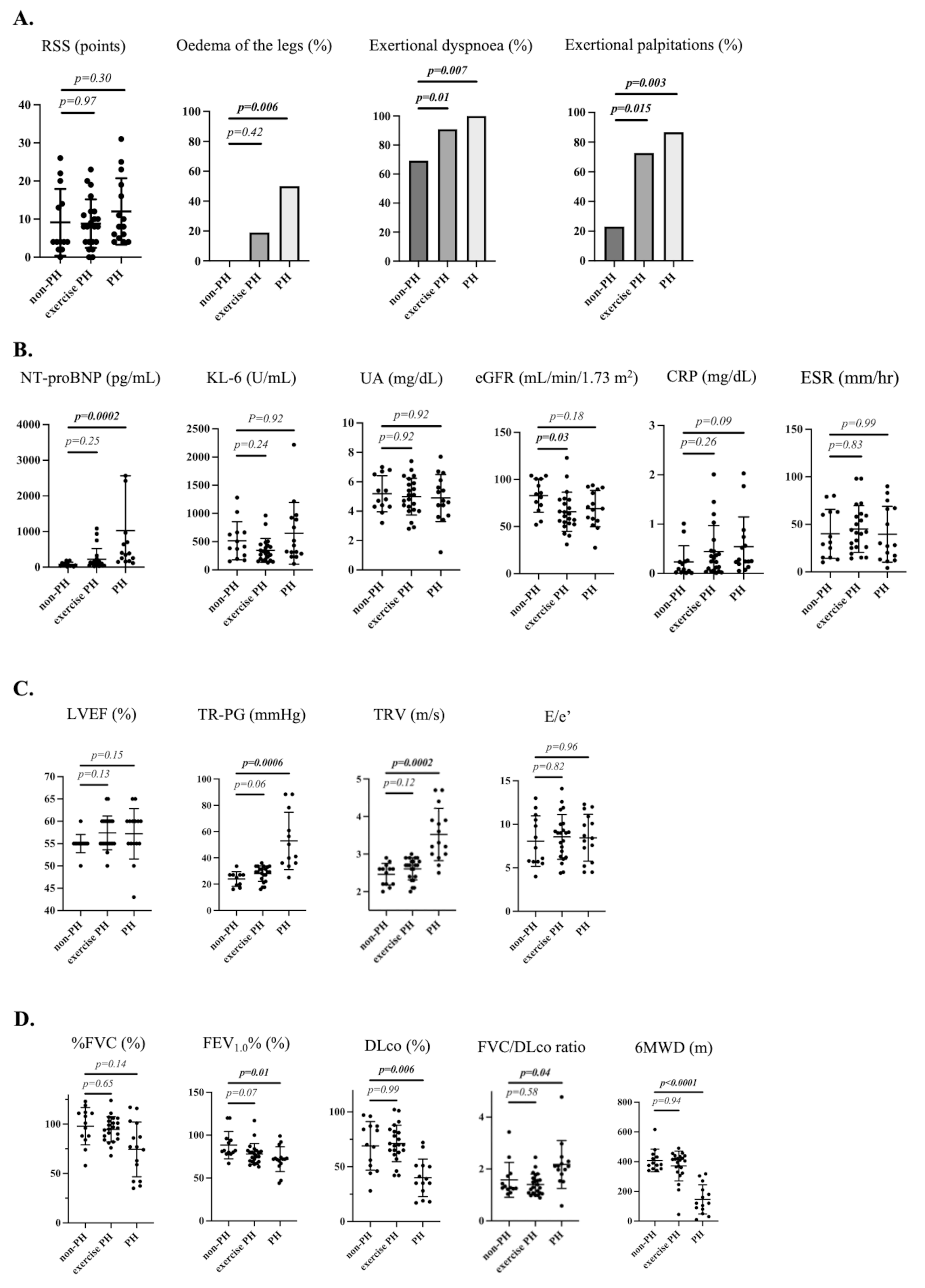
Data are presented as the mean ± standard error. Statistical analysis was performed using a two-tailed, unpaired Student’s t-test for comparisons of two groups. Multiple groups were analysed by one-way ANOVA, followed by post hoc multiple comparisons using GraphPad Prism 10.4.1.


Comments (0)