Cells, media, and reagents
SBR (PubChem CID: 68486, purity = 99.98%) was obtained from Shanghai Haoyuan ChemExpress Co. Ltd. (Shanghai, China; Item No. HY-N1196, CAS number: 581-31-7). RA-FLS and bone marrow-derived macrophages (BMDMs) were grown in Dulbecco’s modified Eagle’s medium (DMEM) (Gibco, USA) with 10% fetal bovine serum (FBS) (Gibco, USA) and 100 U/mL penicillin–streptomycin double antibiotics (HyClone, USA). Roswell Park Memorial Institute (RPMI) 1640 culture medium (Gibco, USA) was used for additional experiments. Type IV collagenase and Escherichia coli bacterial lipopolysaccharide (LPS) were obtained from Sigma-Aldrich (St. Louis, USA). Recombinant mouse IL-4 and interferon-γ (IFN-γ) were procured from PeproTech (Cranbury, USA), while recombinant mouse macrophage colony-stimulating factor (M-CSF) was obtained from BioLegend (San Diego, USA). Recombinant human TNF-α was acquired from Sino Biological (Beijing, China).
Isolation and cultivation of FLS
In a sterile operating room environment, synovial tissue was acquired from individuals diagnosed with RA from West China Hospital, considering the 1987 classification criteria of the American College of Rheumatology (ACR) and the 2010 classification criteria of the ACR/European Alliance against Rheumatism; exclusion of other diseases was also confirmed. The excised synovial tissues underwent meticulous processing, including the elimination of surplus adipose and connective tissue. The synovial tissues were enzymatically digested in an RPMI-1640 medium containing 0.5 mg/mL of type IV collagenase with shaking at 37 °C for 2 h. After filtration and centrifugation, the resultant cell pellet was resuspended in DMEM supplemented with 10% FBS and 1% penicillin–streptomycin. The isolated cells were cultured at 37 °C and 5% CO2. Cells from passages three to five were utilized in the subsequent experiments. This study was approved by the Ethics Committee of West China Hospital, Sichuan University (approval number: 20201151). Informed written consent was secured from all participating individuals.
Polarization of BMDMs
BMDMs were isolated from C57BL/6J wild-type mice, according to a previously established protocol [18]. The retrieved cells were cultured in DMEM supplemented with 20 ng/mL M-CSF, at 37 °C and 5% CO2. The medium was refreshed after 5 d of cultivation. After 7 d, BMDMs were induced to differentiate into M1 macrophages by adding 250 ng/mL LPS and 25 ng/mL IFN-γ. Alternatively, BMDMs differentiated into M2 macrophages after induction with 20 ng/mL IL-4.
Cytotoxicity of SBR
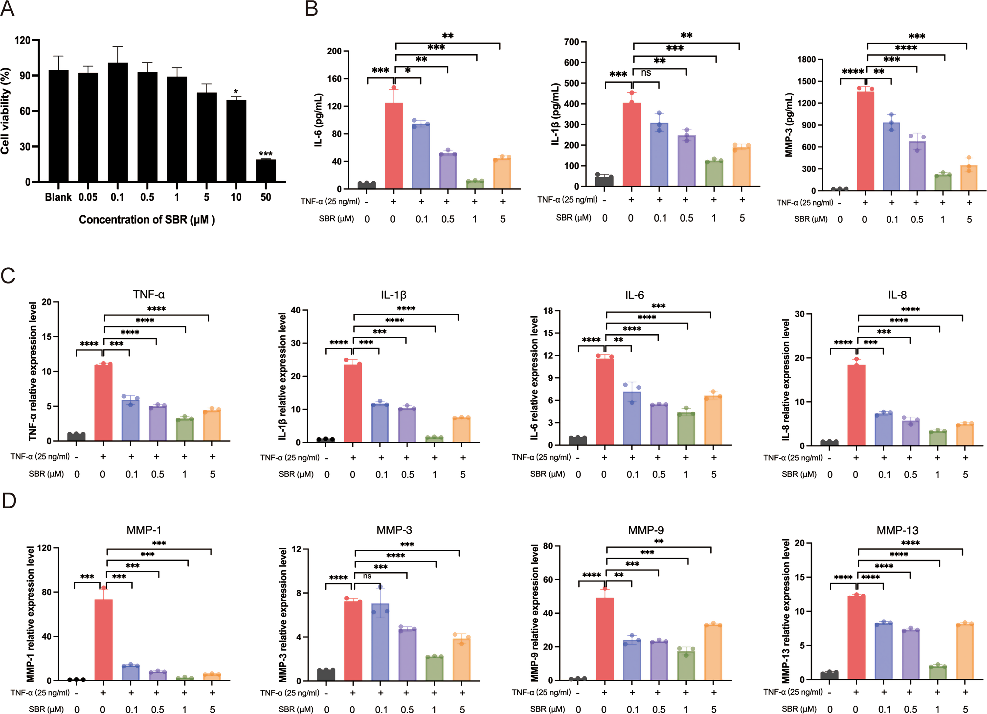
RA-FLS viability was determined by CCK-8 assay as described previously [2]. Approximately 5,000 RA-FLS were cultured in 96-well plates. After 24 h, added in different concentrations SBR. After 24 h, cells were washed and incubated in 90 µL DMEM with 10 µL of CCK8 reagent (GlpBio, USA). After 3 h, the absorbance at 450 nm were measured.
Drug treatment
For cell experiment, SBR was solubilized in dimethyl sulfoxide (DMSO) to achieve a stock concentration of 20 mM and preserved at -80 °C. For RA-FLS, the working concentrations were sequentially 0.1 µM, 0.5 µM, 1µM and 5µM. For macrophage, the working concentrations were 0.1 µM and 1µM. Cells were treated with 20µM Itacitinib (SelleckChem, USA), 100 µM STAT3 inhibitor (S3I-201) (SelleckChem) or 10 µM STAT6 inhibitor (AS1517499) (SelleckChem). For animal experiment, SBR was diluted with PEG300, Tween-80, and normal saline to various concentrations. Starting on the 21st day after the establishment of the CIA model, SBR was administered to mice via daily oral gavage using a syringe. Dosage options for SBR included a low-concentration group (0.5 mg/kg/d) and a high-concentration group (2 mg/kg/d). The model and normal groups received equivalent volumes of solvent.
Inducing and evaluating CIA
For the CIA model, male DBA/1 mice aged eight weeks were procured from Beijing Huafukang Biotechnology Co. Ltd. (Beijing, China). A total of 28 mice were randomly allocated to four groups: control, CIA, 0.5 mg/kg/d SBR, and 2 mg/kg/d SBR, with seven mice in each group. CIA model induction involved anesthetizing the mice via intraperitoneal injection of pentobarbital sodium. Concurrently, chick type II collagen (Chondrex, USA) was emulsified with complete Freund’s adjuvant (Chondrex) at a 1:1 ratio. Subsequently, the emulsion (100 µL) was subcutaneously administered at the base of the tail (1.5–2 cm from the root). Twenty-one days after the first immunization, a newly prepared emulsion (100 µL per mouse) was injected subcutaneously at a site close to the root of the tail. Following enhanced immunization, joint inflammation was assessed every other day, with scores assigned as follows: 0, normal; 1, red or swollen on one finger, 2 red or swollen on two fingers or more, 3 moderate redness and swelling throughout the entire wrist/ankle or all toes; and 4, severe redness and swelling throughout the entire wrist/ankle or all toes. The maximum score for each limb was 4, and the maximum score for each mouse was 16 [13]. On day 42 of modeling, the mice were humanely euthanized and both blood and joint specimens were collected. The animal studies were approved by the Animal Ethics Committee of the West China Hospital, Sichuan University (approval number: 20221103002).
Hematoxylin and eosin (H&E) staining and histological evaluation
The collected joint specimens were fixed with 4% paraformaldehyde (Servicebio, China) at 25 °C for 1 d and then decalcified at 4 °C using 10% EDTA (pH = 7.4). The decalcification solution was replaced every 2 d. After completion of decalcification (one month later), the joint specimens were embedded in paraffin and sectioned into 5-µm-thick slices. Slices were successively deparaffinized in xylene and hydrated in a gradient ethanol solution. Subsequently, H&E staining was performed using an H&E staining kit (Servicebio). The stained sections were sealed with resinene at 25 °C and observed under a light microscope (Leica, Germany). H&E-stained sections of the knee and ankle joints of mice were scanned using a SLIDEVIEW™ VS200 Slide Scanner (Olympus, Japan). Following published scoring criteria [19, 20], H&E-stained sections were evaluated for three aspects: inflammation, cartilage destruction, and bone erosion.
Immunofluorescence
BMDMs were inoculated directly onto round coverslips. After differentiation, cells were treated with 0.1 µM or 1 µM SBR for 1 h. Subsequently, BMDMs were polarized into either M1 or M2 macrophages under different conditions and fixed with 4% paraformaldehyde (Servicebio) at 25 °C for 10 min. For animal tissues, mouse knee tissue sections required antigen retrieval at 95 °C using Tris-EDTA buffer (Servicebio) at pH 9. Following retrieval, sections were blocked with 5% goat serum (Servicebio) at 25 °C for 30 min. M1 macrophages were stained overnight at 4 °C with anti-F4/80 (1:100, ab6640; Abcam, UK) and anti-iNOS (1:100, AF0199; Affinity, China) primary antibodies. For M2 macrophages, overnight staining at 4 °C was performed using anti-F4/80 (1:100, ab6640; Abcam) and anti-CD206 (1:100, DF4149; Affinity) primary antibodies. After three washes with 1× phosphate-buffered saline (PBS) containing Tween® detergent, the sections were exposed to fluorescent secondary antibodies at 37 °C for 1 h. Subsequently, nuclei were stained with 4’,6-diamidino-2-phenylindole (Abcam, USA). Imaging was performed using an inverted fluorescence microscope (Leica, Germany).
Enzyme-linked immunosorbent assay (ELISA)
Serum samples were collected from normal control, model, and SBR treatment groups. The concentrations of TNF-α, IL-1β, IL-6, and MMP-3 were assessed using ELISA kits (Ruixinbio, China). Cell cultural supernatants were collected and centrifuged with 300 g for 10 min, the IL-1β, IL-6, and MMP-3 levels of RA-FLS were tested by human ELISA kits (Multi Sciences, China). In M1 macrophage, the levels of TNF-α and IL-6 were assessed, in M2 macrophage, the levels of IL-10 was detected by mouse ELISA kits (Multi Sciences).
Western blot analysis
RA-FLS were pretreated with 1 µM SBR for 2 h, then stimulated with 25 ng/mL TNF-α for 15, 30, 60, 120, or 240 min, and the cells were finally harvested. BMDM was polarized to M1 and M2 as described above, then treated with different concentrations of SBR for 24 h, and the cells were finally harvested. total protein of cells was isolated using radioimmunoprecipitation assay (RIPA) buffer (Solabio, China) supplemented with a protease and phosphatase inhibitor cocktail (MCE, China). Synovial tissues were collected from the joints of mice, and placed into 1.5 mL centrifuge tube containing Grinding beads, RIPA, protease and phosphatase inhibitor cocktail, and ground for 20 min at 4 °C by a cryogenic tissue grinder (Servicebio, China). Then, centrifuged and the protein-containing supernatant was collected.
Protein concentration was determined using the bicinchoninic acid protein assay kit (Beyotime, China), and the protein samples were mixed with a loading buffer (Beyotime), then were heated in a metal bath at 95 °C for 10 min. Subsequently, western blot was performed as described previously [2]. Equal amounts of the protein samples were subjected to sodium dodecyl sulfate-polyacrylamide gel electrophoresis and then transferred onto polyvinylidene fluoride membranes (0.45 μm). The membranes were blocked with a rapid blocking buffer (EpiZyme Biotech, China) at 25 °C for 20 min and then incubated overnight at 4 °C with the respective primary antibodies. The following primary antibodies were used: anti-JAK1 (1:1000, 66466-1-Ig; Proteintech, Wuhan, China), anti-p-JAK1 (1:1000, 3331; Cell Signaling Technology [CST], USA), anti-signal transducer and activator of transcription (STAT) 3 (1:1000, 60199-1-Ig; Proteintech), anti-p-STAT3 (1:1000, 9145T; CST), anti-STAT6 (1:1000, 5397 S; CST), anti-p-STAT6 (1:1000, 9361T; CST), and anti-GAPDH (1:10000, ET1601-4; Huabio, China). Membranes were washed three times with TBST and subsequently incubated with horseradish peroxidase-conjugated secondary antibodies at 25 °C for 90 min. After incubation, the membranes were washed, incubated with an enhanced chemiluminescence substrate (Abbkine, China), and imaged using an Image Lab system (Bio-Rad Laboratories, USA).
Quantitative real-time-PCR (qRT-PCR)
Total RNA was isolated from cells using the RaPure Total RNA kit (Magen, China) and from joint tissues using the TRIzol reagent (Invitrogen, USA). Subsequently, mRNA was reverse transcribed into cDNA using 5× All-In-One RT Master Mix (Applied Biological Materials, Richmond, Canada). qRT-PCR was conducted on the CFX96 System (Bio-Rad Laboratories) using 2× qPCR MasterMix (Applied Biological Materials). The resulting values were standardized to the expression levels of Ga mRNA and analyzed utilizing the 2-ΔΔCq method. Primer sequences used are listed in Additional File 1: Table S1.
Flow cytometry
Briefly, BMDMs were treated with 0.25% trypsin, and after centrifugation, they were resuspended in PBS supplemented with 1% bovine serum albumin. F4/80-PE (565410; BD Pharmingen, USA) and CD11b-FITC (101206; BioLegend) were used as markers of non-polarized macrophages. CD80-BV711 (104743; BioLegend) was used as a marker of M1 macrophages, whereas CD206-APC (141707; BioLegend) was used as a marker of M2 macrophages. Flow cytometry results were analyzed using FlowJo v10.8.1 software.
Suberosin analysis based on network pharmacology
The SwissTargetPrediction website (http://swisstargetprediction.ch/) was used to predict the prospective targets of SBR. The GeneCards website (https://www.genecards.org/) was used to identify the targets associated with RA. Venny2.1.0 (https://bioinfogp.cnb.csic.es/tools/venny/index.html) was employed to identify the intersection of these target sets, revealing 60 overlapping target genes as candidate targets of SBR in RA treatment. Gene Ontology (GO) and Kyoto Encyclopedia of Genes and Genomes (KEGG) analyses were performed using the DAVID website (https://david.ncifcrf.gov/).
Protein-protein interaction (PPI) analysis was conducted using STRING (http://string-db.org/, ver.10), and the results were visualized using Cytoscape (ver3.8.2; http://cytoscape.org/). The TargetNet website (http://targetnet.scbdd.com) was used to explore the potential combined targets of SBR. Molecular docking studies of drugs and targets were performed using the COACH-D site (https://yanglab.nankai.edu.cn/COACH-D).
Statistical analysis
The experimental results are presented as the mean ± standard deviation (M ± SD). All statistical analyses were performed using Prism 8 software. Statistical significance was assessed using the unpaired two-tailed Student’s t-test for comparisons between two groups, whereas one-way analysis of variance followed by Tukey’s multiple comparison test was used for comparisons among multiple groups. Statistical significance was set at P < 0.05.


Comments (0)