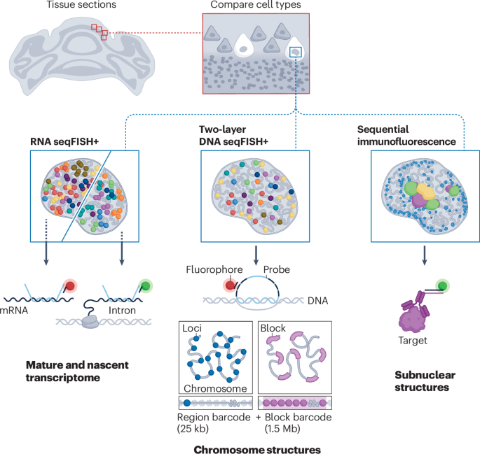
Spatial multi-omics of nuclear architecture with two-layer seqFISH+
The mammalian nucleus is organized into higher-order structures that orchestrate cell-type-specific gene expression programmes. Technologies that directly and simultaneously measure spatial relationships between DNA, RNA and protein molecules within individual nuclei offer unique opportunities to understand how specific nuclear architectures relate to gene regulation at the single-cell level. In recent years, imaging-based multimodal technologies have emerged to profile the subnuclear organization of various molecular species in single cells. However, owing to optical crowding caused by overlapping fluorescent signals in densely labelled regions, these genome-wide technologies have been limited to coarse visualization at the megabase scale, constraining our understanding of spatial relationships between nuclear architecture and gene regulation in sub-megabase regions.
The key technical advancement is the two-layer seqFISH+ barcoding strategy, which extends the capabilities of seqFISH. Previous studies demonstrated that DNA loci within a megabase genome region can be spatially confined within approximately 300 nm in the nucleus, creating optical crowding that prevents individual loci from being resolved by conventional seqFISH barcoding. We overcome this limitation through a two-layer barcoding approach in which we first image 60 25-kb DNA loci in each 1.5-Mb chromosome block across the genome in parallel (region barcode) and then assign unique barcodes to each chromosome block (block barcode). The identity of each 25-kb DNA locus is decoded using the combination of its region and block barcodes, with physical localizations super-resolved in three-dimensional (3D) space. This two-layer DNA seqFISH+ strategy drastically increases genome-wide coverage from thousands to over 100,000 DNA loci.

Comments (0)