Occlusive primary endobronchial amyloid tumour: a rare case
A female aged 69 year never smoker presented with 4 months of non-resolving productive cough and shortness of breath on exertion following mild COVID-19 infection. Her background history was significant for tuberculosis infection in 1996, for which she completed a full course of treatment.
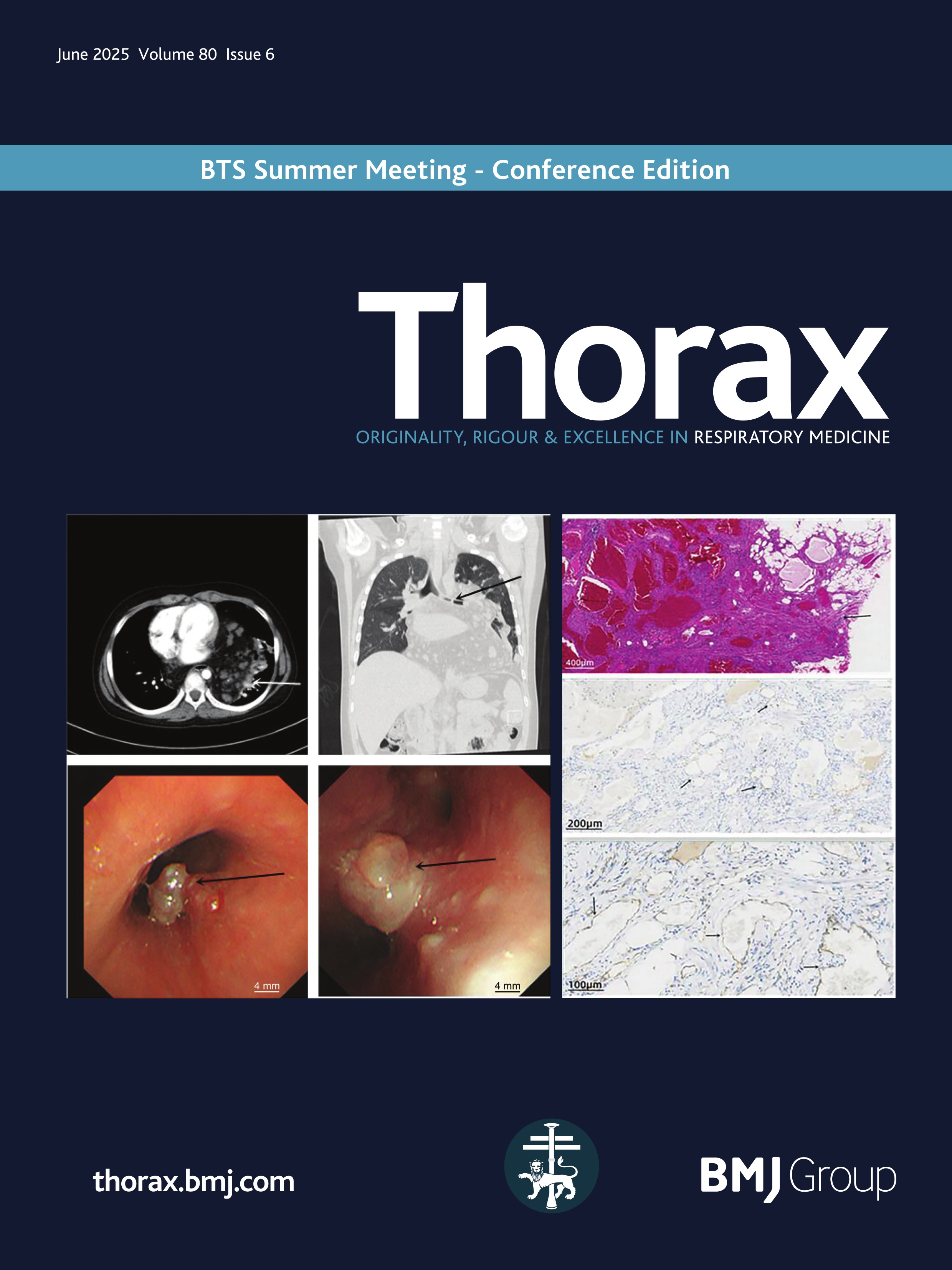
An initial CT scan of the chest found bulky left hilar and mediastinal lymphadenopathy with heterogeneous calcification. A subsequent fluorodeoxyglucose-positron emission tomography CT demonstrated intense metabolic activity in these lymph nodes. Further CT chest with contrast (figure 1a, c) showed interval development of new left upper lobe collapse with stable lymphadenopathy. Lung function was within normal limits. Bronchoscopic examination demonstrated a large obstructing mass in the left upper lobe bronchus (figure 2a), with subtotal occlusion of the left lower lobe bronchus (figure …


Comments (0)