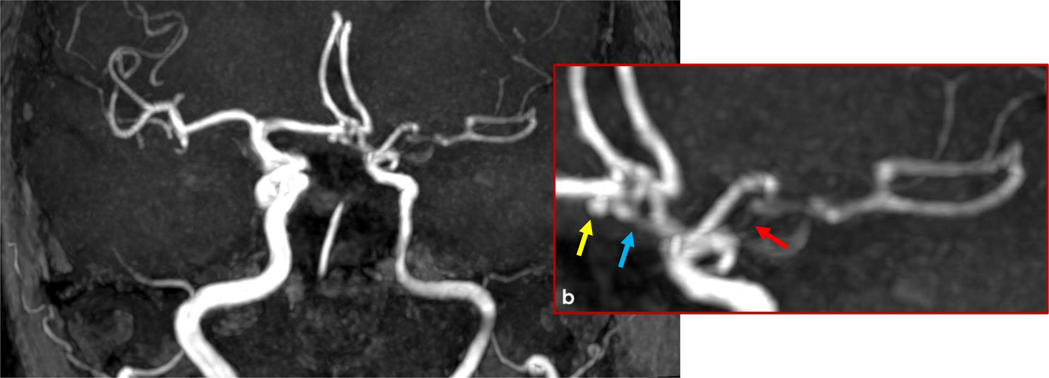
Detailed knowledge of the anatomy of the OA is of utmost importance to avoid potential disastrous complications during therapeutic embolisation of neoplastic or vascular processes fed by the OA [2]. Knowledge of the course and diameter of the OA is also important because the OA is used as a donor vessel in extra-to-intracranial bypasses to the cerebral circulation [2]. As a companion vessel of the PBD, the OA crosses usually over the IJV. The OA does not have a constant vertical level of origin from the ECA, nor a single landmark for a safe identification [3]. To our knowledge, the course of the OA deep to the IJV was not reported previously. However, when the suboccipital segment of the OA is approached, such as in vertebral artery revascularization [7], this modified course may not be of great relevance. The OA can be involved in dural arteriovenous fistulas and is a feasible artery for the embolisation of these [6]. True aneurysms and pseudoaneurysms can develop in the OA and may be managed through either surgical resection or endovascular embolization [6]. Vascular lesions are a significant but often overlooked cause of parotid region masses [10]. An intraparotid OA represents a novel, yet plausible site for such vascular pathologies, and should be included in the differential diagnosis of parotid swellings.
OA pseudoaneurysm secondary to trauma during radical neck dissection is rare but potentially life-threatening, usually presenting as a progressively enlarging pulsatile mass [14]. When the OA originates within the parotid gland, as in our case, a pseudoaneurysm may also occur as a complication following parotid surgery. This anatomical variation increases the risk of iatrogenic vascular injury and may necessitate urgent endovascular embolization to prevent hemorrhagic or neurologic complications. Surgeons should be aware of this possibility, particularly in cases involving unexpected vascular structures within the parotid space.
The OA can also be used for intraoperative angiography [6]. On the other hand, when the carotid triangle is surgically dissected, an OA running beneath the IJV may appear as a falsely absent OA. The distinction between true and false OAs may be determined through imaging examinations. It has also been reported that the sternomastoid branch of the occipital artery (SBOA) demonstrates a consistent size and location within the reflected fascia of the sternomastoid muscle [11]. Notably, the spinal accessory nerve was within 11 mm of the SBOA in 100% of cases [11]. However, in cases where the OA has intraparotid origin, this anatomical variation may compromise the reliability of the SBOA as a visual landmark for locating the spinal accessory nerve, thereby increasing the risk of nerve injury during neck dissection. Surgeons may misidentify small muscular branches or accessory arteries as the SBOA, potentially leading to disorientation and heightened risk during surgical navigation.
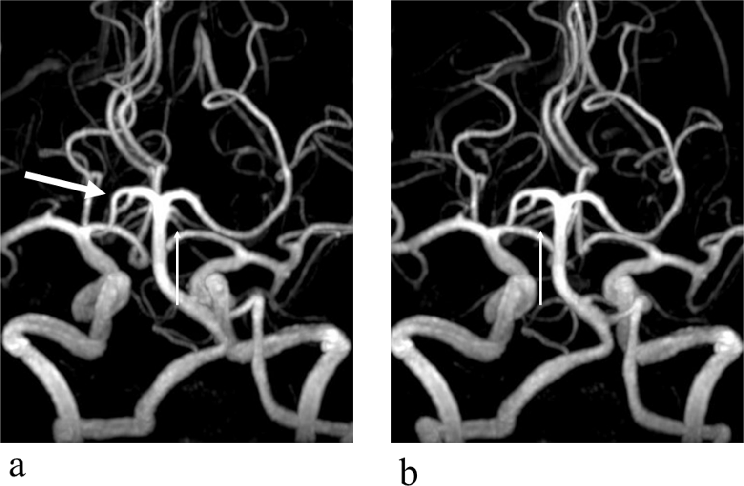
Successive branches of the ECA may form various common trunks of origin, such as the thyrolingual, thyrolinguofacial, linguofacial, and occipitoauricular trunks [8, 15]. They must be carefully distinguished when approaching the carotid axis.
Although the LFT was reported consistently, a recent study demonstrated that its morphology and topography are individually variable and should be evaluated on a case-by-case basis [5]. The unilateral linguofacial trunk was found to have a prevalence of 15.38% [15]. This variant was identified in 2 out of 4 cases reported here.
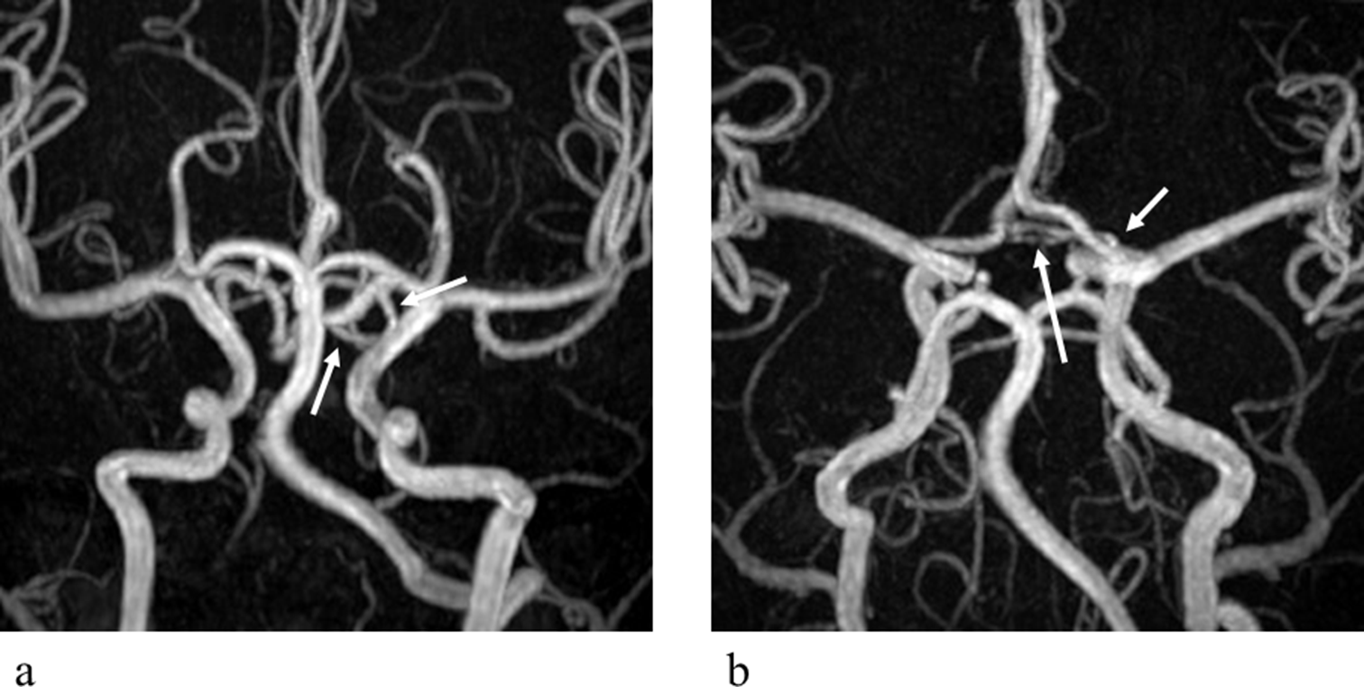
The occipitoauricular trunk occurs in 14% of cases [8]. This report describes a variant that differs from previously documented cases. Specifically, it originates above the PBD, deep to the parotid gland, and arises from the superficial temporal artery rather than the ECA, representing a novel anatomical variation. Consequently, during dissection of the intraparotid portion of the superficial temporal artery, caution should be exercised to identify a possible aberrant origin of an occipitoauricular trunk.


Comments (0)