
Remember me


A 27-year-old female presented to our hospital with a chief complaint of generalized reddish skin papules and vesicles that had persisted for 6 days. The patient reported concurrent onset of fever and pruritus, with the rash appearing uniformly across her body. She disclosed recent contact with a family member diagnosed with VZV infection. Initially, the patient sought care from an infectious disease specialist, who prescribed acetaminophen 500 mg twice daily for symptomatic relief of pain and fever, along with acyclovir 800 mg five times daily. Outpatient management was deemed appropriate at that time. However, despite adhering to the prescribed regimen for 6 days, the patient experienced no significant improvement in her symptoms, prompting her visit to our facility. Upon admission, given the severity of her condition and the lack of response to oral antiviral therapy, intravenous acyclovir therapy was initiated immediately at a dose of 10 mg/kg every 8 h. This decision was made in accordance with current guidelines for the management of severe VZV infections.
Upon presentation, the patient reported a new onset of epigastric abdominal pain that had developed over the preceding 48 h. She characterized the pain as constant and non-colicky, without radiation to other abdominal regions. Accompanying her abdominal discomfort, the patient experienced a significant loss of appetite, coupled with nausea and vomiting. Importantly, she denied experiencing diarrhea, or constipation. She also reported no recent consumption of suspect foods or contact with individuals exhibiting similar gastrointestinal symptoms. Specifically, there was no history of ingestion of fava beans, legumes, or other foods known to trigger hemolysis in individuals with G6PD deficiency.
Review of the patient’s past medical history revealed glucose-6-phosphate dehydrogenase (G6PD) deficiency diagnosed in childhood but was otherwise unremarkable. She denied any history of diabetes mellitus, hypertension, cardiovascular disease, hepatic disorders, or other chronic medical conditions. The patient had no prior surgical interventions and reported no known allergies to foods, medications, or environmental factors. She affirmed abstinence from tobacco, alcohol, and illicit substances.
The patient’s family history was non-contributory, with no reported hereditary or congenital disorders.
The patient reported strict adherence to her prescribed medications. She denied the use of any other prescription medications, over-the-counter drugs, or dietary supplements.
Physical examination revealed a young woman in mild distress. Vital signs were as follows: temperature 38.9 °C, heart rate 92 beats per minute, respiratory rate 18 breaths per minute, blood pressure 118/76 mmHg, and oxygen saturation 98% on room air.
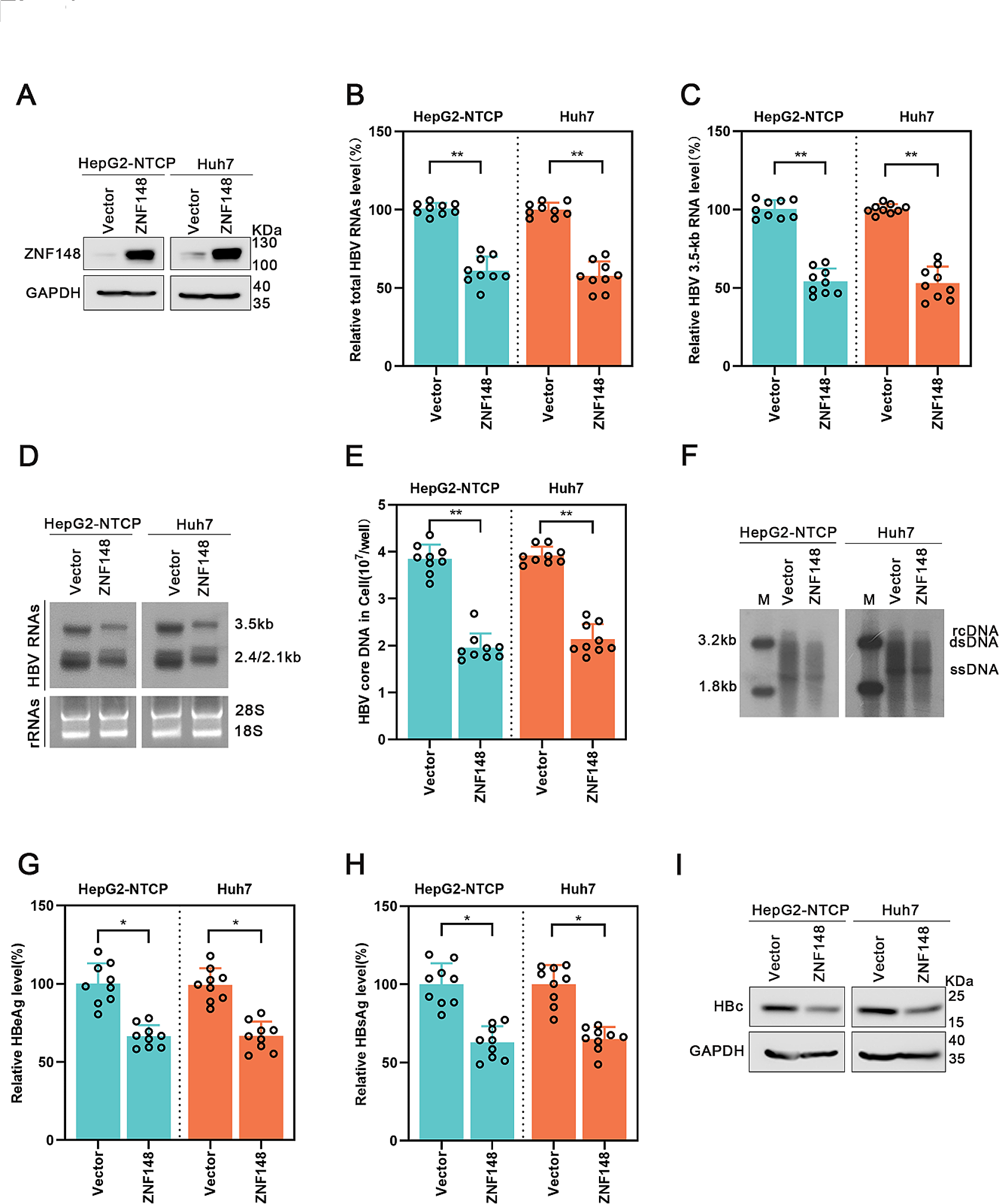
Positive findings included generalized icterus, scleral icterus, conjunctival pallor, mild tenderness in the epigastric region and right upper quadrant, and a diffuse erythematous rash with papules and vesicles in various stages of evolution. The lesions were distributed across the trunk, extremities, face, and scalp.The rest of the systemic examination, including respiratory, cardiovascular, neurological, and musculoskeletal evaluations, was unremarkable (Fig. 1).
Fig. 1
Papules and Vesicles Spread All Over the Body Due to Varicella Zoster Infection
These findings, particularly the evidence of jaundice and abdominal tenderness, suggest a potential hepatobiliary involvement that warrants further investigation in the context of the patient’s presenting symptoms and history.
Following the initial assessment and physical examination, we proceeded with a comprehensive diagnostic workup. Laboratory investigations included a complete blood count, liver function tests, coagulation profile, pancreatic enzymes, and viral hepatitis markers. Testing for Hepatitis A, B, C, and E viruses was performed. The results for Hepatitis A, B, and C were negative. Hepatitis E virus (HEV) serology and PCR testing were also conducted and returned negative, ruling out HEV as a contributing factor in this case. This comprehensive testing confirmed that the acute hepatitis was not attributable to other common viral pathogens, further substantiating the diagnosis of VZV-associated fulminant hepatitis. The results of laboratory exams are summarized in Table 1.
Table 1 Laboratory examination results at admissionAn abdominal ultrasound was performed to evaluate the hepatobiliary system. The imaging revealed increased echogenicity of the liver parenchyma with slight enlargement, measuring 16 cm in the midclavicular line. Mild periportal echogenicity was noted, suggesting edema around the portal tracts. The gallbladder appeared normal without evidence of stones or wall thickening, and the common bile duct measured 5 mm in diameter with no signs of obstruction. The pancreas was unremarkable, while the spleen showed mild enlargement at 13 cm in length.
These findings, combining laboratory results and imaging studies, strongly suggest acute hepatitis, likely associated with the recent VZV infection. While drug-induced liver injury is a potential consideration, the minimal dosage of acetaminophen (500 mg twice daily) prescribed to the patient makes this an unlikely contributor to the observed fulminant hepatitis. Instead, the clinical presentation and findings strongly support varicella zoster virus infection as the primary etiology, compounded by the patient’s underlying G6PD deficiency.
Following the initial assessment and diagnostic workup, a polymerase chain reaction (PCR) test for VZV was performed. The test revealed a strongly positive result with a high viral load, indicating active and severe VZV infection. This finding confirmed the diagnosis of VZV-associated fulminant hepatitis.
A liver biopsy was also performed to further evaluate the etiology of the acute liver failure. Autoimmune hepatitis (AIH) was considered as a potential differential diagnosis due to the rapid progression of the patient’s condition. Although AIH-related serology was not performed due to logistical constraints, histopathological examination of the liver tissue showed no lymphocytic infiltration or features suggestive of AIH, effectively ruling out this diagnosis. The absence of serological and histological markers of autoimmunity, combined with the positive PCR result for VZV, confirmed VZV-associated fulminant hepatitis as the etiology. Histopathological examination of the liver tissue showed extensive hepatocellular necrosis without significant lymphocytic infiltration, granulomas, or features suggestive of autoimmune hepatitis. Additionally, PCR analysis of the liver biopsy specimen confirmed the presence of VZV DNA. Although serum PCR had already established systemic VZV infection, the liver biopsy PCR was performed to directly demonstrate the presence of VZV DNA within the liver tissue. This step was critical in confirming the liver as the site of active viral replication and damage, thereby substantiating the diagnosis of VZV-associated fulminant hepatitis. The absence of histological and serological markers of autoimmunity, combined with the positive PCR result for VZV, excluded autoimmune hepatitis as the underlying cause of liver failure in this patient.
Given the severity of the patient’s condition and the potential for infectious complications, she was promptly isolated and admitted to the Intensive Care Unit (ICU) for close monitoring and aggressive management. This decision was made to ensure optimal care and to prevent potential nosocomial spread of the VZV infection.
Upon admission to the ICU, the patient was immediately placed on a comprehensive conservative management protocol. This included the initiation of intravenous acyclovir therapy at a dose of 10 mg/kg every 8 h, in addition to intravenous hydration with balanced crystalloid solutions to maintain adequate fluid balance and support renal function. The decision to escalate to intravenous antiviral therapy was based on the severity of her condition and the lack of response to oral acyclovir therapy during the preceding 6 days. Vital signs were monitored closely, with assessments performed every six hours to detect any early signs of clinical deterioration or hemodynamic instability.
N-acetyl cysteine (NAC) was not administered in this case, as the primary etiology of the acute liver failure was identified as varicella zoster virus (VZV)-associated fulminant hepatitis, compounded by the patient’s underlying G6PD deficiency [13]. The prescribed dosage of acetaminophen (500 mg twice daily) was minimal and well below the threshold for hepatotoxicity, making acetaminophen-induced liver injury an unlikely contributor. Additionally, there was no clinical or laboratory evidence of oxidative liver injury that would have necessitated the use of NAC.
A rigorous laboratory monitoring schedule was implemented to track the progression of hepatic dysfunction and assess for potential complications. This included:
1.Complete blood count (CBC) performed daily to monitor for signs of bone marrow suppression or developing pancytopenia.
2.Liver function tests (LFTs) conducted twice daily (BID) to closely track the progression of hepatocellular damage and synthetic function.
3.Coagulation profile, including prothrombin time (PT), international normalized ratio (INR), and activated partial thromboplastin time (aPTT), assessed daily to monitor for developing coagulopathy.
4.Renal function tests, electrolytes, and arterial blood gases performed daily to assess for signs of multi-organ dysfunction.
As the patient’s LFTs continued to deteriorate, her blood pressure was closely monitored. Transient episodes of borderline hypotension were observed, but her blood pressure remained largely within a manageable range and did not necessitate vasopressor support. These hemodynamic changes were attributed to the systemic inflammatory response and evolving multi-organ dysfunction.
Despite supportive measures, the patient’s condition continued to decline precipitously. Approximately 48 h post-admission, she began to exhibit signs of altered mental status, progressing rapidly to frank confusion. This neurological deterioration was consistent with the development of hepatic encephalopathy, signaling the onset of acute liver failure.
Concurrently, coagulation studies demonstrated significant abnormalities, with marked elevations in PT, INR, and aPTT. These findings were indicative of developing coagulopathy, a hallmark of severe hepatic dysfunction.
Further complicating the clinical picture, the patient’s renal function parameters showed a sharp decline. Elevated serum creatinine and blood urea nitrogen (BUN) levels signaled the onset of acute kidney injury, likely due to hepatorenal syndrome or direct viral-induced nephropathy.
During the progression of her condition, the patient developed oliguria as part of the evolving acute kidney injury (AKI) associated with multi-organ failure. Renal replacement therapy (RRT) was not initiated, as the oliguria was managed conservatively with aggressive intravenous hydration and close monitoring of fluid balance. Despite the reduced urine output, the patient’s metabolic parameters, including electrolyte levels and acid-base status, remained largely stable, and there were no signs of severe volume overload that would necessitate urgent RRT.
The constellation of multi-organ dysfunction—encompassing acute liver failure, coagulopathy, hepatic encephalopathy, and acute kidney injury—was consistent with a diagnosis of multi-organ failure syndrome. This rapid progression from isolated hepatitis to multi-organ failure underscores the potential severity of VZV infection in adults, particularly in the context of underlying G6PD deficiency and possible drug-induced liver injury.
In response to the patient’s critical condition, a multidisciplinary team was assembled, comprising hepatologists, infectious disease specialists, critical care physicians, hematologists, and transplant surgeons. After careful consideration of the limited therapeutic options available in the face of fulminant hepatic failure, the team implemented a multi-faceted approach to management.
Firstly, intravenous immunoglobulin (IVIG) therapy was initiated as a potential immunomodulatory intervention, given its reported efficacy in severe viral infections. Following IVIG administration, a transient improvement in liver function tests was observed, with decreases in both aspartate aminotransferase (AST) and alanine aminotransferase (ALT) levels. However, this biochemical improvement did not correlate with the patient’s clinical status, which continued to deteriorate. This discordance between laboratory values and clinical presentation likely indicated the completion of hepatic failure, suggesting that the remaining functional hepatic tissue had been exhausted.
Concurrently, the worsening coagulopathy necessitated hematology consultation. The hematologist recommended the administration of cryoprecipitate to ameliorate the clotting dysfunction. This intervention aimed to mitigate the risk of spontaneous bleeding and to prepare the patient for potential invasive procedures.
Given the rapid progression of hepatic failure, an urgent surgical consultation was sought to evaluate the patient’s candidacy for liver transplantation. Following a comprehensive assessment, the patient was placed on the urgent liver transplant list. The decision to pursue transplantation underscored the severity of the patient’s condition and the team’s recognition of the limited efficacy of conservative management in the face of fulminant hepatic failure.
Despite these aggressive interventions, including IVIG therapy, correction of coagulopathy with cryoprecipitate, and initiation of the transplant evaluation process, the patient’s multi-organ failure continued to progress. The deterioration of the patient’s clinical condition, despite the transient improvement in liver function tests following IVIG administration, highlighted the irreversible nature of the hepatic damage and the limitations of current therapeutic modalities in managing fulminant liver failure.
Tragically, before a suitable donor liver could be procured for transplantation, the patient succumbed to the complications of fulminant hepatic failure and multi-organ dysfunction. Despite the initiation of IVIG and maximal supportive care, including management of cerebral edema, correction of coagulopathy, and renal replacement therapy, the patient’s condition continued to deteriorate. Tragically, despite these aggressive interventions, the patient succumbed to her illness and died.
Comments (0)