The case report of Nicolì P. with the intriguing and short sentence "Intuitively, the earlier the diagnosis, the better the outcome," emphasizes the real picture and seriousness of the TRAP. As it is known, intrauterine cardiovascular circulation differs from postnatal circulation [5]. Having sufficient knowledge in this topic ensures correct approaches to fetal cardiovascular diseases and contributes to a precise determination of postnatal prognosis. Predicting the outcome of pregnancy complicated by TRAP is challenging due to the rarity of this disease and the heterogeneity of its clinical presentation. Sullivan et al. found a less dramatic rate of fetal/neonatal mortality in TRAP pregnancies, with only one case of ten TRAP-diagnosed patients (10%) resulting in the death of the pump fetus, compared with rates of 50% to 75% previously reported [6]. What are the real predictors of good fetal/neonatal outcomes in TRAP pregnancies?
To the best of our knowledge, this is the first reported case combining these three findings: TRAP, SUA, and multiple cystic placenta appearances without pathological abnormalities. The placenta is a pregnancy-related organ developed at implantation of the blastocyst and loses function immediately after delivery of the baby. Proper implantation, vascularization, and placenta development are essential in embryogenesis and fetal growth. In our patient, one of the main findings first noticed upon evaluation by the initial sonographer and perinatology team was the placenta's multicystic appearance and SUA's. The multiple cystic placenta images we found in the US may be related to molar degeneration or PMD [7]. In our case, we sonographically identified aneurysmal dilatation of the vessels on the fetal surface of the placenta, but the biopsy sample does not confirm this PMD finding. In this report, the widespread cystic appearance of the placenta during US screening can be explained by abnormal vascular anastomosis formation due to TRAP disorder. Malone FD et al. defined the twin-to-twin transfusion syndrome as an abnormality of the placental vascular architecture with arteriovenous communications that are not sufficiently balanced by arterioarterial and venovenous anastomoses [8]. Understanding this abnormal vascular formation that occurs in TRAP allows obstetricians and perinatologists to manage this condition with a less invasive method, such as radiofrequency thermal ablation of the umbilical cord of the acardiac fetus, rather than hysterotomy and removal of the acardiac twin [9, 10].
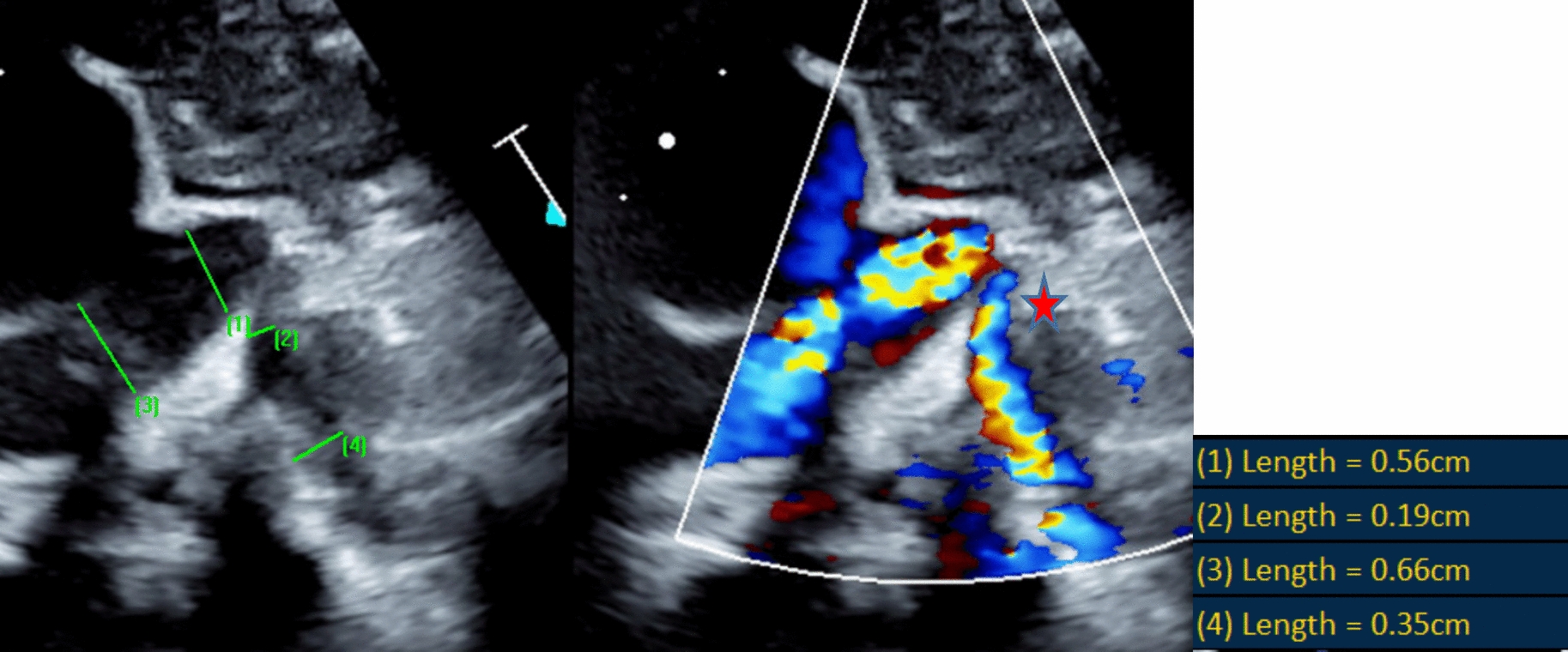
SUA occurs in approximately 0.5–5% of pregnancies screened antenatally [11]. SUA is considered a nonspecific marker, and when isolated, detection of SUA is associated with increased perinatal morbidity, such as fetal growth restriction, polyhydramnios, and oligohydramnios [12]. In addition, SUA may be associated with fetal chromosomal defects [13]. In TRAP, when there is reverse perfusion, the parasitic-twin steals poor oxygen blood from a provider twin through the umbilical artery (or arteries) and moves away through the umbilical vein, which is in the opposite direction to what usually occurs. There are no data on comparative analysis of TRAP cases with and without SUA. We hypothesized lower complication rates—such as FHF and preterm delivery—in pump twins when SUA is present. In our TRAP case, the presence of only one umbilical artery may limit the volume of blood reaching the acardiac twin, thus reducing the hemodynamic load on the pump twin's heart. Previously, the hemodynamic features of blood circulation in TRAP pregnancies have been discussed by Dashe et al., who revealed that the pump twins with poor pregnancy outcomes had a small resistive index between the pump and acardiac twin. Authors also found out in their TRAP series that poor outcomes were associated with larger acardiac twins (≥ 1100 g), smaller pump twins (≤ 2300 g), and higher twin weight ratios (≥ 48%) [14]. Additionally, one study has shown that a larger acardiac twin (often associated with normal two-artery cords) increases the hemodynamic burden on the pump twin [8]. Undesirable circulatory disorders that can develop in pump twins (cardiac failure, polyhydramnios, and hydrops, up to fetal loss) are not observed in the perinatal period in our case. Even in the 3-month-old and 6-month-old postpartum period, the child shows no cardiovascular pathologies. Normally, as we know, the umbilical cord consists of 2 umbilical arteries and 1 vein. In our case, postnatal macroscopic placental evaluation shows 2-vessel umbilical cord detection, later confirmed by the placenta's postpartum pathological examination. Notably, detecting an absent umbilical artery may indicate an additional screening for fetal anomaly due to association with concomitant fetal abnormalities. However, in the present report, an observation generates a hypothesis that the nonexistence of an impacted intrauterine fetal development and FHF of the provider twin may be explained by the presence of one umbilical artery, resulting in less blood flow to the parasitic twin, which can be accepted as a protective factor-effect for pump twin circulation in TRAP pregnancies.
MCMA pregnancies, especially if complicated by TRAP, require particular multidisciplinary approaches by perinatology and pediatric cardiology. Malone emphasized a vital need for large prospective studies for monochorionic gestation with late pediatric outcomes [8]. Prospective designed studies may potentially be helpful in the identification of incidence, natural history, long-term outcomes, and complications of monochorionicity. Our observation association between SUA and reduced hemodynamic load on the pump twin, which came across in our case report, is based on limited data and should be considered a preliminary finding.


Comments (0)